Advertisement

Main menu

  • Home
  • Content
    • Current Issue
    • Accepted Manuscripts
    • Article Preview
    • Past Issue Archive
    • Video Articles
    • AJNR Case Collection
    • Case of the Week Archive
    • Case of the Month Archive
    • Classic Case Archive
  • Special Collections
    • AJNR Awards
    • Low-Field MRI
    • Alzheimer Disease
    • ASNR Foundation Special Collection
    • Photon-Counting CT
    • View All
  • Multimedia
    • AJNR Podcasts
    • AJNR SCANtastic
    • Trainee Corner
    • MRI Safety Corner
    • Imaging Protocols
  • For Authors
    • Submit a Manuscript
    • Submit a Video Article
    • Submit an eLetter to the Editor/Response
    • Manuscript Submission Guidelines
    • Statistical Tips
    • Fast Publishing of Accepted Manuscripts
    • Graphical Abstract Preparation
    • Imaging Protocol Submission
    • Author Policies
  • About Us
    • About AJNR
    • Editorial Board
    • Editorial Board Alumni
  • More
    • Become a Reviewer/Academy of Reviewers
    • Subscribers
    • Permissions
    • Alerts
    • Feedback
    • Advertisers
    • ASNR Home

User menu

  • Alerts
  • Log in

Search

  • Advanced search
American Journal of Neuroradiology

American Journal of Neuroradiology

ASHNR American Society of Functional Neuroradiology ASHNR American Society of Pediatric Neuroradiology ASSR
  • Alerts
  • Log in

Advanced Search

  • Home
  • Content
    • Current Issue
    • Accepted Manuscripts
    • Article Preview
    • Past Issue Archive
    • Video Articles
    • AJNR Case Collection
    • Case of the Week Archive
    • Case of the Month Archive
    • Classic Case Archive
  • Special Collections
    • AJNR Awards
    • Low-Field MRI
    • Alzheimer Disease
    • ASNR Foundation Special Collection
    • Photon-Counting CT
    • View All
  • Multimedia
    • AJNR Podcasts
    • AJNR SCANtastic
    • Trainee Corner
    • MRI Safety Corner
    • Imaging Protocols
  • For Authors
    • Submit a Manuscript
    • Submit a Video Article
    • Submit an eLetter to the Editor/Response
    • Manuscript Submission Guidelines
    • Statistical Tips
    • Fast Publishing of Accepted Manuscripts
    • Graphical Abstract Preparation
    • Imaging Protocol Submission
    • Author Policies
  • About Us
    • About AJNR
    • Editorial Board
    • Editorial Board Alumni
  • More
    • Become a Reviewer/Academy of Reviewers
    • Subscribers
    • Permissions
    • Alerts
    • Feedback
    • Advertisers
    • ASNR Home
  • Follow AJNR on Twitter
  • Visit AJNR on Facebook
  • Follow AJNR on Instagram
  • Join AJNR on LinkedIn
  • RSS Feeds

AJNR Awards, New Junior Editors, and more. Read the latest AJNR updates

Research ArticleAdult Brain

Improving Multiple Sclerosis Plaque Detection Using a Semiautomated Assistive Approach

J. van Heerden, D. Rawlinson, A.M. Zhang, R. Chakravorty, M.A. Tacey, P.M. Desmond and F. Gaillard
American Journal of Neuroradiology August 2015, 36 (8) 1465-1471; DOI: https://doi.org/10.3174/ajnr.A4375
J. van Heerden
aFrom the Department of Radiology (J.v.H., P.M.D., F.G.), The Royal Melbourne Hospital and University of Melbourne, Parkville, Victoria, Australia
  • Find this author on Google Scholar
  • Find this author on PubMed
  • Search for this author on this site
D. Rawlinson
bDepartment of Electrical and Electronic Engineering (D.R., A.M.Z.), School of Engineering, University of Melbourne, Parkville, Victoria, Australia
  • Find this author on Google Scholar
  • Find this author on PubMed
  • Search for this author on this site
A.M. Zhang
bDepartment of Electrical and Electronic Engineering (D.R., A.M.Z.), School of Engineering, University of Melbourne, Parkville, Victoria, Australia
  • Find this author on Google Scholar
  • Find this author on PubMed
  • Search for this author on this site
R. Chakravorty
cIBM Research (R.C.), Melbourne, Victoria, Australia
  • Find this author on Google Scholar
  • Find this author on PubMed
  • Search for this author on this site
  • ORCID record for R. Chakravorty
M.A. Tacey
dMelbourne EpiCentre (M.A.T.), The Royal Melbourne Hospital and Department of Medicine, University of Melbourne, Parkville, Victoria, Australia.
  • Find this author on Google Scholar
  • Find this author on PubMed
  • Search for this author on this site
P.M. Desmond
aFrom the Department of Radiology (J.v.H., P.M.D., F.G.), The Royal Melbourne Hospital and University of Melbourne, Parkville, Victoria, Australia
  • Find this author on Google Scholar
  • Find this author on PubMed
  • Search for this author on this site
F. Gaillard
aFrom the Department of Radiology (J.v.H., P.M.D., F.G.), The Royal Melbourne Hospital and University of Melbourne, Parkville, Victoria, Australia
  • Find this author on Google Scholar
  • Find this author on PubMed
  • Search for this author on this site
  • Article
  • Figures & Data
  • Supplemental
  • Info & Metrics
  • Responses
  • References
  • PDF
Loading

Article Figures & Data

Figures

  • Tables
  • Fig 1.
    • Download figure
    • Open in new tab
    • Download powerpoint
    Fig 1.

    Annotated capture of the software reporting screen. A, Axial FLAIR with superimposed change map shows the new occipital white matter lesion in orange. Coregistered and resectioned FLAIR sequences comparing axial of new study (B) with axial of old study (C); and sagittal of new study (E) with sagittal old study (F)—thus confirming that the lesion is real and consistent with a new demyelinating plaque. D, Each lesion is marked with 3D coordinates.

  • Fig 2.
    • Download figure
    • Open in new tab
    • Download powerpoint
    Fig 2.

    Preprocessing for change-detection on receipt of a new study. A pair of old and new studies are required, each containing a volumetric series used for change detection. In our case, this series uses the FLAIR protocol. Due to significant deformation in soft tissues outside the cranium, it is preferable to register the studies by using only the brain tissue. To this end, a brain-surface extraction tool (BrainSuite from the University of Southern California)13 is fitted (1) and then used to mask the brain in the new study (2). Next, the equivalent series in the old study is retrieved and coregistered to the new study (3) by using the Mutual Information algorithm. The recovered transformation is stored in the PACS data base. Note that it is only necessary to mask the new study during registration and that rigid registration yielded sufficient accuracy after exclusion of the masked areas. DOF indicates degrees of freedom.
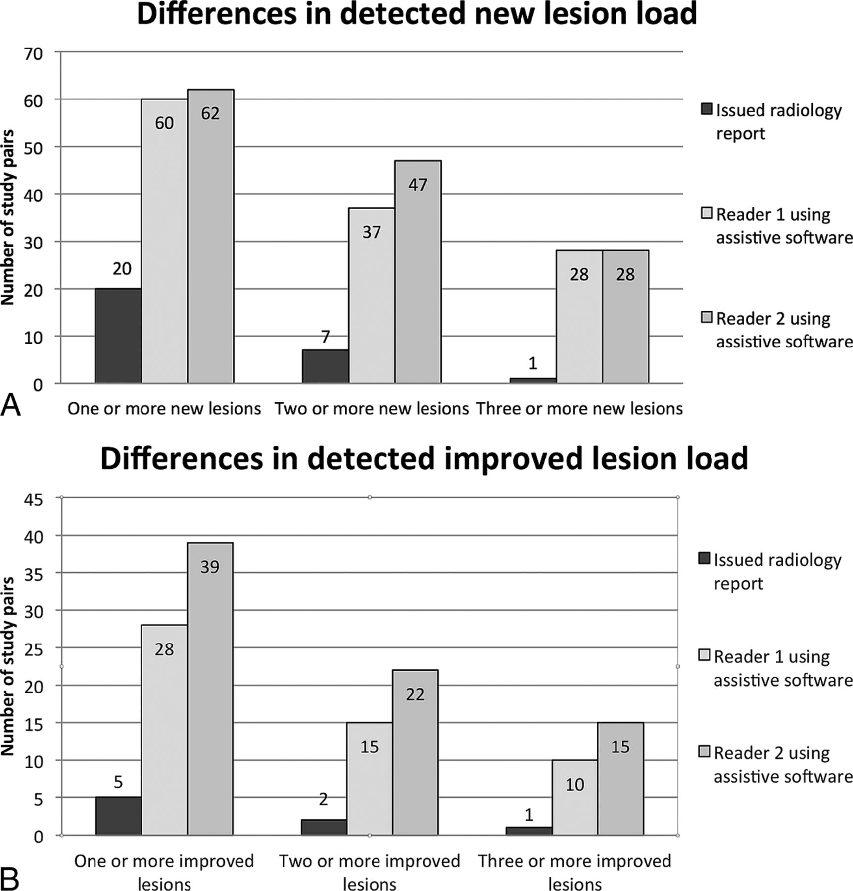
  • Fig 3.
    • Download figure
    • Open in new tab
    • Download powerpoint
    Fig 3.

    A, Comparative graphic representation of the number of study pairs with new lesions detected by both readers when using the software compared to the issued radiology report. B, Comparing the number of study pairs improved with demyelinating lesions detected by both readers when using the newly developed assistive software to the issued radiology report.

Tables

  • Figures
    • View popup
    Table 1:

    Demonstrating the number of study pairs showing a change in lesion load as identified using conventional side-by-side comparison and the software

    Change in Lesion LoadIssued Radiology ReportReader 1Reader 2
    Study pairs with new lesions (No.)2060 (P < .001)62 (P < .001)
    Study pairs with improved lesions (No.)528 (P < .001)39 (P < .001)
    • View popup
    Table 2:

    Interreader agreement demonstrated with binary groupings of new and improved lesions when using the software

    Change in Lesion Load, Binary Groupingκ95% CI
    New lesions (0, 1+)0.870.79–0.95
    New lesions (0–1, 2+)0.810.71–0.91
    New lesions (0–2, 3+)0.960.90–1.00
    Improved lesions (0, 1+)0.720.59–0.85
    Improved lesions (0–1, 2+)0.790.64–0.94
    Improved lesions (0–2, 3+)0.700.49–0.91
    • View popup
    Table 3:

    Intrareader agreement demonstrated with binary groupings of new and improved lesions using both conventional side-by-side comparison and the softwarea

    New Lesions (κ) (95% CI)Improved Lesions (κ) (95% CI)
    One or more lesions
        VTS 1st vs VTS 2nd read1.0000.937 (0.815–1.000)
        CSSC 1st vs CSSC 2nd read0.941 (0.826–1.000)0.462 (0.039–0.886)
    Two or more lesions
        VTS 1st vs VTS 2nd read1.0000.731 (0.448–1.000)
        CSSC 1st vs CSSC 2nd read0.846 (0.640–1.000)0.482 (−0.118–1.000)
    Three or more lesions
        VTS 1st vs VTS 2nd read1.0000.774 (0.472–1.000)
        CSSC 1st vs CSSC 2nd read0.724 (0.361–1.000)0.482 (−0.118–1.000)
    • ↵a Correlations demonstrated substantial intrareader agreement. The software generally outperformed conventional side-by-side comparison without, however, reaching statistical significance.

PreviousNext
Back to top

In this issue

American Journal of Neuroradiology: 36 (8)
American Journal of Neuroradiology
Vol. 36, Issue 8
1 Aug 2015
  • Table of Contents
  • Index by author
  • Complete Issue (PDF)
Advertisement
Print
Download PDF
Email Article

Thank you for your interest in spreading the word on American Journal of Neuroradiology.

NOTE: We only request your email address so that the person you are recommending the page to knows that you wanted them to see it, and that it is not junk mail. We do not capture any email address.

Enter multiple addresses on separate lines or separate them with commas.
Improving Multiple Sclerosis Plaque Detection Using a Semiautomated Assistive Approach
(Your Name) has sent you a message from American Journal of Neuroradiology
(Your Name) thought you would like to see the American Journal of Neuroradiology web site.
CAPTCHA
This question is for testing whether or not you are a human visitor and to prevent automated spam submissions.
Cite this article
J. van Heerden, D. Rawlinson, A.M. Zhang, R. Chakravorty, M.A. Tacey, P.M. Desmond, F. Gaillard
Improving Multiple Sclerosis Plaque Detection Using a Semiautomated Assistive Approach
American Journal of Neuroradiology Aug 2015, 36 (8) 1465-1471; DOI: 10.3174/ajnr.A4375

Citation Manager Formats

  • BibTeX
  • Bookends
  • EasyBib
  • EndNote (tagged)
  • EndNote 8 (xml)
  • Medlars
  • Mendeley
  • Papers
  • RefWorks Tagged
  • Ref Manager
  • RIS
  • Zotero
0 Responses
Respond to this article
Share
Bookmark this article
Improving Multiple Sclerosis Plaque Detection Using a Semiautomated Assistive Approach
J. van Heerden, D. Rawlinson, A.M. Zhang, R. Chakravorty, M.A. Tacey, P.M. Desmond, F. Gaillard
American Journal of Neuroradiology Aug 2015, 36 (8) 1465-1471; DOI: 10.3174/ajnr.A4375
del.icio.us logo Twitter logo Facebook logo Mendeley logo
  • Tweet Widget
  • Facebook Like
  • Google Plus One
Purchase

Jump to section

  • Article
    • Abstract
    • ABBREVIATIONS:
    • Materials and Methods
    • Results
    • Discussion
    • Conclusions
    • Acknowledgments
    • Footnotes
    • References
  • Figures & Data
  • Supplemental
  • Info & Metrics
  • Responses
  • References
  • PDF

Related Articles

  • PubMed
  • Google Scholar

Cited By...

  • Interest of structured reporting and combined automated co-registration and lesion color-coding maps for longitudinal magnetic resonance imaging analysis in patients with multiple sclerosis : the MS-LOBI-SR study protocol
  • PACS Integration of Semiautomated Imaging Software Improves Day-to-Day MS Disease Activity Detection
  • Improved Detection of New MS Lesions during Follow-Up Using an Automated MR Coregistration-Fusion Method
  • Fast and Robust Unsupervised Identification of MS Lesion Change Using the Statistical Detection of Changes Algorithm
  • Neuroradiologists Compared with Non-Neuroradiologists in the Detection of New Multiple Sclerosis Plaques
  • Crossref (17)
  • Google Scholar

This article has been cited by the following articles in journals that are participating in Crossref Cited-by Linking.

  • A Novel Digital Care Management Platform to Monitor Clinical and Subclinical Disease Activity in Multiple Sclerosis
    Wim Van Hecke, Lars Costers, Annabel Descamps, Annemie Ribbens, Guy Nagels, Dirk Smeets, Diana M. Sima
    Brain Sciences 2021 11 9
  • Improved Detection of New MS Lesions during Follow-Up Using an Automated MR Coregistration-Fusion Method
    A. Galletto Pregliasco, A. Collin, A. Guéguen, M.A. Metten, J. Aboab, R. Deschamps, O. Gout, L. Duron, J.C. Sadik, J. Savatovsky, A. Lecler
    American Journal of Neuroradiology 2018 39 7
  • Neuroradiologists Compared with Non-Neuroradiologists in the Detection of New Multiple Sclerosis Plaques
    W. Wang, J. van Heerden, M.A. Tacey, F. Gaillard
    American Journal of Neuroradiology 2017 38 7
  • Follow-up MRI in multiple sclerosis patients: automated co-registration and lesion color-coding improves diagnostic accuracy and reduces reading time
    David Zopfs, Kai R. Laukamp, Stefanie Paquet, Simon Lennartz, Daniel Pinto dos Santos, Christoph Kabbasch, Alexander Bunck, Marc Schlamann, Jan Borggrefe
    European Radiology 2019 29 12
  • Diagnostic accuracy of semiautomatic lesion detection plus quantitative susceptibility mapping in the identification of new and enhancing multiple sclerosis lesions
    Shun Zhang, Thanh D. Nguyen, Yize Zhao, Susan A. Gauthier, Yi Wang, Ajay Gupta
    NeuroImage: Clinical 2018 18
  • Association between urinary symptom severity and white matter plaque distribution in women with multiple sclerosis
    Ariana L. Smith, Steven J. Weissbart, Siobhán M. Hartigan, Michel Bilello, Diane K. Newman, Alan J. Wein, Anna P. Malykhina, Guray Erus, Yong Fan
    Neurourology and Urodynamics 2020 39 1
  • FLAIRfusion Processing with Contrast Inversion
    M. A. Schmidt, R. A. Linker, S. Lang, H. Lücking, T. Engelhorn, S. Kloska, M. Uder, A. Cavallaro, A. Dörfler, P. Dankerl
    Clinical Neuroradiology 2018 28 3
  • Health Economic Impact of Software-Assisted Brain MRI on Therapeutic Decision-Making and Outcomes of Relapsing-Remitting Multiple Sclerosis Patients—A Microsimulation Study
    Diana M. Sima, Giovanni Esposito, Wim Van Hecke, Annemie Ribbens, Guy Nagels, Dirk Smeets
    Brain Sciences 2021 11 12
  • Computer-Aided Detection Can Bridge the Skill Gap in Multiple Sclerosis Monitoring
    Ariel Dahan, Wayland Wang, Frank Gaillard
    Journal of the American College of Radiology 2018 15 1
  • Subtraction Maps Derived from Longitudinal Magnetic Resonance Imaging in Patients with Glioma Facilitate Early Detection of Tumor Progression
    Nico Sollmann, Magaly Gutbrod-Fernandez, Egon Burian, Isabelle Riederer, Bernhard Meyer, Andreas Hock, Jens Gempt, Claus Zimmer, Jan S. Kirschke
    Cancers 2020 12 11

More in this TOC Section

  • Diagnostic Neuroradiology of Monoclonal Antibodies
  • Clinical Outcomes After Chiari I Decompression
  • Segmentation of Brain Metastases with BLAST
Show more ADULT BRAIN

Similar Articles

Advertisement

Indexed Content

  • Current Issue
  • Accepted Manuscripts
  • Article Preview
  • Past Issues
  • Editorials
  • Editor's Choice
  • Fellows' Journal Club
  • Letters to the Editor
  • Video Articles

Cases

  • Case Collection
  • Archive - Case of the Week
  • Archive - Case of the Month
  • Archive - Classic Case

More from AJNR

  • Trainee Corner
  • Imaging Protocols
  • MRI Safety Corner
  • Book Reviews

Multimedia

  • AJNR Podcasts
  • AJNR Scantastics

Resources

  • Turnaround Time
  • Submit a Manuscript
  • Submit a Video Article
  • Submit an eLetter to the Editor/Response
  • Manuscript Submission Guidelines
  • Statistical Tips
  • Fast Publishing of Accepted Manuscripts
  • Graphical Abstract Preparation
  • Imaging Protocol Submission
  • Evidence-Based Medicine Level Guide
  • Publishing Checklists
  • Author Policies
  • Become a Reviewer/Academy of Reviewers
  • News and Updates

About Us

  • About AJNR
  • Editorial Board
  • Editorial Board Alumni
  • Alerts
  • Permissions
  • Not an AJNR Subscriber? Join Now
  • Advertise with Us
  • Librarian Resources
  • Feedback
  • Terms and Conditions
  • AJNR Editorial Board Alumni

American Society of Neuroradiology

  • Not an ASNR Member? Join Now

© 2025 by the American Society of Neuroradiology All rights, including for text and data mining, AI training, and similar technologies, are reserved.
Print ISSN: 0195-6108 Online ISSN: 1936-959X

Powered by HighWire