
Article Figures & Data
Figures
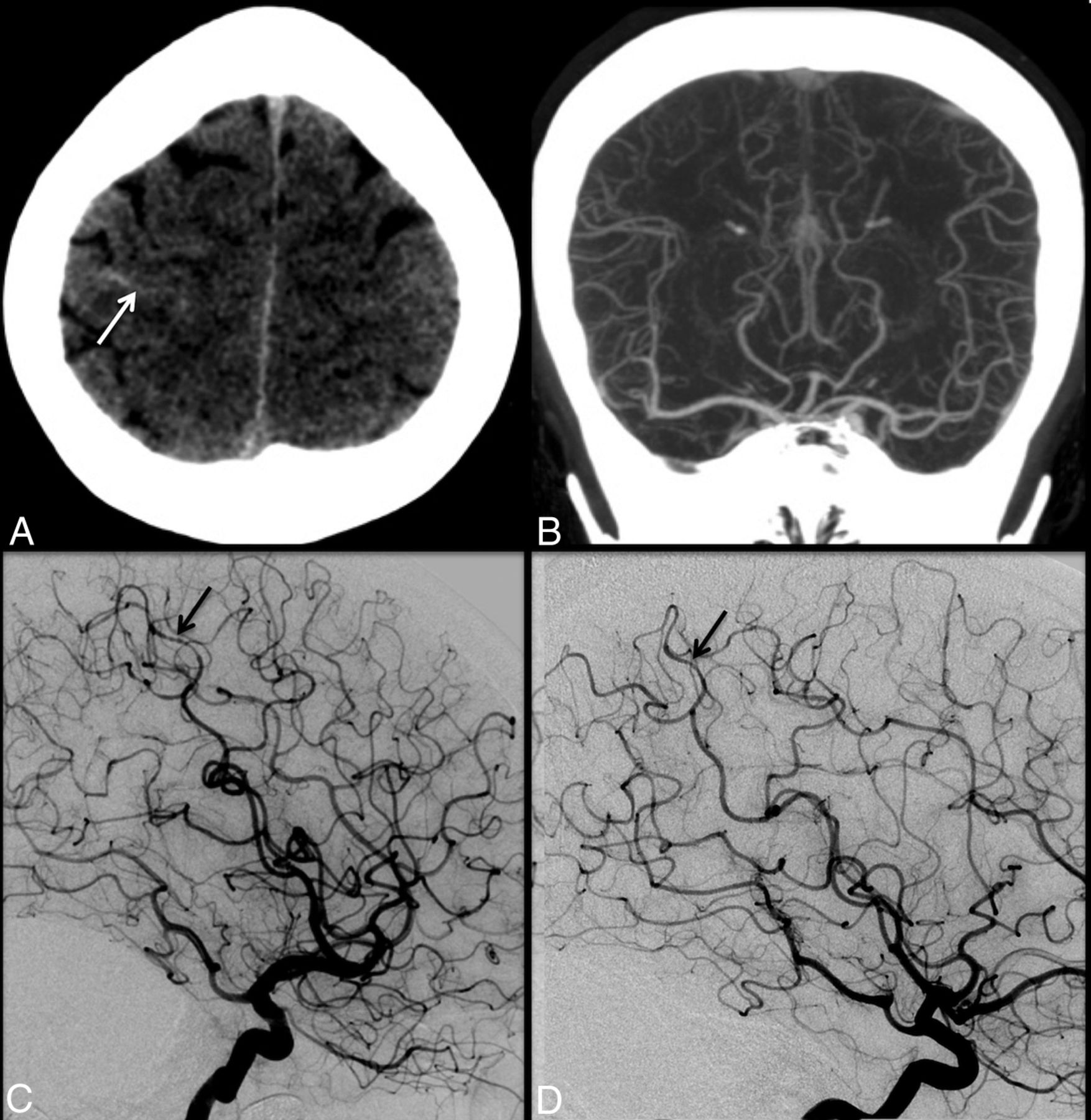
- Fig 1.
A 47-year-old woman with the sudden onset of severe headache. Initial noncontrast head CT (A) demonstrates trace sulcal subarachnoid hemorrhage (white arrow) near the vertex. CT angiography performed at the same time (B) is interpreted as having unremarkable findings. Conventional angiography (C) demonstrates mild diffuse irregularity with multifocal narrowings throughout the cerebral vasculature with a beaded appearance, most pronounced in distal right middle cerebral artery cortical branches (black arrow). Findings are most consistent with RCVS. Follow-up catheter angiogram performed 1 month later (D) demonstrates complete resolution of cerebral vasoconstriction (black arrow).
- Fig 2.
A 42-year-old woman who presented with altered mental status and lethargy. FLAIR imaging (A) demonstrates signal hyperintensity involving the cortex and underlying subcortical white matter in the parietal and occipital lobes (white arrows), consistent with PRES. There is no evidence of associated diffusion restriction. Trace sulcal subarachnoid hemorrhage was also noted overlying the right frontal lobe (not shown). Note subtle irregularity and multifocal narrowings involving distal cortical branches of the bilateral middle and anterior cerebral arteries (black arrows) on cerebral angiography (B), suggestive of RCVS. The patient made a full recovery, with complete resolution of cerebral areas of abnormal FLAIR hyperintensity (C) and cerebral vasoconstriction (not shown).
- Fig 3.
A 19-year-old man with a 2-day history of recurrent headaches and prior marijuana use. Noncontrast CT was negative for acute hemorrhage (not shown). Conventional angiography (A) reveals multifocal areas of moderate narrowing and irregularity involving the cerebral vasculature (white arrows, A). These areas resolved following intra-arterial administration of verapamil (white arrows, B). Clinical course and imaging findings are consistent with RCVS.


Tables
Prior Terms Migrainous vasospasm Benign angiopathy of the central nervous system Postpartum angiopathy Thunderclap headache with reversible vasospasm Sexual headache Drug-induced angiopathy Call-Fleming syndrome Criteria Severe, acute headaches, with or without additional neurologic signs or symptoms Uniphasic disease course with no new symptoms after 1 month of onset No evidence for aneurysmal SAH Normal or near-normal findings on CSF analysis (protein level, <80 mg/dL; leukocyte level, <10/mm3; normal glucose level) Multifocal segmental cerebral artery vasoconstriction demonstrated on either catheter angiography or indirectly on CTA/MRA Reversibility of angiographic abnormalities within 12 weeks after onset. If death occurs before the follow-up studies are completed, postmortem rules out such conditions as vasculitis, intracranial atherosclerosis, and aneurysmal SAH, which can also manifest with headache and stroke Triggers of Secondary RCVS Vasoactive medications Sympathomimetic drugs, bromocriptine, ergotamine, pseudoephedrine, selective serotonin-uptake inhibitors, interferon, triptans, diet pills, nonsteroidal anti-inflammatory drugs Vasoactive recreational drugs Alcohol, amphetamines, cannabis, cocaine, ecstasy, nicotine Pregnancy and postpartum states Blood products Blood transfusions, erythropoietin, intravenous immunoglobulin Headache disorders Migraines Tumors Pheochromocytoma Paraganglioma Trauma Carotid dissection, unruptured cerebral aneurysm Head and neck surgery Various medical conditions Hemolysis, elevated liver enzymes, low platelets Antiphospholipid antibody syndrome Thrombotic thrombocytopenic purpura


Jump to section
Related Articles
Cited By...
- Reversible Cerebral Vasoconstriction Syndrome Secondary to Escitalopram
- Reversible Cerebral Vasoconstriction Syndrome: Symptoms, Incidence, and Resource Utilization in a Population-Based US Cohort
- Quantifying Intra-Arterial Verapamil Response as a Diagnostic Tool for Reversible Cerebral Vasoconstriction Syndrome
- Reversible cerebral vasoconstriction syndrome during caesarean section
- OnabotulinumtoxinA injections: treatment of reversible cerebral vasoconstriction syndrome chronic daily headaches
- Regarding "Cerebral Angiography for Evaluation of Patients with CT Angiogram-Negative Subarachnoid Hemorrhage: An 11-Year Experience"