Advertisement

Main menu

  • Home
  • Content
    • Current Issue
    • Accepted Manuscripts
    • Article Preview
    • Past Issue Archive
    • Video Articles
    • AJNR Case Collection
    • Case of the Week Archive
    • Case of the Month Archive
    • Classic Case Archive
  • Special Collections
    • AJNR Awards
    • Low-Field MRI
    • Alzheimer Disease
    • ASNR Foundation Special Collection
    • Photon-Counting CT
    • View All
  • Multimedia
    • AJNR Podcasts
    • AJNR SCANtastic
    • Trainee Corner
    • MRI Safety Corner
    • Imaging Protocols
  • For Authors
    • Submit a Manuscript
    • Submit a Video Article
    • Submit an eLetter to the Editor/Response
    • Manuscript Submission Guidelines
    • Statistical Tips
    • Fast Publishing of Accepted Manuscripts
    • Graphical Abstract Preparation
    • Imaging Protocol Submission
    • Author Policies
  • About Us
    • About AJNR
    • Editorial Board
    • Editorial Board Alumni
  • More
    • Become a Reviewer/Academy of Reviewers
    • Subscribers
    • Permissions
    • Alerts
    • Feedback
    • Advertisers
    • ASNR Home

User menu

  • Alerts
  • Log in

Search

  • Advanced search
American Journal of Neuroradiology

American Journal of Neuroradiology

ASHNR American Society of Functional Neuroradiology ASHNR American Society of Pediatric Neuroradiology ASSR
  • Alerts
  • Log in

Advanced Search

  • Home
  • Content
    • Current Issue
    • Accepted Manuscripts
    • Article Preview
    • Past Issue Archive
    • Video Articles
    • AJNR Case Collection
    • Case of the Week Archive
    • Case of the Month Archive
    • Classic Case Archive
  • Special Collections
    • AJNR Awards
    • Low-Field MRI
    • Alzheimer Disease
    • ASNR Foundation Special Collection
    • Photon-Counting CT
    • View All
  • Multimedia
    • AJNR Podcasts
    • AJNR SCANtastic
    • Trainee Corner
    • MRI Safety Corner
    • Imaging Protocols
  • For Authors
    • Submit a Manuscript
    • Submit a Video Article
    • Submit an eLetter to the Editor/Response
    • Manuscript Submission Guidelines
    • Statistical Tips
    • Fast Publishing of Accepted Manuscripts
    • Graphical Abstract Preparation
    • Imaging Protocol Submission
    • Author Policies
  • About Us
    • About AJNR
    • Editorial Board
    • Editorial Board Alumni
  • More
    • Become a Reviewer/Academy of Reviewers
    • Subscribers
    • Permissions
    • Alerts
    • Feedback
    • Advertisers
    • ASNR Home
  • Follow AJNR on Twitter
  • Visit AJNR on Facebook
  • Follow AJNR on Instagram
  • Join AJNR on LinkedIn
  • RSS Feeds

AJNR Awards, New Junior Editors, and more. Read the latest AJNR updates

Research ArticleBrain

Enhanced Axonal Metabolism during Early Natalizumab Treatment in Relapsing-Remitting Multiple Sclerosis

O.T. Wiebenga, A.M. Klauser, M.M. Schoonheim, G.J.A. Nagtegaal, M.D. Steenwijk, J.A. van Rossum, C.H. Polman, F. Barkhof, P.J.W. Pouwels and J.J.G. Geurts
American Journal of Neuroradiology June 2015, 36 (6) 1116-1123; DOI: https://doi.org/10.3174/ajnr.A4252
O.T. Wiebenga
aFrom the Departments of Radiology and Nuclear Medicine (O.T.W., G.J.A.N.†, M.D.S., F.B.)
bAnatomy and Neurosciences (O.T.W., A.M.K., M.M.S., G.J.A.N.†, J.J.G.G.)
  • Find this author on Google Scholar
  • Find this author on PubMed
  • Search for this author on this site
A.M. Klauser
bAnatomy and Neurosciences (O.T.W., A.M.K., M.M.S., G.J.A.N.†, J.J.G.G.)
  • Find this author on Google Scholar
  • Find this author on PubMed
  • Search for this author on this site
M.M. Schoonheim
bAnatomy and Neurosciences (O.T.W., A.M.K., M.M.S., G.J.A.N.†, J.J.G.G.)
  • Find this author on Google Scholar
  • Find this author on PubMed
  • Search for this author on this site
G.J.A. Nagtegaal
aFrom the Departments of Radiology and Nuclear Medicine (O.T.W., G.J.A.N.†, M.D.S., F.B.)
bAnatomy and Neurosciences (O.T.W., A.M.K., M.M.S., G.J.A.N.†, J.J.G.G.)
  • Find this author on Google Scholar
  • Find this author on PubMed
  • Search for this author on this site
M.D. Steenwijk
aFrom the Departments of Radiology and Nuclear Medicine (O.T.W., G.J.A.N.†, M.D.S., F.B.)
  • Find this author on Google Scholar
  • Find this author on PubMed
  • Search for this author on this site
J.A. van Rossum
cNeurology (J.A.v.R., C.H.P.)
  • Find this author on Google Scholar
  • Find this author on PubMed
  • Search for this author on this site
C.H. Polman
cNeurology (J.A.v.R., C.H.P.)
  • Find this author on Google Scholar
  • Find this author on PubMed
  • Search for this author on this site
F. Barkhof
aFrom the Departments of Radiology and Nuclear Medicine (O.T.W., G.J.A.N.†, M.D.S., F.B.)
  • Find this author on Google Scholar
  • Find this author on PubMed
  • Search for this author on this site
P.J.W. Pouwels
dPhysics and Medical Technology (P.J.W.P.), Neuroscience Campus Amsterdam and VU University Medical Center, Amsterdam, the Netherlands.
  • Find this author on Google Scholar
  • Find this author on PubMed
  • Search for this author on this site
J.J.G. Geurts
bAnatomy and Neurosciences (O.T.W., A.M.K., M.M.S., G.J.A.N.†, J.J.G.G.)
  • Find this author on Google Scholar
  • Find this author on PubMed
  • Search for this author on this site
  • Article
  • Figures & Data
  • Supplemental
  • Info & Metrics
  • Responses
  • References
  • PDF
Loading

Article Figures & Data

Figures

  • Tables
  • Fig 1.
    • Download figure
    • Open in new tab
    • Download powerpoint
    Fig 1.

    A, The illustration shows axial T2-weighted and sagittal T1-weighted MR images. The pituitary-fastigium axis (dashed line) is used to position the grid (in red) with 64 voxels. B, An example of the WM, GM, and lesion PVE calculated on the MRSI grid and superimposed with the T2 image of the center of the slab. The yellow in the lesion PVE image represents the projection of the lesion content of the slab. C, The allocation of the different regions of GM, NAWM, and LWM based on the rows of the grid and the lesion content is seen.
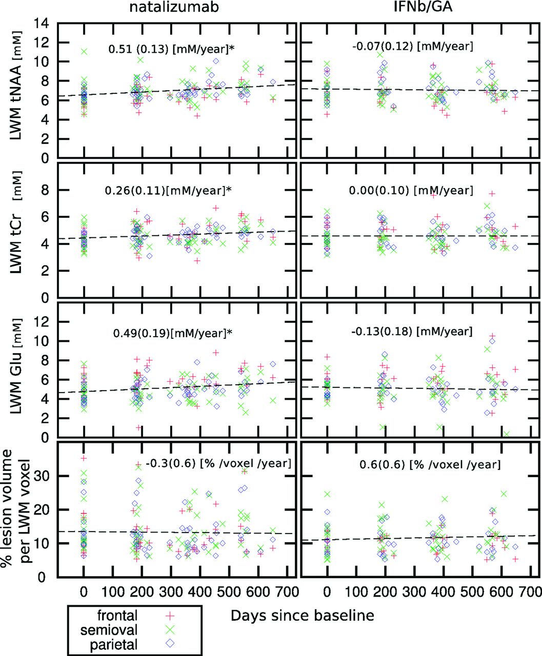
  • Fig 2.
    • Download figure
    • Open in new tab
    • Download powerpoint
    Fig 2.

    Scatterplot of individual measurements of tNAA, tCr, and Glu in LWM (mmol/L) (upper 3 rows) and percentage of lesion volume per LWM voxel (lower row) as a function of the days elapsed since the baseline for the patients treated with natalizumab and IFNb/GA. The asterisk indicates that patients treated with natalizumab show a significant increase of tNAA, tCr, and Glu in the LWM (mmol/L/year ± SD). At each time point (baseline, month 6, month 12, and month 18), the number of subjects in the natalizumab group was, respectively, 18, 25, 23, and 15; in the IFNb/GA group, it was 15, 17, 18, and 16; and in the healthy control group, it was 11, 12, 11, and 8.

Tables

  • Figures
    • View popup
    Table 1:

    Baseline variables of patients and controlsa

    Natalizumab (n = 25)IFNb/GA (n = 18)Healthy Controls (n = 12)P Value
    Age (yr)36.0 ± 8.938.2 ± 5.037.6 ± 8.7.619
    Sex (male/female)9:169:93:9.368
    EDSSb,c3.0 (1.5–6.5)2.5 (1.0–6.5).589
    Duration since onset (yr)7.9 ± 6.18.8 ± 5.3.550
    Prior IFNb/GA duration at baseline (yr)2.6 ± 3.04.4 ± 3.9.121
    Brain volumes
        NGMV (L)0.75 ± 0.040.73 ± 0.050.77 ± 0.04.063
        NWMV (L)0.70 ± 0.030.69 ± 0.030.72 ± 0.04.061
        NBV (L)1.45 ± 0.051.42 ± 0.081.49 ± 0.07.020d
    T2 lesion volume (mL)e6.1 (2.4–14.3)4.9 (2.4–11.9).599
    • Note:—EDSS indicates Expanded Disability Status Scale; NGMV, normalized total GM volume; NWMV, normalized total WM volume; NBV, normalized whole-brain volume.

    • ↵a Data are mean ± SD. When normally distributed, a multivariate general linear model was used with age and sex included as covariates. Nonparametric testing was performed using Kruskal-Wallis and post hoc Mann-Whitney U tests.

    • ↵b χ2 test.

    • ↵c Median and range.

    • ↵d Only significant between patients treated with IFNb/GA and healthy controls.

    • ↵e Median and interquartile range.

    • View popup
    Table 2:

    Baseline absolute metabolite concentrations in NAWM, LWM, and GMa

    Natalizumab (n = 25)IFNb/GA (n = 18)Controls (n = 12)
    NAWMLWMGMNAWMLWMGMWMGM
    tCr
        Frontal4.86 ± 0.714.35c ± 0.588.70 ± 1.894.93 ± 0.924.95 ± 0.908.70 ± 1.414.93 ± 0.648.48 ± 1.31
        Semiovale4.67 ± 0.634.30c ± 0.814.83 ± 0.754.41b ± 0.774.72 ± 0.60
        Parietal4.52 ± 0.624.42 ± 0.527.78 ± 0.884.72 ± 0.694.57 ± 0.938.24 ± 1.024.58 ± 0.457.48 ± 0.99
    tNAA
        Frontal7.17 ± 1.205.89c ± 0.8111.1 ± 1.747.06 ± 1.166.75 ± 1.5410.93 ± 1.617.62 ± 0.9611.66 ± 1.37
        Semiovale7.96 ± 0.876.70c ± 1.518.25 ± 1.567.05c ± 1.178.51 ± 0.94
        Parietal7.43d ± 1.056.55c ± 0.7410.51 ± 1.087.59 ± 0.987.08 ± 1.2411.52 ± 1.468.22 ± 0.8110.89 ± 1.30
    Cho
        Frontal1.50 ± 0.201.51 ± 0.252.16 ± 0.491.59 ± 0.281.64 ± 0.292.11 ± 0.451.62 ± 0.252.08 ± 0.39
        Semiovale1.44 ± 0.191.42 ± 0.281.53 ± 0.211.49 ± 0.281.49 ± 0.16
        Parietal1.13 ± 0.241.36 ± 0.191.31 ± 0.221.34 ± 0.231.39 ± 0.271.36 ± 0.241.37 ± 0.171.13 ± 0.20
    mIns
        Frontal4.82e ± 1.185.29 ± 1.017.49 ± 1.574.44 ± 0.925.45c ± 1.517.30 ± 1.113.90 ± 0.657.00 ± 1.16
        Semiovale4.47e ± 1.184.76 ± 0.914.27 ± 0.694.71 ± 1.013.56 ± 0.44
        Parietal4.79e ± 0.645.59b ± 1.076.47 ± 0.914.98 ± 0.935.32 ± 1.426.47 ± 0.834.01 ± 0.625.85 ± 0.52
    Glu
        Frontal6.34 ± 1.465.36b ± 1.1312.96 ± 2.985.86 ± 1.186.03 ± 1.4812.71 ± 2.576.25 ± 0.9614.01 ± 2.40
        Semiovale5.38 ± 1.084.73b ± 1.165.50 ± 0.915.11 ± 0.965.49 ± 0.72
        Parietal5.17 ± 1.344.22b ± 0.9412.99 ± 3.875.37 ± 1.574.80 ± 0.6812.68 ± 1.445.31 ± 0.6711.81 ± 1.85
    • ↵a In mmol/L tissue, mean ± SD. Absolute metabolite concentrations (mmol/L) of NAWM, GM, WM, and LWM in the frontal, centrum semiovale, and parietal regions averaged over the subjects at the baseline measurement.

    • ↵b Significant difference within the group between NAWM and LWM in the same region of the baseline measurement (P < .05).

    • ↵c Significant difference within the group between NAWM and LWM in the same region of the baseline (P < .01).

    • ↵d Significant differences between patients treated with natalizumab and healthy controls including all the time points (P < .05).

    • ↵e Significant differences between patients treated with natalizumab and healthy controls including all the time points (P < .01).

PreviousNext
Back to top

In this issue

American Journal of Neuroradiology: 36 (6)
American Journal of Neuroradiology
Vol. 36, Issue 6
1 Jun 2015
  • Table of Contents
  • Index by author
  • Complete Issue (PDF)
Advertisement
Print
Download PDF
Email Article

Thank you for your interest in spreading the word on American Journal of Neuroradiology.

NOTE: We only request your email address so that the person you are recommending the page to knows that you wanted them to see it, and that it is not junk mail. We do not capture any email address.

Enter multiple addresses on separate lines or separate them with commas.
Enhanced Axonal Metabolism during Early Natalizumab Treatment in Relapsing-Remitting Multiple Sclerosis
(Your Name) has sent you a message from American Journal of Neuroradiology
(Your Name) thought you would like to see the American Journal of Neuroradiology web site.
CAPTCHA
This question is for testing whether or not you are a human visitor and to prevent automated spam submissions.
Cite this article
O.T. Wiebenga, A.M. Klauser, M.M. Schoonheim, G.J.A. Nagtegaal, M.D. Steenwijk, J.A. van Rossum, C.H. Polman, F. Barkhof, P.J.W. Pouwels, J.J.G. Geurts
Enhanced Axonal Metabolism during Early Natalizumab Treatment in Relapsing-Remitting Multiple Sclerosis
American Journal of Neuroradiology Jun 2015, 36 (6) 1116-1123; DOI: 10.3174/ajnr.A4252

Citation Manager Formats

  • BibTeX
  • Bookends
  • EasyBib
  • EndNote (tagged)
  • EndNote 8 (xml)
  • Medlars
  • Mendeley
  • Papers
  • RefWorks Tagged
  • Ref Manager
  • RIS
  • Zotero
0 Responses
Respond to this article
Share
Bookmark this article
Enhanced Axonal Metabolism during Early Natalizumab Treatment in Relapsing-Remitting Multiple Sclerosis
O.T. Wiebenga, A.M. Klauser, M.M. Schoonheim, G.J.A. Nagtegaal, M.D. Steenwijk, J.A. van Rossum, C.H. Polman, F. Barkhof, P.J.W. Pouwels, J.J.G. Geurts
American Journal of Neuroradiology Jun 2015, 36 (6) 1116-1123; DOI: 10.3174/ajnr.A4252
del.icio.us logo Twitter logo Facebook logo Mendeley logo
  • Tweet Widget
  • Facebook Like
  • Google Plus One
Purchase

Jump to section

  • Article
    • Abstract
    • ABBREVIATIONS:
    • Materials and Methods
    • Results
    • Discussion
    • Conclusions
    • Footnotes
    • References
  • Figures & Data
  • Supplemental
  • Info & Metrics
  • Responses
  • References
  • PDF

Related Articles

  • PubMed
  • Google Scholar

Cited By...

  • Natalizumab for Multiple Sclerosis: A Case in Point for the Impact of Translational Neuroimmunology
  • Crossref (17)
  • Google Scholar

This article has been cited by the following articles in journals that are participating in Crossref Cited-by Linking.

  • Quantifying the Metabolic Signature of Multiple Sclerosis by in vivo Proton Magnetic Resonance Spectroscopy: Current Challenges and Future Outlook in the Translation From Proton Signal to Diagnostic Biomarker
    Kelley M. Swanberg, Karl Landheer, David Pitt, Christoph Juchem
    Frontiers in Neurology 2019 10
  • Exploring potential mechanisms of action of natalizumab in secondary progressive multiple sclerosis
    Finn Sellebjerg, Diego Cadavid, Deborah Steiner, Luisa Maria Villar, Richard Reynolds, Daniel Mikol
    Therapeutic Advances in Neurological Disorders 2016 9 1
  • Molecular Effects of FDA-Approved Multiple Sclerosis Drugs on Glial Cells and Neurons of the Central Nervous System
    Kim M. A. De Kleijn, Gerard J. M. Martens
    International Journal of Molecular Sciences 2020 21 12
  • Application of advanced MRI techniques to monitor pharmacologic and rehabilitative treatment in multiple sclerosis: current status and future perspectives
    Maria A. Rocca, Paolo Preziosa, Massimo Filippi
    Expert Review of Neurotherapeutics 2019 19 9
  • The Role of Advanced Magnetic Resonance Imaging Techniques in Multiple Sclerosis Clinical Trials
    Kedar R. Mahajan, Daniel Ontaneda
    Neurotherapeutics 2017 14 4
  • Occurrence and microstructural features of slowly expanding lesions on fingolimod or natalizumab treatment in multiple sclerosis
    Paolo Preziosa, Elisabetta Pagani, Lucia Moiola, Mariaemma Rodegher, Massimo Filippi, Maria A Rocca
    Multiple Sclerosis Journal 2021 27 10
  • Metabolites predict lesion formation and severity in relapsing-remitting multiple sclerosis
    Antoine M Klauser, Oliver T Wiebenga, Anand JC Eijlers, Menno M Schoonheim, Bernard MJ Uitdehaag, Frederik Barkhof, Petra JW Pouwels, Jeroen JG Geurts
    Multiple Sclerosis Journal 2018 24 4
  • Natalizumab for Multiple Sclerosis: A Case in Point for the Impact of Translational Neuroimmunology
    Afsaneh Shirani, Olaf Stüve
    The Journal of Immunology 2017 198 4
  • Grey matter atrophy is associated with disability increase in natalizumab-treated patients
    Ethel Ciampi, Deborah Pareto, Jaume Sastre-Garriga, Angela Vidal-Jordana, Carmen Tur, Jordi Río, Mar Tintoré, Cristina Auger, Alex Rovira, Xavier Montalban
    Multiple Sclerosis Journal 2017 23 4
  • Proton MR spectroscopy of lesion evolution in multiple sclerosis: Steady‐state metabolism and its relationship to conventional imaging
    Ivan I. Kirov, Shu Liu, Assaf Tal, William E. Wu, Matthew S. Davitz, James S. Babb, Henry Rusinek, Joseph Herbert, Oded Gonen
    Human Brain Mapping 2017 38 8

More in this TOC Section

  • Multimodal CT Provides Improved Performance for Lacunar Infarct Detection
  • Optimal MRI Sequence for Identifying Occlusion Location in Acute Stroke: Which Value of Time-Resolved Contrast-Enhanced MRA?
  • Evaluating the Effects of White Matter Multiple Sclerosis Lesions on the Volume Estimation of 6 Brain Tissue Segmentation Methods
Show more BRAIN

Similar Articles

Advertisement

Indexed Content

  • Current Issue
  • Accepted Manuscripts
  • Article Preview
  • Past Issues
  • Editorials
  • Editor's Choice
  • Fellows' Journal Club
  • Letters to the Editor
  • Video Articles

Cases

  • Case Collection
  • Archive - Case of the Week
  • Archive - Case of the Month
  • Archive - Classic Case

More from AJNR

  • Trainee Corner
  • Imaging Protocols
  • MRI Safety Corner
  • Book Reviews

Multimedia

  • AJNR Podcasts
  • AJNR Scantastics

Resources

  • Turnaround Time
  • Submit a Manuscript
  • Submit a Video Article
  • Submit an eLetter to the Editor/Response
  • Manuscript Submission Guidelines
  • Statistical Tips
  • Fast Publishing of Accepted Manuscripts
  • Graphical Abstract Preparation
  • Imaging Protocol Submission
  • Evidence-Based Medicine Level Guide
  • Publishing Checklists
  • Author Policies
  • Become a Reviewer/Academy of Reviewers
  • News and Updates

About Us

  • About AJNR
  • Editorial Board
  • Editorial Board Alumni
  • Alerts
  • Permissions
  • Not an AJNR Subscriber? Join Now
  • Advertise with Us
  • Librarian Resources
  • Feedback
  • Terms and Conditions
  • AJNR Editorial Board Alumni

American Society of Neuroradiology

  • Not an ASNR Member? Join Now

© 2025 by the American Society of Neuroradiology All rights, including for text and data mining, AI training, and similar technologies, are reserved.
Print ISSN: 0195-6108 Online ISSN: 1936-959X

Powered by HighWire