
Article Figures & Data
Figures
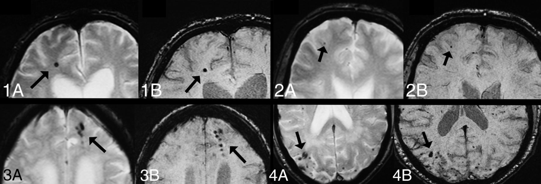
- Fig 1.
CMBs on tSWI but not T2*. For all images: A, T2*. B, tSWI. 1, Only Rater 1 identified the CMBs on tSWI. 2, Raters 1 and 2 identified the CMBs on tSWI.
- Fig 2.
CMBs on both T2* and tSWI. For all images: A, T2*. B, tSWI. 1, All raters identified the single CMB on both T2* and tSWI. 2, The pale CMB on T2* was only identified by Rater 2. On tSWI, the CMB was clearly delineated and was identified by all raters. 3 and 4, Disagreement on the exact number of CMBs occurred on T2*. On tSWI, the CMBs are more clearly outlined.


Tables
All Raters 1 2 3 Prevalence of CMBs on T2* (No.) (%) 43 (17)b 42 (17)b 45 (18) 38 (15)b,c Prevalence of CMBs on TSWI (No.) (%) 50 (20) 49 (20) 46 (19) 51 (21)c Prevalence of CMBs on tSWI (No.) (%) 51 (21)b 51 (21)b 50 (20) 51 (21)b T2* sum CMBs (No.) 365b,c 373b,c 461b 260b,c TSWI sum CMBs (No.) 528c 570c 450d 576c tSWI sum CMBs (No.) 699b 619b 806b,d 672b Times more CMBs on TSWI than T2* 1.5 1.5 1.0 2.2 Times more CMBs on tSWI than T2* 1.9 1.7 1.7 2.6 Times more CMBs on tSWI than TSWI 1.3 1.1 1.8 1.2 ↵a McNemar and Wilcoxon signed rank tests were used to determine the difference in prevalence and sum of CMBs, respectively; analysis was done separately between T2* and TSWI, T2* and tSWI, and TSWI and tSWI. “All Raters” represents the median value of all separate ratings for all patients (ie, not the final values). The whole cohort is included in the analysis (N = 246). For “Prevalence,” values are given as the number of patients (percentage). All significant values are designated in the Table. All P values have been Bonferroni-corrected.
↵b P < .05 between T2* and tSWI.
↵c P < .05 between T2* and TSWI.
↵d P < .05 between TSWI and tSWI.
Diagnosis AD (n = 62) MCI (n = 80) Other (n = 33) SCI (n = 71) Prevalence of CMBs on T2* (No.) (%) 20 (32) 13 (16) 6 (16) 4 (6) Prevalence of CMBs on TSWI (No.) (%) 22 (32) 16 (20) 7 (22) 5 (7) Prevalence of CMBs on tSWI (No.) (%) 22 (35) 16 (20) 7 (22) 6 (8) T2* sum CMBs (No.) 258b,c 69c 47 6 TSWI sum CMBs (No.) 344b 101 73 10 tSWI sum CMBs (No.) 459c 116c 79 12 Times more CMBs on TSWI than T2* 1.3 1.5 1.6 1.7 Times more CMBs on tSWI than T2* 1.8 1.7 1.7 2.0 Times more CMBs on tSWI than TSWI 1.3 1.1 1.1 1.2 Note:—AD indicates Alzheimer disease; Other, other dementias; MCI, mild cognitive impairment; SCI, subjective cognitive impairment.
↵a Differences in prevalence and sum of CMBs were determined with the McNemar and Wilcoxon signed rank test, respectively; analysis was made between T2* and TSWI, T2* and tSWI, and TSWI and tSWI. Ratings represent the median value of all separate ratings for all patients. All significant values are designated in the Table. All P values have been Bonferroni-corrected.
↵b P < .05 between T2* and TSWI.
↵c P < .05 between T2* and tSWI.
Sequenceb Patients (No.) All Ratersc Raters 1 and 2 Raters 1 and 3 Raters 2 and 3 Analysis 1 T2* 55 0.942 (0.915–0.961) 0.990 (0.981–0.994) 0.858 (0.758–0.917) 0.818 (0.689–0.894) TSWI 55 0.982 (0.974–0.988) 0.963 (0.942–0.977) 0.994 (0.990–0.996) 0.959 (0.936–0.974) tSWI 55 0.991 (0.986–0.994) 0.979 (0.964–0.988) 0.997 (0.995–0.998) 0.984 (0.972–0.991) Analysis 2 T2* 25 0.959 (0.795–0.944) 0.992 (0.982–0.996) 0.911 (0.798–0.961) 0.882 (0.733–0.948) TSWI 37 0.984 (0.972–0.991) 0.968 (0.938–0.984) 0.994 (0.988–0.997) 0.963 (0.929–0.981) tSWI 33 0.991 (0.985–0.995) 0.983 (0.966–0.992) 0.997 (0.994–0.999) 0.983 (0.966–0.992) ↵a Interrater agreement was determined with the intraclass correlation coefficient, and the 95% confidence intervals are given in parenthesis.
↵b Analysis was made twice: 1) for all patients (n = 55) with CMBs, on the basis of the initial KIDS CMB analysis; 2) for all patients with >1 CMB according to Rater 1.
↵c “All Raters” represents analysis among Raters 1, 2, and 3.
Clinical Parameters T2* CMB+ (n = 43) T2* CMB− (n = 203) TSWI CMB+ (n = 50) TSWI CMB− (n = 196) tSWI CMB+ (n = 51) tSWI CMB− (n = 195) Women (No.) (%) 19 (44) 113 (56) 22 (44) 110 (56) 22 (43) 109 (56) Age (yr) (mean) (SD) 68 (11)b 63 (10)b 68 (11)b 63 (10)b 68 (11) 63 (10) Hypertension (No.) (%) 20 (47) 74 (37) 23 (46) 71 (36) 24 (47) 70 (36) Hyperlipidemia (No.) (%) 6 (14) 31 (16) 8 (16) 29 (15) 9 (18) 28 (15) Diabetes (No.) (%) 7 (16) 21 (10) 8 (16) 20 (10) 7 (14) 21 (11) Current smoking (No.) (%) 4 (12) 35 (23) 6 (15) 33 (22) 6 (15) 33 (23) Alcohol (No.) (%) 30 (91) 131 (82) 35 (90) 126 (82) 36 (92) 125 (82) Heredity (No.) (%) 21 (72) 82 (56) 23 (67) 80 (57) 23 (68) 80 (57) Anticoagulant medication (No.) (%) 11 (26) 36 (18) 14 (28) 33 (17) 13 (26) 34 (18) MMSE (mean) (SD) 25 (4) 25 (5) 25 (4) 25 (5) 25 (4) 25 (5) BMI (mean) (SD) 27 (5) 25 (3) 25 (4) 27 (5) 25 (4) 27 (5) WMH ≥1 (No.) (%) 38 (88)b 138 (68)b 45 (90)b 131 (74)b 45 (88)b 130 (67)b WMH ≥2 (No.) (%) 19 (44)c 20 (10)c 21 (42)c 18 (9)c 21 (41)c 18 (9)c WMH = 3 (No.) (%) 12 (28)c 8 (4)c 14 (28)c 6 (3)c 14 (28)c 6 (3)c Note:—MMSE indicates Mini-Mental State Examination; BMI, body mass index; CMB+, patients with CMBs; CMB−, patients without CMBs.
↵a χ2 and Mann-Whitney U tests were done to determine in-sequence differences; significance testing was only done within sequences, and significant values are designated in the Table. All analyses were made on the basis of the median rating value of all raters on all patients. All P values have been Bonferroni-corrected.
↵b P < .05.
↵c P < .001.


Jump to section
Related Articles
Cited By...
- Automated Quantification of Cerebral Microbleeds in SWI: Association with Vascular Risk Factors, White Matter Hyperintensity Burden, and Cognitive Function
- Imaging protocol suggested by large language model depends on language: preliminary experiments using GPT-4
- Using Transfer Learning for Automated Microbleed Segmentation
- Cerebral Microbleeds, Cerebral Amyloid Angiopathy, and Their Relationships to Quantitative Markers of Neurodegeneration
- Automated Detection of Cerebral Microbleeds on MR images using Knowledge Distillation Framework
- Cerebral microbleeds: from depiction to interpretation
- Incidence, Locations, and Longitudinal Course of Cerebral Microbleeds in European Moyamoya
- Clinicoradiologic Correlations of Cerebral Microbleeds in Advanced Age
- Recurrent stroke risk and cerebral microbleed burden in ischemic stroke and TIA: A meta-analysis
- Prevalence of Brain Microbleeds in Alzheimer Disease: A Systematic Review and Meta-Analysis on the Influence of Neuroimaging Techniques