
Article Figures & Data
Figures
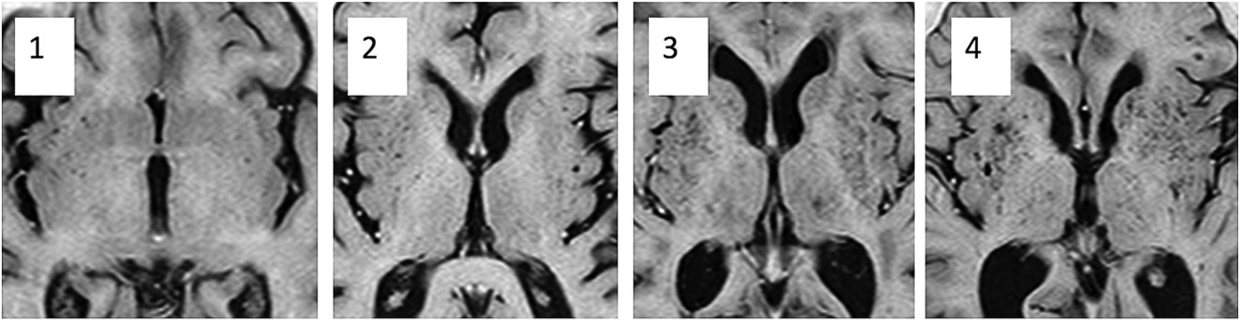
- Fig 1.
Images show dilated perivascular spaces in the basal ganglia corresponding to PVS-2BG scores of 1–4.
- Fig 2.
A, Boxplot shows the Scheltens white matter scores for the 3 subject groups. B, Boxplot shows the PVS-2BG scores for the basal ganglia in the 3 subject groups.


Tables
Subscale Range of Scores Definition of Scores 1) Periventricular Frontal 0/1/2 0 = absent Occipital 0/1/2 1 ≤ 5 mm Bands on lateral ventricle 0/1/2 2 ≥ 5 and <10 mm Subtotal score ≤6 2) Deep white matter Frontal 0/1/2/3/4/5/6 Parietal 0/1/2/3/4/5/6 Occipital 0/1/2/3/4/5/6 Temporal 0/1/2/3/4/5/6 0 = none Subtotal score 0–24 1 ≤3 mm, n ≤ 5 3) Basal ganglia 2 ≤ 3 mm, n > 5 Caudate 0/1/2/3/4/5/6 3 = 4–10 mm, n ≤ 5 Lentiform nucleus 0/1/2/3/4/5/6 4 = 4–10 mm, n > 6 Thalamus 0/1/2/3/4/5/6 5 ≥ 11 mm, n > 1 Subtotal score 0–18 6 = confluent 4) Subtentorial Cerebellum 0/1/2/3/4/5/6 Mesencephalon 0/1/2/3/4/5/6 Medulla 0/1/2/3/4/5/6 Pons 0/1/2/3/4/5/6 Subtotal score 0–24 Total score 0–72 Group Healthy AD VaD No. 65 47 39 Age (yr) 78 (5.6) 74.1 (8.5) 76.9 (7.7) Modified Scheltens score 12.50 (2.5) 13.16 (2.34) 22.76 (2.84) PVS-1 5.87 (0.21) 6.03 (0.14) 6.34 (0.16) PVS-2BG 2.1 (0.5) 2.22 (0.09) 2.471 (0.87) PVS-2CSOV 2.51 (0.13) 2.61 (0.10) 2.53 (0.15) ↵a Data are mean values (SD).
Modified Scheltens Score PVS-1 PVS-2 PVS-2BG Interobserver Agreement Rater 1 vs 2 0.82 0.79 0.92 0.91 Rater 1 vs 3 0.91 0.84 0.89 0.86 Rater 2 vs 3 0.88 0.82 0.83 0.90 Intraobserver agreement Rater 1 0.82 0.87 0.92 0.90 Rater 2 0.86 0.96 0.84 0.86 ↵a Data represent weighted Cohen κ statistics. All ratings were very good, except 0.70, which as good.
Model Healthy vs Dementia Healthy vs AD Healthy vs VaD AD vs VaD Variables entered in the model PVS-1 PVS-1 PVS-1 PVS-1 PVS-2BG PVS-2BG PVS-2BG PVS-2BG Modified Scheltens Modified Scheltens Modified Scheltens Modified Scheltens Variables included in the model PVS-2BG PVS-2BG PVS-2BG PVS-2BG Modified Scheltens Modified Scheltens Modified Scheltens Modified Scheltens PVS-2BG z score/β coefficient (significance) −3.074/−1.094 (<.001) −3.161/−1.002 (<.01) −2.623/−1.956 (<.01) 2.212/0.757 (<.05) Modified Scheltens z score/β coefficient (significance) −1.928/−1.221 (NS) −1.82/−0.951 (NS) −2.537/−2.727 (<.05) 1.674/0.498 (NS) Area under ROC curve 0.855 0.774 0.928 0.7135 Sensitivity (%) 94.1 78.9 91.8 65.3 Specificity (%) 71.1 73.6 84.6 71.4 Positive predictive value (%) 83.1 75.0 83.8 80.0 Negative predictive value (%) 88.9 77.8 91.7 54.1 Note:—NS indicates not significant.
↵a Diagnoses were treated as categories (AD = 1, VaD = 2, and Healthy = 3). Patient age and sex were entered as covariates in each model. All imaging variables were standardized by calculation of z scores (β/standard error) and were entered into the model if they showed a baseline correlation with the diagnosis with significance < .05. Individual z score, β coefficients, and significance are given for each imaging variable in the final model.


Jump to section
Related Articles
Cited By...
- Association of MRI Indices of Glymphatic System With Amyloid Deposition and Cognition in Mild Cognitive Impairment and Alzheimer Disease
- MRI-Visible Perivascular Spaces and Risk of Incident Dementia: The Framingham Heart Study
- Physiology and Clinical Relevance of Enlarged Perivascular Spaces in the Aging Brain
- Prevalence of hippocampal enlarged perivascular spaces in a sample of patients with hypertension and their relation with vascular risk factors and cognitive function
- Perivascular Spaces in Old Age: Assessment, Distribution, and Correlation with White Matter Hyperintensities
- Topography and Determinants of Magnetic Resonance Imaging (MRI)-Visible Perivascular Spaces in a Large Memory Clinic Cohort