
Article Figures & Data
Figures
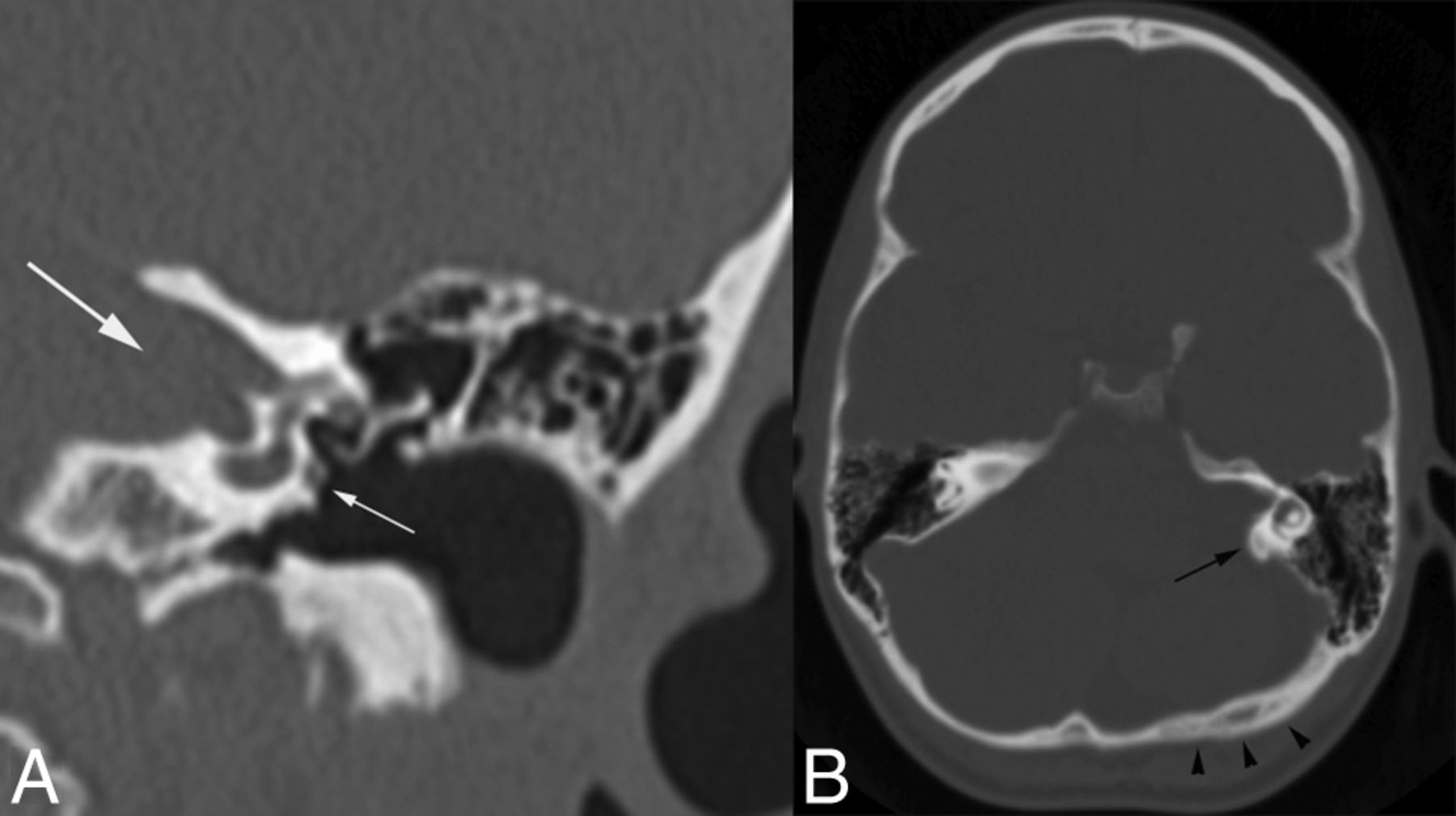
- Fig 1.
Coronal CT image of the left temporal bone (A) shows a prominent IAC (large white arrow) and a persistent stapedial artery (small white arrow). Axial CT image of the head (B) shows prominence of the left posterior petrous ridge (black arrow) and a relatively flattened, slightly thickened ipsilateral occipital calvarium (arrowheads).
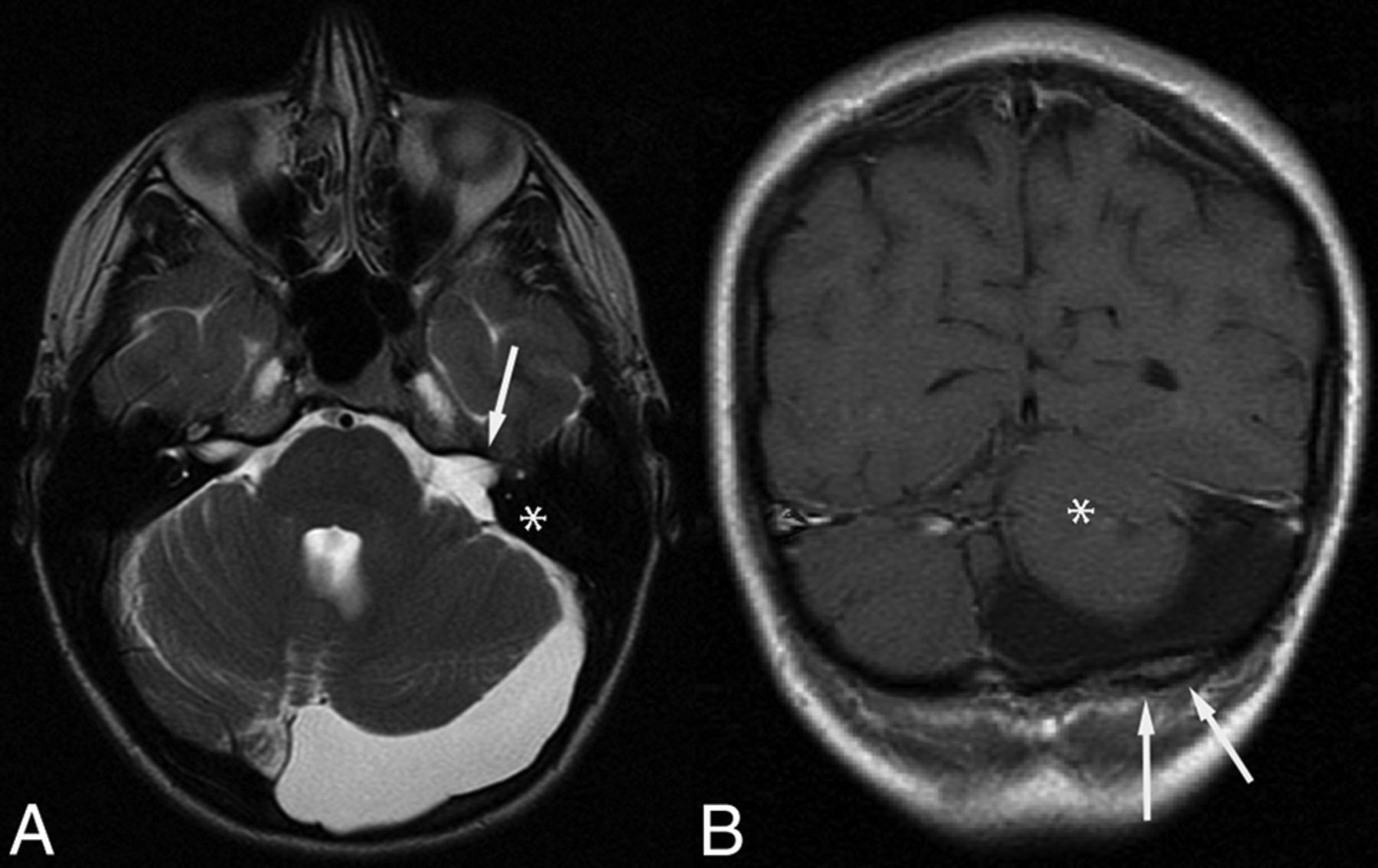
- Fig 2.
MR imaging of the same adult patient as in Fig 1. Axial T2-weighted image (A) shows prominent CSF in the left posterior fossa, enlarged left IAC (white arrow), and prominence of the posterior petrous ridge (asterisk in A). B, Coronal postcontrast T1-weighted image. The left cerebellum is hypoplastic (asterisk in B), and there is subtle calvarial deformity with focal thickening of the diploic space (arrows in B).
- Fig 3.
A 6-month-old girl. Axial (A) and coronal (B) T2-weighted images show unilateral enlargement and downsloping of the right IAC (white arrows). Note ipsilateral cerebellar hypoplasia (asterisk in A) and prominence of the posterior petrous ridge (black arrow). Additional coronal T2-weighted image (C) shows mild occipital calvarial flattening (arrowheads) ipsilateral to the cerebellar hypoplasia.
- Fig 4.
A 26-month-old girl. Axial T2-weighted image shows a right facial hemangioma (white arrow), ipsilateral to the enlarged right IAC (black arrow), and cerebellar hypoplasia (asterisk).
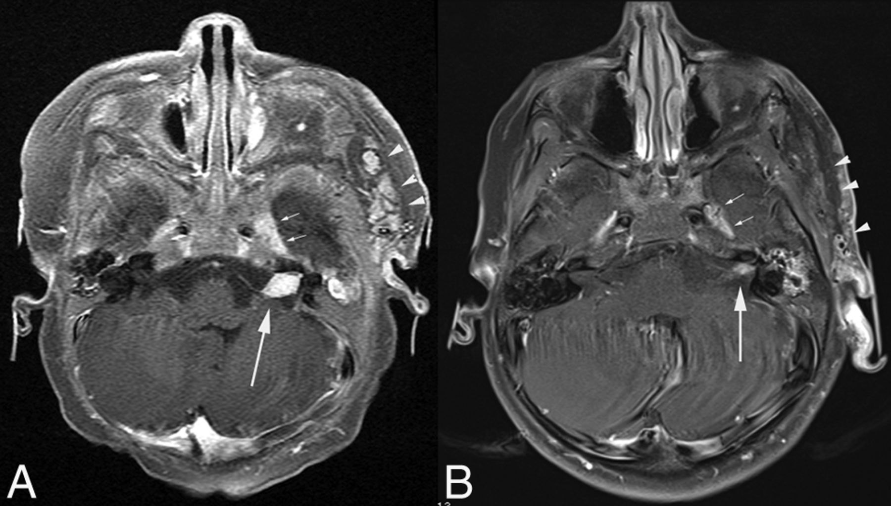
- Fig 5.
A 4-month-old girl. Axial postcontrast fat-suppressed T1-weighted images show an enhancing mass in the left IAC (arrow in A), which markedly diminishes in size on follow-up imaging at 3 years of age (arrow in B). Note the ipsilateral left facial and preauricular hemangioma, which similarly involutes (arrowheads in A and B). Note also mild prominence of the left cavernous sinus, thought to harbor an additional small hemangioma (small arrows in A and B). Minimal left cerebellar hypoplasia was better demonstrated on T2-weighted images (not shown).
- Fig 6.
A 3-month-old boy. A, Axial postcontrast T1-weighted image shows an enhancing mass in the right IAC (white arrow). There is also abnormal enhancement in the fourth ventricle (black arrow), suspicious for an additional hemangioma. B, Fifteen-month follow-up imaging of the same patient. Fat-suppressed T1-weighted image shows diminished enhancement in the right IAC (white arrow). The previously seen enhancement in the fourth ventricle is also less conspicuous, beginning to resemble normal choroidal enhancement (black arrow). The cerebellum appears normal.


Tables
Posterior fossa anomalies in 19 patients with PHACES with IAC enlargement
Age at Initial MRI Sex IAC Enhancing Posterior Petrous Prominence Cerebellar Hypoplasia Occipital Bone Side of Facial Hemangioma 3 mo F Yesa Yes Yes Smaller Ipsilateral 1 mo F Yesa Yes No No Ipsilateral 2 mo F Yesa Yes No No Ipsilateral 4 mo F Yesa Yes Yes Smaller Ipsilateral 38 d M Yes No Yes Larger Ipsilateral 9 mo F Yes Yes Yes Smaller Ipsilateralb 29 mo M Yes Yes Yes Smaller Ipsilateral 24 mo F No Yes Yes Smaller Ipsilateral 18 mo M No Yes Yes Smaller Ipsilateral 15 mo F No Yes Yes Deformed Contralateral 4 mo F No Yes Yes Smaller Ipsilateral 24 mo F No Yes Yes Smaller Ipsilateral 21 d F No Yes Yes Larger Ipsilateral 32 d F No Yes Yes Larger Ipsilateral 18 y F No Yes Yes Smaller Ipsilateral 8 y F NA No Yes Smaller Ipsilateral 13 y F NA Yes Yes Deformed Ipsilateral 17 y F NA No No No Ipsilateral 8 d F NA Yes Yes Larger Ipsilateral


