Advertisement

Main menu

  • Home
  • Content
    • Current Issue
    • Accepted Manuscripts
    • Article Preview
    • Past Issue Archive
    • Video Articles
    • AJNR Case Collection
    • Case of the Week Archive
    • Case of the Month Archive
    • Classic Case Archive
  • Special Collections
    • AJNR Awards
    • Low-Field MRI
    • Alzheimer Disease
    • ASNR Foundation Special Collection
    • Photon-Counting CT
    • View All
  • Multimedia
    • AJNR Podcasts
    • AJNR SCANtastic
    • Trainee Corner
    • MRI Safety Corner
    • Imaging Protocols
  • For Authors
    • Submit a Manuscript
    • Submit a Video Article
    • Submit an eLetter to the Editor/Response
    • Manuscript Submission Guidelines
    • Statistical Tips
    • Fast Publishing of Accepted Manuscripts
    • Graphical Abstract Preparation
    • Imaging Protocol Submission
    • Author Policies
  • About Us
    • About AJNR
    • Editorial Board
    • Editorial Board Alumni
  • More
    • Become a Reviewer/Academy of Reviewers
    • Subscribers
    • Permissions
    • Alerts
    • Feedback
    • Advertisers
    • ASNR Home

User menu

  • Alerts
  • Log in

Search

  • Advanced search
American Journal of Neuroradiology

American Journal of Neuroradiology

ASHNR American Society of Functional Neuroradiology ASHNR American Society of Pediatric Neuroradiology ASSR
  • Alerts
  • Log in

Advanced Search

  • Home
  • Content
    • Current Issue
    • Accepted Manuscripts
    • Article Preview
    • Past Issue Archive
    • Video Articles
    • AJNR Case Collection
    • Case of the Week Archive
    • Case of the Month Archive
    • Classic Case Archive
  • Special Collections
    • AJNR Awards
    • Low-Field MRI
    • Alzheimer Disease
    • ASNR Foundation Special Collection
    • Photon-Counting CT
    • View All
  • Multimedia
    • AJNR Podcasts
    • AJNR SCANtastic
    • Trainee Corner
    • MRI Safety Corner
    • Imaging Protocols
  • For Authors
    • Submit a Manuscript
    • Submit a Video Article
    • Submit an eLetter to the Editor/Response
    • Manuscript Submission Guidelines
    • Statistical Tips
    • Fast Publishing of Accepted Manuscripts
    • Graphical Abstract Preparation
    • Imaging Protocol Submission
    • Author Policies
  • About Us
    • About AJNR
    • Editorial Board
    • Editorial Board Alumni
  • More
    • Become a Reviewer/Academy of Reviewers
    • Subscribers
    • Permissions
    • Alerts
    • Feedback
    • Advertisers
    • ASNR Home
  • Follow AJNR on Twitter
  • Visit AJNR on Facebook
  • Follow AJNR on Instagram
  • Join AJNR on LinkedIn
  • RSS Feeds

AJNR Awards, New Junior Editors, and more. Read the latest AJNR updates

Research ArticleEXTRACRANIAL VASCULAR

Diagnostic Accuracy of 4 Commercially Available Semiautomatic Packages for Carotid Artery Stenosis Measurement on CTA

J. Borst, H.A. Marquering, M. Kappelhof, T. Zadi, A.C. van Dijk, P.J. Nederkoorn, R. van den Berg, A. van der Lugt and C.B.L.M. Majoie
American Journal of Neuroradiology October 2015, 36 (10) 1978-1987; DOI: https://doi.org/10.3174/ajnr.A4400
J. Borst
aFrom the Departments of Radiology (J.B., H.A.M., M.K., R.v.d.B., C.B.L.M.M.)
  • Find this author on Google Scholar
  • Find this author on PubMed
  • Search for this author on this site
H.A. Marquering
aFrom the Departments of Radiology (J.B., H.A.M., M.K., R.v.d.B., C.B.L.M.M.)
bBiomedical Engineering and Physics (H.A.M.)
  • Find this author on Google Scholar
  • Find this author on PubMed
  • Search for this author on this site
M. Kappelhof
aFrom the Departments of Radiology (J.B., H.A.M., M.K., R.v.d.B., C.B.L.M.M.)
  • Find this author on Google Scholar
  • Find this author on PubMed
  • Search for this author on this site
T. Zadi
dDepartment of Radiology (T.Z., A.C.v.D., A.v.d.L.), Erasmus University Medical Center Rotterdam, Rotterdam, the Netherlands.
  • Find this author on Google Scholar
  • Find this author on PubMed
  • Search for this author on this site
A.C. van Dijk
dDepartment of Radiology (T.Z., A.C.v.D., A.v.d.L.), Erasmus University Medical Center Rotterdam, Rotterdam, the Netherlands.
  • Find this author on Google Scholar
  • Find this author on PubMed
  • Search for this author on this site
P.J. Nederkoorn
cNeurology (P.J.N.), Academic Medical Center, Amsterdam, the Netherlands
  • Find this author on Google Scholar
  • Find this author on PubMed
  • Search for this author on this site
R. van den Berg
aFrom the Departments of Radiology (J.B., H.A.M., M.K., R.v.d.B., C.B.L.M.M.)
  • Find this author on Google Scholar
  • Find this author on PubMed
  • Search for this author on this site
A. van der Lugt
dDepartment of Radiology (T.Z., A.C.v.D., A.v.d.L.), Erasmus University Medical Center Rotterdam, Rotterdam, the Netherlands.
  • Find this author on Google Scholar
  • Find this author on PubMed
  • Search for this author on this site
C.B.L.M. Majoie
aFrom the Departments of Radiology (J.B., H.A.M., M.K., R.v.d.B., C.B.L.M.M.)
  • Find this author on Google Scholar
  • Find this author on PubMed
  • Search for this author on this site
  • Article
  • Figures & Data
  • Supplemental
  • Info & Metrics
  • Responses
  • References
  • PDF
Loading

Article Figures & Data

Figures

  • Tables
  • Fig 1.
    • Download figure
    • Open in new tab
    • Download powerpoint
    Fig 1.

    Scatterplots of the manual-versus-semiautomatic assessment of the degree of stenosis (percentage).

  • Fig 2.
    • Download figure
    • Open in new tab
    • Download powerpoint
    Fig 2.

    Bland-Altman plots of the degree of stenosis determined by manual and semiautomatic assessment. The black lines represent the mean paired difference and 95% limits of agreement. The characteristic V-shape in the Bland-Altman plot is caused by 1 of the 2 measurements being zero with the other measurement being nonzero. These measurements happened particularly when the degree of stenosis was small (<30%).
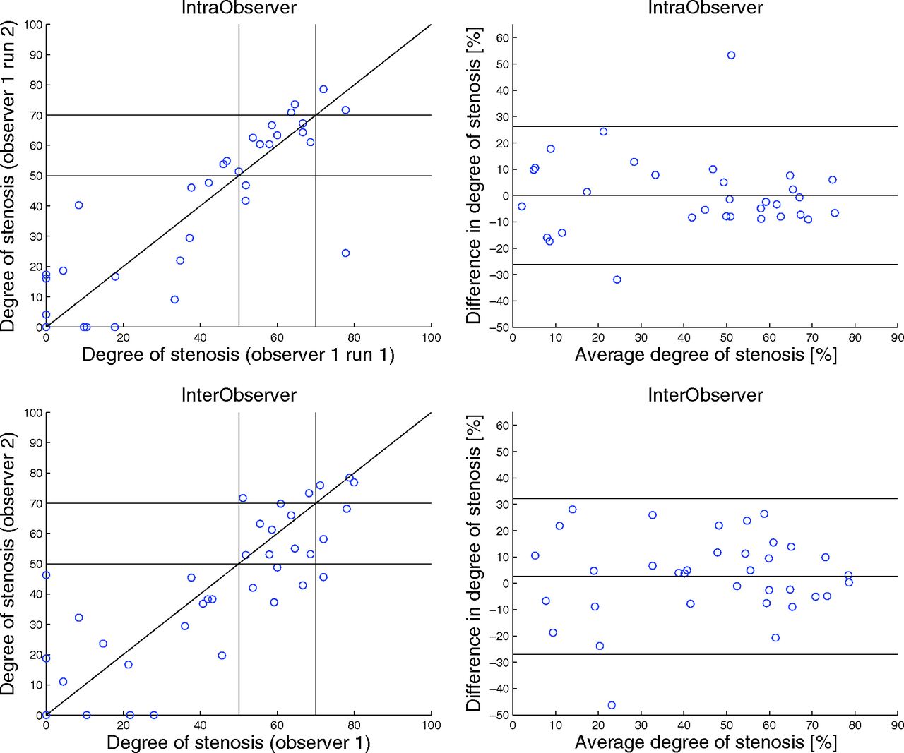
  • Fig 3.
    • Download figure
    • Open in new tab
    • Download powerpoint
    Fig 3.

    Scatterplot (upper left corner) and Bland-Altman plot (upper right corner) of the repeated manual stenosis measurement (percentage) (intraobserver). Scatterplot (lower left corner) and Bland-Altman plot (lower right corner) of the manual assessment of the degree of stenosis (percentage) measured by observer 1 and observer 2 (interobserver). The black lines in the right figures represent the mean paired difference and 95% limits of agreement.
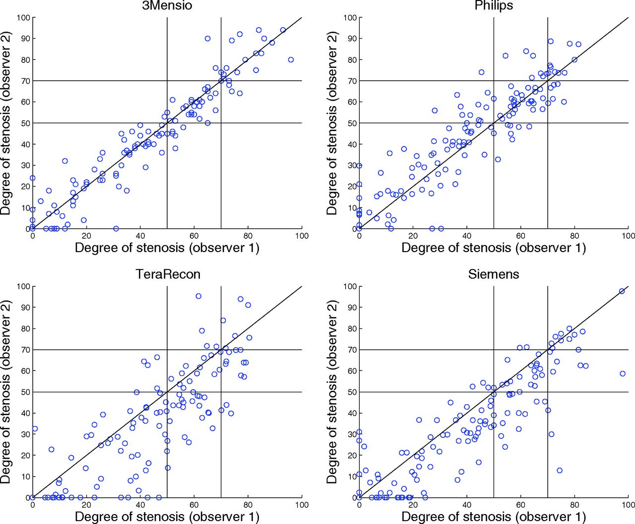
  • Fig 4.
    • Download figure
    • Open in new tab
    • Download powerpoint
    Fig 4.

    Scatterplots of the repeated semiautomatic assessment of the degree of stenosis (percentage) measured by observers 1 and 2.

  • Fig 5.
    • Download figure
    • Open in new tab
    • Download powerpoint
    Fig 5.

    Bland-Altman plots of the repeated semiautomatic assessment of the degree of stenosis measured by observers 1 and 2. The black lines represent the mean paired difference and 95% limits of agreement.

  • Fig 6.
    • Download figure
    • Open in new tab
    • Download powerpoint
    Fig 6.

    Example of ulcerative plaque. On the left side how 3mensio segments the artery is shown, and in the right upper corner how 3mensio segments the lumen and the ulcerative plaque is shown. The turquoise line is the minimal stenosis diameter as determined by 3mensio (3.5 mm); the white-with-red line is a measurement of the true lumen (1.2 mm). The right lower corner shows a sagittal view of the ulceration. This image also shows 3mensio fitting an ellipse (yellow) on the segmented lumen of the artery (turquoise).

Tables

  • Figures
    • View popup
    Table 1:

    Average processing time

    Average Processing Time ± SD (seconds)
    Manual measurements138 ± 31
    3mensio86 ± 42
    Philips115 ± 77
    TeraRecon84 ± 64
    Siemens89 ± 86
    • View popup
    Table 2:

    Agreement manual vs semiautomatic stenosis measurement

    Average Difference Degree of Stenosis ± SD (%) (Manual, Semiautomatic)Bland-Altman 95% Limits of Agreement (%)ICC for Degree of Stenosis (95% CI)
    3mensio (observer 1)3.8 ± 14 (P = .002)−24–310.86 (0.80–0.90)
    Philips (observer 1)2.1 ± 13 (P = .049)−23–270.88 (0.83–0.91)
    TeraRecon (observer 1)3.1 ± 13 (P = .007)−23–290.87 (0.82–0.90)
    Siemens (observer 1)3.5 ± 13 (P = .002)−22–290.88 (0.83–0.91)
    • View popup
    Table 3:

    Diagnostic performance of semiautomatic measurement in detecting a stenosis degree of ≥70% and ≥50%

    Sensitivity (95% CI)Specificity (95% CI)PPV (95% CI)NPV (95% CI)Accuracy (95% CI)
    70% Cutoff
        3mensio (observer 1)62 (43–78)96 (91–98)76 (53–92)92 (85–96)89 (82–93)
        Philips (observer 1)46 (29–65)96 (91–99)75 (48–93)88 (82–94)87 (80–92)
        TeraRecon (observer 1)58 (37–77)97 (93–99)83 (59–96)91 (85–95)90 (84–94)
        Siemens (observer 1)62 (40–81)97 (91–99)80 (56–94)92 (85–96)90 (84–94)
    50% Cutoff
        3mensio (observer 1)77 (65–86)93 (86–97)91 (80–97)83 (73–90)86 (79–91)
        Philips (observer 1)82 (70–89)93 (86–97)91 (81–97)86 (76–92)88 (81–93)
        TeraRecon (observer 1)76 (65–86)92 (84–97)89 (78–96)82 (73–90)85 (78–90)
        Siemens (observer 1)77 (65–86)95 (87–99)93 (82–98)83 (73–9087 (80–91)
    • Note:—PPV indicates positive predictive value; NPV, negative predictive value.

    • View popup
    Table 4:

    Observer reproducibility

    Average Difference Degree of Stenosis ± SD (%)Bland-Altman Limits of Agreement (%)ICC (95% CI) for Degree of Stenosis
    Manual intraobserver (n = 38)0.083 ± 13−26–260.88 (0.79–0.94)
    Manual interobserver (n = 37)2.6 ± 15−28–320.81 (0.70–0.90)
    3mensio interobserver (n = 141)0.94 ± 7.5a (P = .007)−14–160.96 (0.95–0.97a (P < .001)
    Philips interobserver (n = 141)−2.8 ± 11−23–180.90 (0.86–0.93)a (P = .0041)
    TeraRecon interobserver (n = 141)7.0 ± 14−20–340.83 (0.70–0.90)
    Siemens interobserver (n = 141)6.5 ± 12−17–300.86 (0.73–0.92)
    • ↵a Significant difference with manual interobserver measurements.

    • View popup
    Table 5:

    Observer reproducibility by statitical κ values

    50% κa (95% CI)70% κb (95% CI)
    Manual intraobserver (n = 38)0.73 (0.50–0.95)0.53 (0.02–1)
    Manual interobserver (n = 37)0.73 (0.52–0.95)0.47 (0.03–0.90)
    3mensio interobserver (n = 141)0.88 (0.80–0.96)0.86 (0.73–0.98)
    Philips interobserver (n = 141)0.71 (0.60–0.83)0.53 (0.30–0.77)
    TeraRecon interobserver (n = 141)0.56 (0.41–0.71)0.37 (0.08–0.66)
    Siemens Interobserver (n = 141)0.67 (0.54–0.80)0.63 (0.42–0.84)
    • ↵a κ values on 50% cutoff.

    • ↵b κ values on 70% cutoff.

PreviousNext
Back to top

In this issue

American Journal of Neuroradiology: 36 (10)
American Journal of Neuroradiology
Vol. 36, Issue 10
1 Oct 2015
  • Table of Contents
  • Index by author
  • Complete Issue (PDF)
Advertisement
Print
Download PDF
Email Article

Thank you for your interest in spreading the word on American Journal of Neuroradiology.

NOTE: We only request your email address so that the person you are recommending the page to knows that you wanted them to see it, and that it is not junk mail. We do not capture any email address.

Enter multiple addresses on separate lines or separate them with commas.
Diagnostic Accuracy of 4 Commercially Available Semiautomatic Packages for Carotid Artery Stenosis Measurement on CTA
(Your Name) has sent you a message from American Journal of Neuroradiology
(Your Name) thought you would like to see the American Journal of Neuroradiology web site.
CAPTCHA
This question is for testing whether or not you are a human visitor and to prevent automated spam submissions.
Cite this article
J. Borst, H.A. Marquering, M. Kappelhof, T. Zadi, A.C. van Dijk, P.J. Nederkoorn, R. van den Berg, A. van der Lugt, C.B.L.M. Majoie
Diagnostic Accuracy of 4 Commercially Available Semiautomatic Packages for Carotid Artery Stenosis Measurement on CTA
American Journal of Neuroradiology Oct 2015, 36 (10) 1978-1987; DOI: 10.3174/ajnr.A4400

Citation Manager Formats

  • BibTeX
  • Bookends
  • EasyBib
  • EndNote (tagged)
  • EndNote 8 (xml)
  • Medlars
  • Mendeley
  • Papers
  • RefWorks Tagged
  • Ref Manager
  • RIS
  • Zotero
0 Responses
Respond to this article
Share
Bookmark this article
Diagnostic Accuracy of 4 Commercially Available Semiautomatic Packages for Carotid Artery Stenosis Measurement on CTA
J. Borst, H.A. Marquering, M. Kappelhof, T. Zadi, A.C. van Dijk, P.J. Nederkoorn, R. van den Berg, A. van der Lugt, C.B.L.M. Majoie
American Journal of Neuroradiology Oct 2015, 36 (10) 1978-1987; DOI: 10.3174/ajnr.A4400
del.icio.us logo Twitter logo Facebook logo Mendeley logo
  • Tweet Widget
  • Facebook Like
  • Google Plus One
Purchase

Jump to section

  • Article
    • Abstract
    • ABBREVIATION:
    • Materials and Methods
    • Results
    • Discussion
    • Conclusions
    • Footnotes
    • REFERENCES
  • Figures & Data
  • Supplemental
  • Info & Metrics
  • Responses
  • References
  • PDF

Related Articles

  • PubMed
  • Google Scholar

Cited By...

  • No citing articles found.
  • Crossref (12)
  • Google Scholar

This article has been cited by the following articles in journals that are participating in Crossref Cited-by Linking.

  • Medical Image Data and Datasets in the Era of Machine Learning—Whitepaper from the 2016 C-MIMI Meeting Dataset Session
    Marc D. Kohli, Ronald M. Summers, J. Raymond Geis
    Journal of Digital Imaging 2017 30 4
  • Extracranial Carotid Disease and Effect of Intra-arterial Treatment in Patients With Proximal Anterior Circulation Stroke in MR CLEAN
    Olvert A. Berkhemer, Jordi Borst, Manon Kappelhof, Albert J. Yoo, Lucie A. van den Berg, Puck S.S. Fransen, Debbie Beumer, Wouter J. Schonewille, Paul J. Nederkoorn, Marieke J.H. Wermer, Henk A. Marquering, Hester F. Lingsma, Yvo B.W.E.M. Roos, Robert J. van Oostenbrugge, Diederik W.J. Dippel, Wim H. van Zwam, Charles B.L.M. Majoie, Bart J. Emmer, Aad van der Lugt
    Annals of Internal Medicine 2017 166 12
  • Carotid near-occlusion frequently has high peak systolic velocity on Doppler ultrasound
    Simon R. Khangure, Hadas Benhabib, Matylda Machnowska, Allan J. Fox, Christer Grönlund, Wendy Herod, Robert Maggisano, Anders Sjöberg, Per Wester, Seyed-Parsa Hojjat, Julia Hopyan, Richard I. Aviv, Elias Johansson
    Neuroradiology 2018 60 1
  • Comparability of semiautomatic tortuosity measurements in the carotid artery
    Evelien E. de Vries, Vanessa E. C. Pourier, Constance J. H. C. M. van Laarhoven, Evert J. Vonken, Joost A. van Herwaarden, Gert J. de Borst
    Neuroradiology 2019 61 2
  • Comparison of Measurement and Grading of Carotid Stenosis with Computed Tomography Angiography and Doppler Ultrasound
    Panagiota Birmpili, Laura Porter, Usman Shaikh, Francesco Torella
    Annals of Vascular Surgery 2018 51
  • Evaluation of Technical Success, Efficacy, and Safety of Portomesenteric Venous Intervention following Nontransplant Hepatobiliary or Pancreatic Surgery
    Vamshi K. Mugu, Scott M. Thompson, Chad J. Fleming, Lavanya Yohanathan, Mark J. Truty, Michael L. Kendrick, James C. Andrews
    Journal of Vascular and Interventional Radiology 2020 31 3
  • Overcoming calcium blooming and improving the quantification accuracy of percent area luminal stenosis by material decomposition of multi-energy computed tomography datasets
    Zhoubo Li, Shuai Leng, Ahmed F. Halaweish, Zhicong Yu, Lifeng Yu, Erik L. Ritman, Cynthia H. McCollough
    Journal of Medical Imaging 2020 7 05
  • Stenosis diagnosis based on peripheral arterial and artificial neural network
    Zheming Li, Wei He
    Network Modeling Analysis in Health Informatics and Bioinformatics 2021 10 1
  • Comparison of Manual versus Semi-Automatic Segmentations of the Stenotic Carotid Artery Bifurcation
    Benjamin Csippa, Zsuzsanna Mihály, Zsófia Czinege, Márton Bence Németh, Gábor Halász, György Paál, Péter Sótonyi
    Applied Sciences 2021 11 17
  • AI-Based Automated Quantification of Arterial Stenosis in Head and Neck CT Angiography: A Comparison with Manual Measurements from Digital Subtraction Angiography and CT Angiography
    Xinyue Huan, Yang Yang, Shengwen Niu, Yongwei Yang, Bitong Tian, Dajing Guo, Kunhua Li
    Clinical Neuroradiology 2024

More in this TOC Section

  • Proximal Vertebral Artery Variants and Embryology
  • High-Risk Plaque Features in Carotid MRI
  • Nonstenotic Carotid Plaques and Stroke Review
Show more EXTRACRANIAL VASCULAR

Similar Articles

Advertisement

Indexed Content

  • Current Issue
  • Accepted Manuscripts
  • Article Preview
  • Past Issues
  • Editorials
  • Editor's Choice
  • Fellows' Journal Club
  • Letters to the Editor
  • Video Articles

Cases

  • Case Collection
  • Archive - Case of the Week
  • Archive - Case of the Month
  • Archive - Classic Case

More from AJNR

  • Trainee Corner
  • Imaging Protocols
  • MRI Safety Corner
  • Book Reviews

Multimedia

  • AJNR Podcasts
  • AJNR Scantastics

Resources

  • Turnaround Time
  • Submit a Manuscript
  • Submit a Video Article
  • Submit an eLetter to the Editor/Response
  • Manuscript Submission Guidelines
  • Statistical Tips
  • Fast Publishing of Accepted Manuscripts
  • Graphical Abstract Preparation
  • Imaging Protocol Submission
  • Evidence-Based Medicine Level Guide
  • Publishing Checklists
  • Author Policies
  • Become a Reviewer/Academy of Reviewers
  • News and Updates

About Us

  • About AJNR
  • Editorial Board
  • Editorial Board Alumni
  • Alerts
  • Permissions
  • Not an AJNR Subscriber? Join Now
  • Advertise with Us
  • Librarian Resources
  • Feedback
  • Terms and Conditions
  • AJNR Editorial Board Alumni

American Society of Neuroradiology

  • Not an ASNR Member? Join Now

© 2025 by the American Society of Neuroradiology All rights, including for text and data mining, AI training, and similar technologies, are reserved.
Print ISSN: 0195-6108 Online ISSN: 1936-959X

Powered by HighWire