
Article Figures & Data
Figures
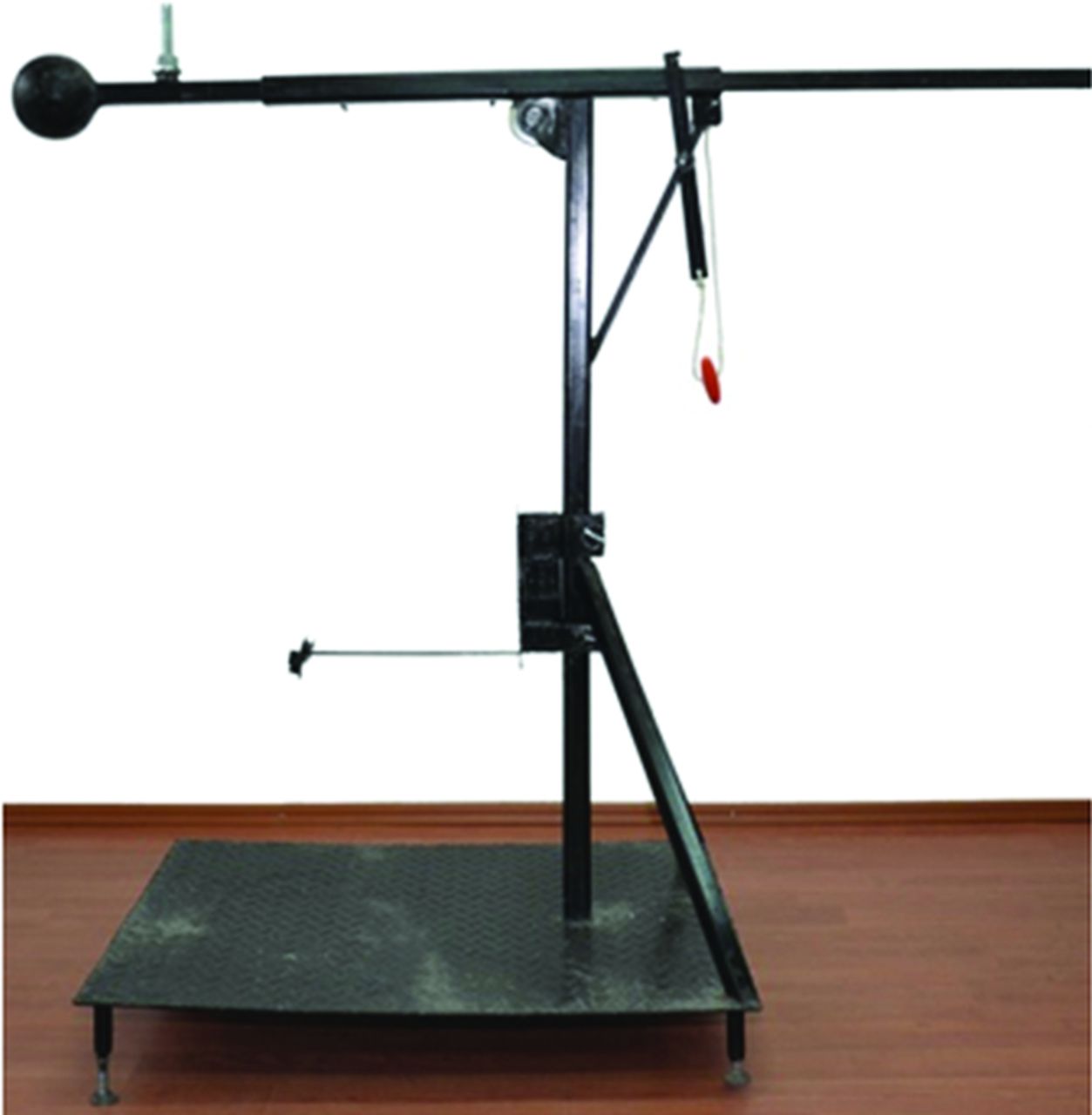
- Fig 1.
The impacting system.
- Fig 2.
Impaction of sample M1 of the control group.
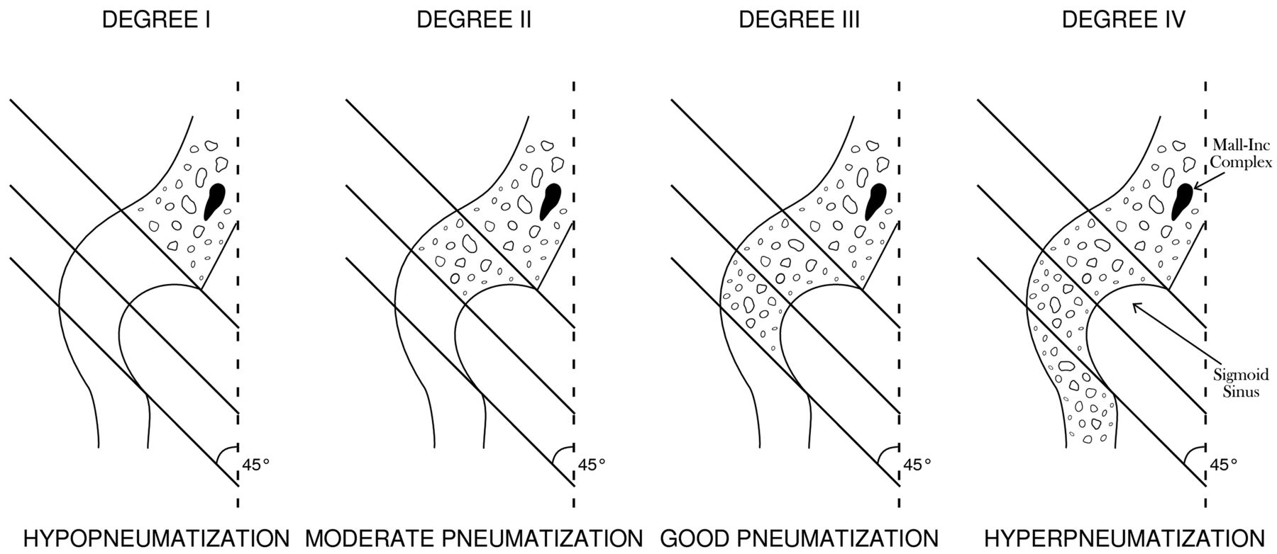
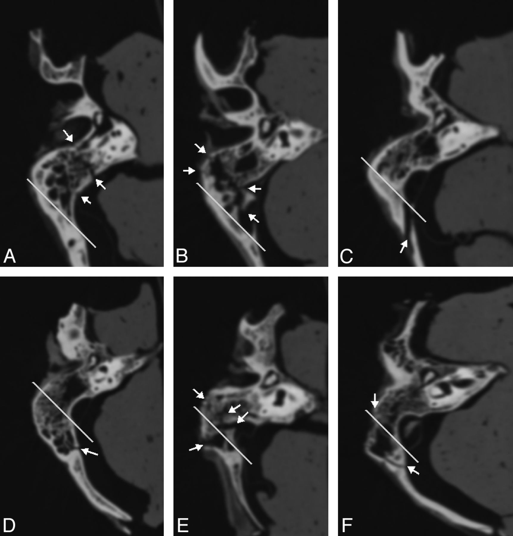
- Fig 3.
Degrees of mastoid pneumatization according to the descriptions of Han et al.6
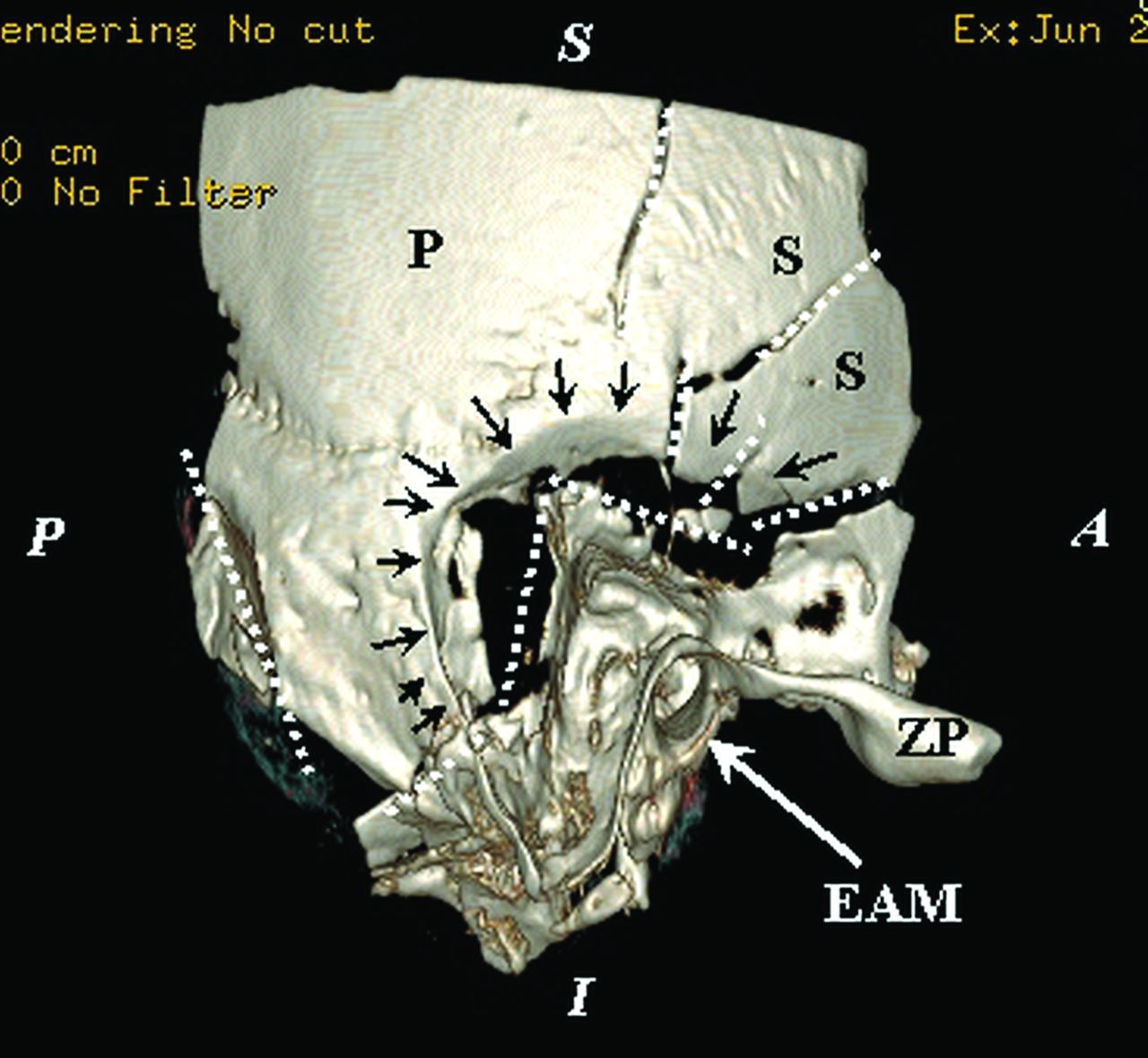
- Fig 4.
Temporal squama comminuted fracture: 3D reconstruction of the exocranial surface of the right temporal bone of sample S1 with a volume-rendering technique. Comminuted fracture of the mastoid with extension in the petrous bone and important depression. In the center of the image is highlighted the aspect of bone depression (black arrows) that keeps the ball contour impaction at the junction with the mastoid and temporal scales; the main fracture lines are marked by dashed white lines. The sample position is indicated by marginal marks with white letters: S indicates superior; I, inferior; A, anterior; and P, posterior; black letter marks: ZP, zygomatic process of the temporal bone; P, parietal portion; S, scaly portion of the temporal bone. EAM, the external acoustic meatus, is indicated by the white arrow.
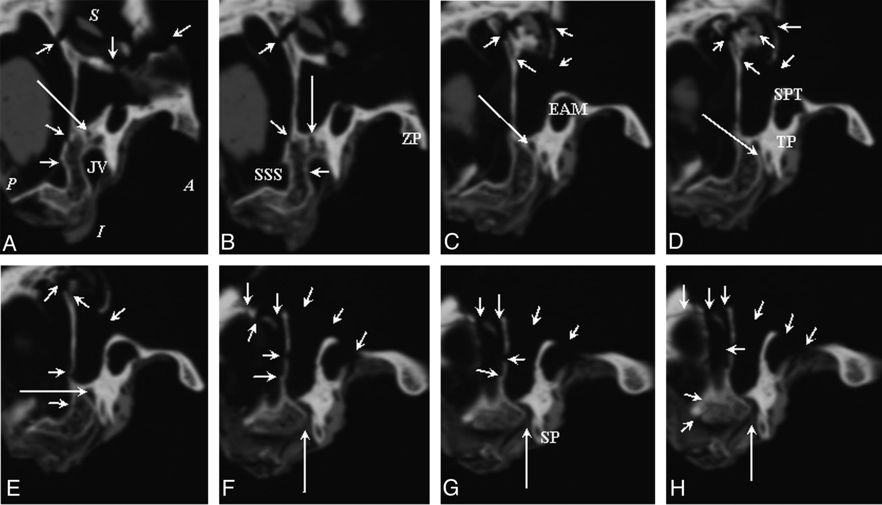
- Fig 5.
CT image of the mastoid segment of the facial nerve of sample S1 (A–H): successive CT sections reconstructed in the sagittal plane in the distal segment of the mastoid canal up to the stylomastoid foramen. In the first image (A), the position of the sample in the sagittal section is marked as follows: A indicates anterior; P, posterior; S, superior; I, inferior. The fracture lines are marked by short arrows; the facial nerve canal is marked by a long arrow. A, JV, jugular vein. B, SSS, sigmoid sinus sulcus; ZP, zygomatic process. C, EAM, external auditory meatus. D and E, The long arrow marks the descending segment of the facial nerve. D, TP, tympanic portion; SPT, squamous portion of the temporal bone (it forms the roof and the posterosuperior wall of the external auditory meatus). F–H, The long arrow indicates the stylomastoid foramen. G, SP, styloid process.
- Fig 6.
Axial CT sections involving vital structures of the temporal bone. The first images (A–C) belong to the control group, and the last images (D–F) belong to the study group. The position of the samples in the axial plane is marked as follows: A indicates anterior; P, posterior; L, lateral; M, medial on the first image. Short white arrows indicate fracture lines with different orientations in different parts of the temporal bone. Long white arrows indicate fractures of walls of vascular and nerve structures: carotid canal in images A and D (double-tipped arrow), and F; sigmoid sinus sulcus and jugular vein bulb in images B and C; the mastoid segment of facial nerve in the image D; the tympanic segment of the facial nerve in the image E; the stylomastoid foramen in the image F. The black arrows indicate the different portions of the facial nerve: the mastoid portion in C and D, tympanic in E, and the stylomastoid foramen in F. IAM indicates internal auditory meatus; SSS, sigmoid sinus sulcus; JB, jugular bulb; JV, jugular vein; EAM, external auditory meatus. A, M3 sample section at the level of the internal auditory meatus. The long white arrow indicates a fracture line at the medial wall of the carotid canal. B, M4 sample section at the level of epitympanic portion. The long white arrow indicates a longitudinal fracture line at the lateral wall of the sigmoid sulcus. The short white arrows indicate a comminuted fracture of the petrous apex. C, M8 sample section at the level of the jugular bulb: The long white arrow indicates a longitudinal fracture line at the lateral wall of the jugular bulb sulcus. The short white arrows indicate a comminuted fracture of the mastoid. D, S4 sample section at the level of the external auditory meatus. The long white arrow indicates a longitudinal fracture line involving the mastoid segment of the facial nerve; the white double-tipped arrow indicates a comminuted fracture of the lateral wall of the carotid canal. E, S6 sample section at the level of the tympanic portion of the facial nerve. The long white arrow indicates a fracture line involving the facial nerve. F, S5 sample section at the level of the stylomastoid foramen. The long white arrow indicates a bone fragment in the roof of the carotid canal.
- Fig 7.
Axial CT section at the level of the sigmoid sinus and epitympanum in samples M1 (A), M5 (B), M6 (C), M7 (D), M8 (E), and M10 (F). The white line marks the separation between pneumatization degree III and IV. Samples from images A–C show pneumatization degree III, and the images D–F show pneumatization degree IV. Short white arrows mark the fracture lines highlighted in these sections.


Tables
Type of Fracture M1–M10 S1–S10 Transverse Fracture, No Fracture Lines Longitudinal Fracture, No Fracture Lines Oblique Fracture, No Fracture Lines Transverse Fracture, No Fracture Lines Longitudinal Fracture, No Fracture Lines Oblique Fracture No, Fracture Lines Petrous fracture 4 0 0 11 4 8 Non-petrous fracture 5 8 9 23 18 29 Type of Fracture Samples M1–M10 Pneumatization Degree Petrous Fracture Nonpetrous Fracture Transverse Fracture, No Fracture Lines Longitudinal Fracture, No Fracture Lines Oblique Fracture, No Fracture Lines Transverse Fracture, No Fracture Lines Longitudinal Fracture, No Fracture Lines Oblique Fracture, No Fracture Lines M1 IV 1 – – 1 – 3 M2 III – – – – 1 1 M3 IV – – – – – 1 M4 IV 1 – – 1 1 – M5 IV 1 – – 3 1 1 M6 III – – – – 1 – M7 IV – – – – 1 – M8 IV – – – – 1 1 M9 III 1 – – – 1 – M10 IV – – – – 1 2 Note:—–indicates no fracture lines.


Jump to section
Related Articles
Cited By...
- No citing articles found.