
Article Figures & Data
Figures
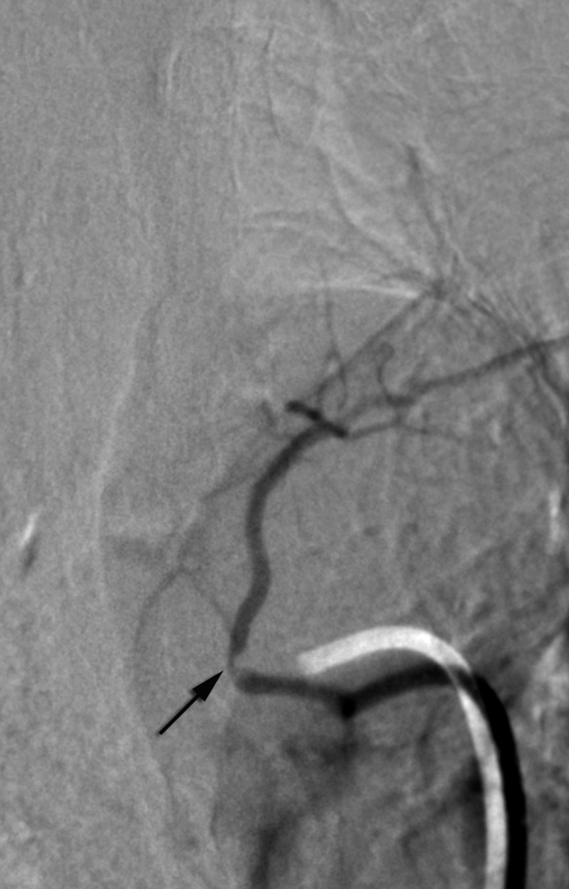
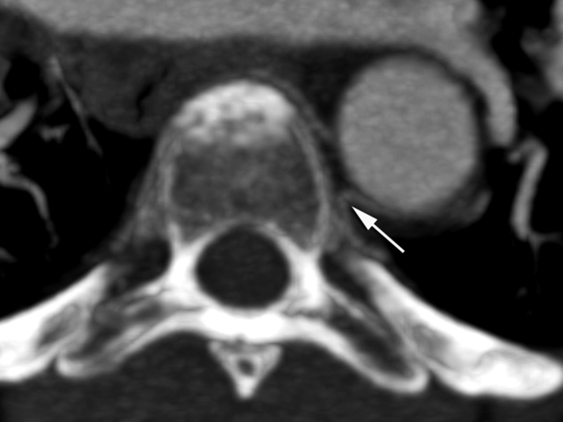
- Fig 1.
Non-ostial proximal intersegmental artery stenosis at left T4 in a 60-year-old woman (arrow).
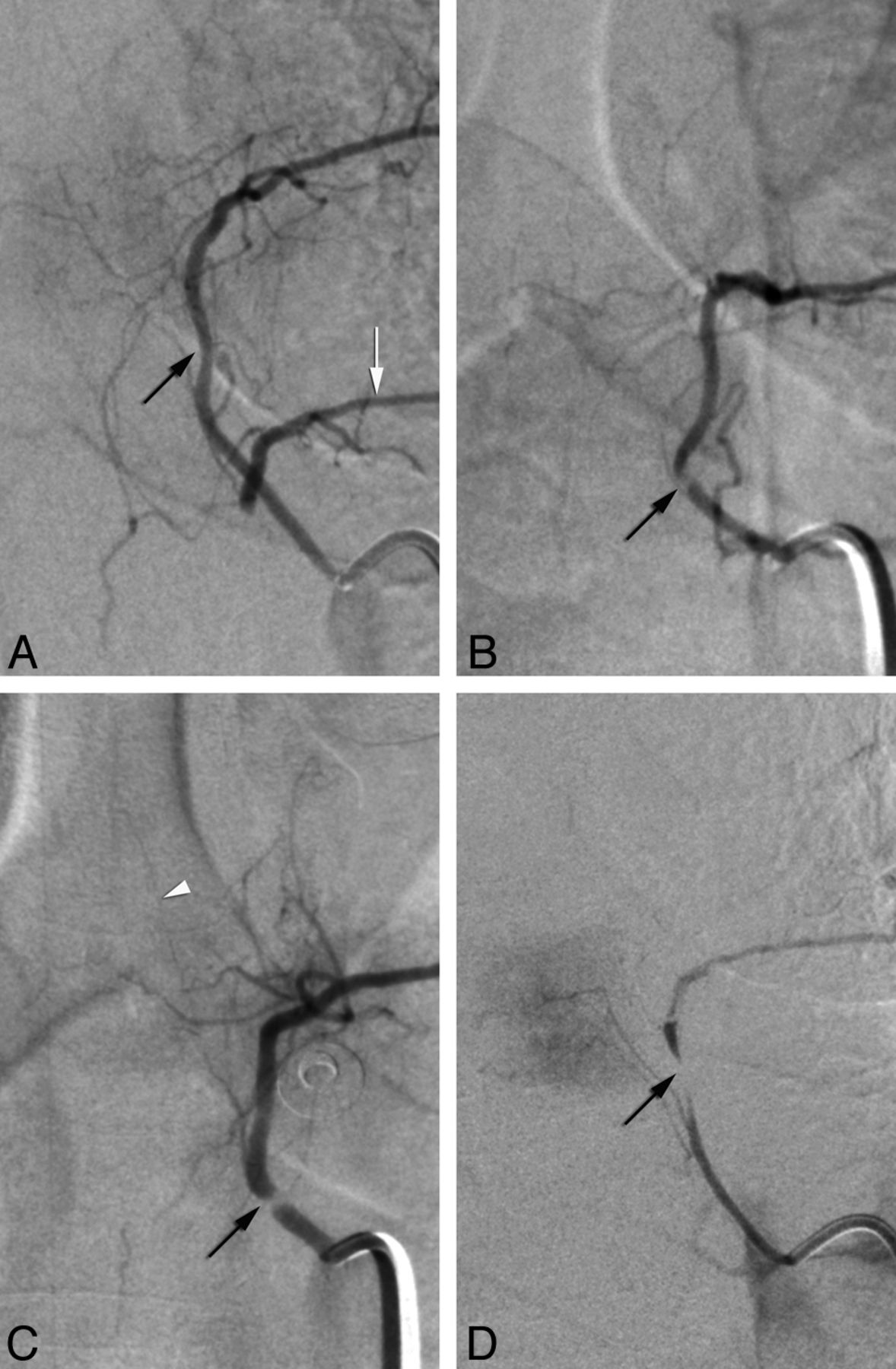
- Fig 2.
Categorization of stenoses as mild/moderate (A and B) or severe (C and D) (from studied cohort). A, Mild stenosis at left T5 in a 69-year-old woman (black arrow) illustrates the mildest form of recorded narrowing. Note collateral flow to left T6 (white arrow), which harbors a near-occlusive lesion. B, Moderate stenosis at left T5 in a 41-year-old woman (arrow); a continuous column of contrast is seen across the narrowing. C, Severe stenosis at left T5 in a 40-year-old woman (arrow); although close in severity to the case shown in B, this narrowing shows a completely unopacified segment. White arrowhead points to an anterior radiculomedullary artery. D, Near-occlusion at left T6 in the patient shown in A (arrow).
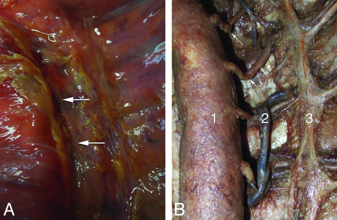
- Fig 3.
Anatomic observations in 2 human specimens. A, Anterior view of the endothoracic fascia in a fresh specimen after resection of the pleura; note the predominantly longitudinal bands of reinforcements (arrows) near the vertebral column. B, Anatomic dissection of an embalmed specimen documenting the proximal kinking of the left upper thoracic intersegmental arteries (arrows); 1, aorta (gently retracted to the right); 2, accessory hemiazygos vein; 3, sympathetic trunk.
- Fig 4.
Mirror intersegmental anatomy in a 50-year-old woman with a right-sided aortic arch. A, Arch aortograms, postero-anterior projection; note that the deflection of the descending aorta from the midline is much less pronounced than in patients with a left-sided arch. B, Right T6 injection shows the typical course of a “normal” left-sided upper thoracic intersegmental artery (ISA), with a short recurrent course followed by a lateral bend. C, Left T6 injection shows the typical course of a “normal” right-sided upper thoracic ISA, with a smooth course across the midline, over the vertebral column.
- Fig 5.
Thoracic CT angiography in a 54-year-old woman, axial image through the T5 level, illustrates the difference in trajectories between left and right upper thoracic intersegmental arteries, whereas the right-sided artery smoothly curves around the vertebral body to cross the midline; the left one, after a short initial recurrent segment, sharply bends dorsally to continue its course along the corresponding rib. This “fixed” point in the vessel path (arrow), which results from the obligatory turn the artery must take around the medial attachment of the endothoracic fascia, corresponds to the site of stenosis.
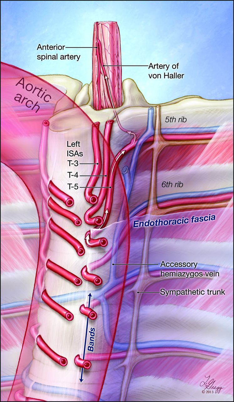
- Fig 6.
Graphic representation of the study findings. This illustration depicts the course of the upper thoracic intersegmental arteries (ISAs) in relation to the vertebral column, the endothoracic fascia, and the position of the thoracic aorta. Note the difference in trajectory between the right-sided ISAs, smoothly curving around the spine to pass under the fascia, and the left-sided ones, which have to adopt a recurrent course to reach the fixed paramedian points imposed by the attachment of the fascia to the lateral aspect of the vertebral column. The reinforced bands of connective tissue found within the paramedian endothoracic fascia are indicated by an arrow at T7 and T8. Note that the length of the recurrent segments and the sharpness of the bends increase with the leftward deviation of the upper thoracic aorta. In this example, the fifth thoracic ISA provides a prominent anterior radiculomedullary branch, the artery of von Haller.
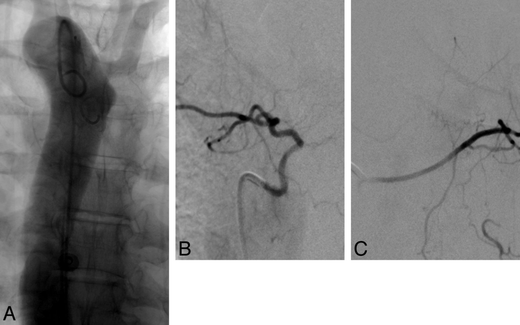
- Fig 7.
Angiographic findings investigating for progressive myelopathy in a 52-year-old woman. A, Non-ostial occlusion of the left T6 intersegmental artery. B, Left T7 injection shows collateral supply to left T6 distal to the site of occlusion (arrow), with opacification of the anterior spinal artery (arrowhead) through a left T6 anterior radiculomedullary artery (artery of von Haller, small arrow).


Tables
Angiographic severity of proximal (non-ostial) stenoses affecting the upper thoracic intersegmental arteries
Levels Left Right Mild/Moderate Severe/Occlusions Mild/Moderate Severe/Occlusions T3 3 1 T4 8 1 1 T5 7 3 T6 3 2 T7 1 1 2 T8 4


