
Article Figures & Data
Figures
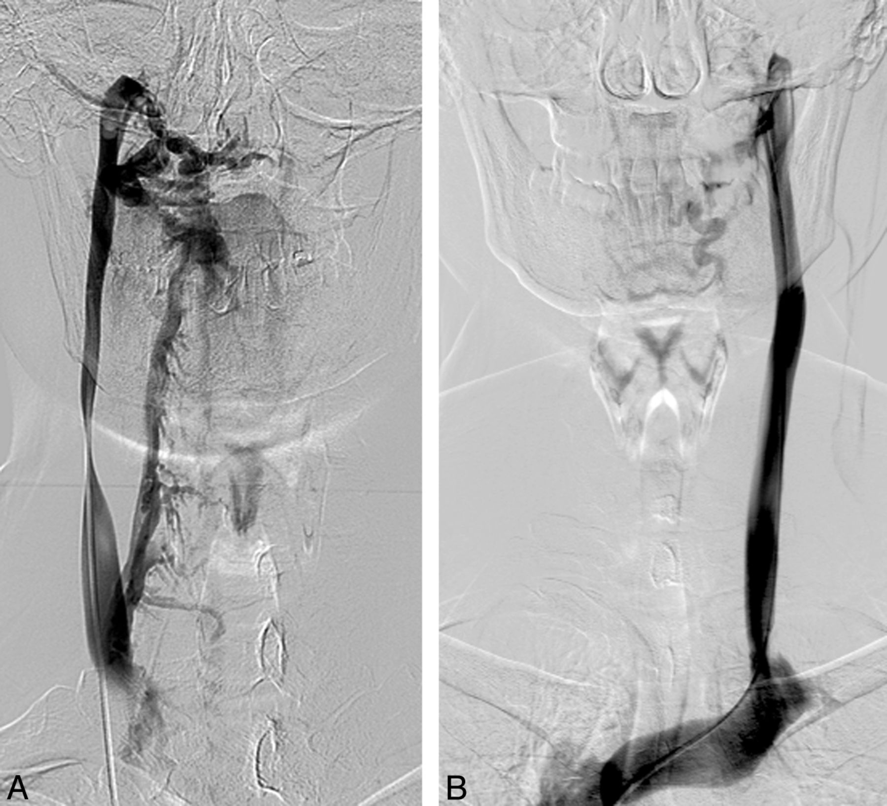
- Fig 1.
Selective catheterization of the azygos vein in a 37-year-old patient with secondary-progressive MS in which the injection of contrast medium showed the presence of a twisting of the AV associated with a shunt with the hemiazygous venous system. Note the opacification of the intraspinal venous plexus.
- Fig 2.
Selective catheterization of the left and right internal jugular veins in a 35-year-old patient with relapsing-remitting MS. Venous stenosis at the level of the section J2 of the right IJV and of its origin is shown, with associated drainage in spinal venous plexus through a collateral branch. The left IJV does not present alterations in size and drainage.
- Fig 3.
Selective catheterization of the left internal jugular vein in a 41-year-old patient with primary-progressive MS. After injection of contrast medium, stenosis of segment J1 with reflux in both vertebral veins and opacification of the vertebral venous plexus is documented.
- Fig 4.
Selective catheterization of the ileo-lumbar plexus in a 33-year-old patient affected by relapsing-remitting MS. After injection of the contrast medium, opacification of the intraspinal venous plexus without opacification of the inferior vena cava is documented.
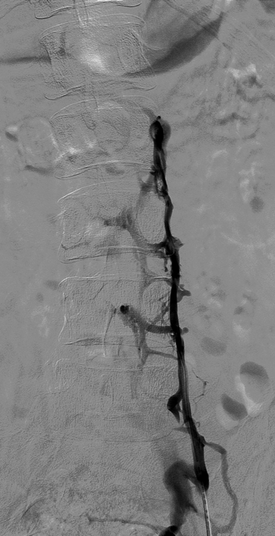
- Fig 5.
Selective catheterization of the azygos vein in a 45-year-old patient affected by relapsing-remitting MS. At the confluence of the azygos vein after administration of contrast medium, a valve is evident.
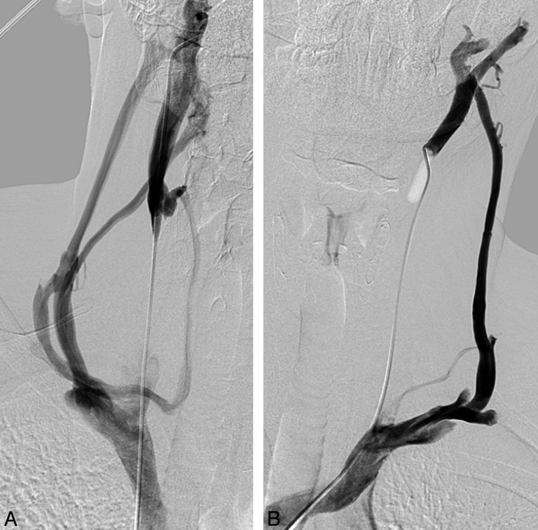
- Fig 6.
Selective catheterization of the internal jugular veins bilaterally (A, -B) in a 27-year-old control subject. Complete obstruction of the IJVs bilaterally with evidence of venous outflow through collateral circulation in the external jugular veins is shown.


Tables
Study Group Control Group P Value No. 29 15 Sex, M/F 10/19 11/4 .03 Age, y 46.4 ± 9.5 42.6 ± 7.1 .18 Disease duration, y 12.6 ± 6.7 NA Disease course (RR/SP/PP) 17/8/4 NA EDSS 3.6 ± 2.1 (1.0–7.0) NA Note:—NA indicates not applicable; RR, relapsing-remitting; SP, secondary-progressive; PP, primary-progressive.
Coefficient SE OR 95% CI P Value Venography 0.77 1.24 2.16 0.18–24.91 .53 Stenosis, n −0.01 0.59 0.98 0.30–3.19 .98 Age 0.06 0.04 1.06 0.97–1.15 .16 Sex, male −1.57 0.72 0.20 0.05–0.86 .03 Note:—SE indicates standard error.
Type Study Group Control Group RR MS SP MS PP MS A 4 2 3 1 0 B 1 0 0 1 0 C 10 4 8 1 1 D 1 0 0 1 0 Note:—RR indicates relapsing-remitting; SP, secondary-progressive; PP, primary-progressive.
Coefficient SE OR 95% CI P Value Venography −2.26 1.60 0.10 0.00–2.38 .15 Stenosis, n 0.76 0.76 2.14 0.48–9.56 .31 Age 0.10 0.05 1.12 1.00–1.24 .04 Sex, male 0.83 0.92 2.29 0.37–14.19 .37 Note:—SE indicates standard error.


Jump to section
Related Articles
Cited By...
- No citing articles found.