
Article Figures & Data
Figures
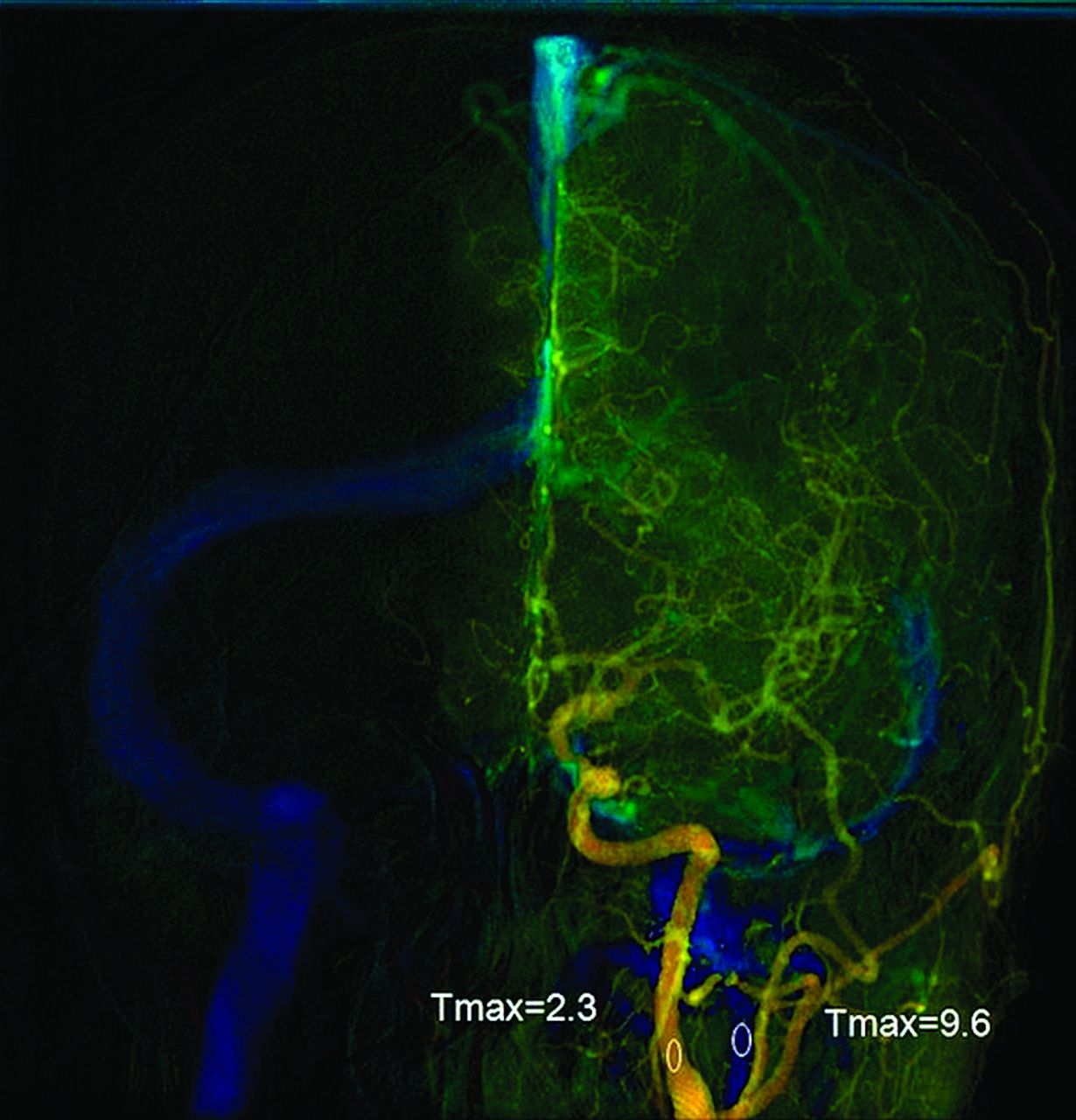
- Fig 1.
Anteroposterior view of quantitative brain DSA of a 51-year-old woman. The midpoint of the cervical portion of the internal carotid artery was selected for the arterial region of interest. The midpoint of the ipsilateral internal jugular vein was selected for venous region of interest. Cerebral circulation time was the time difference between the 2 ROIs.
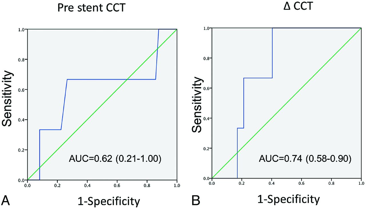
- Fig 2.
Receiver operating characteristic analysis of prestent CCT (A) and ΔCCT (B).
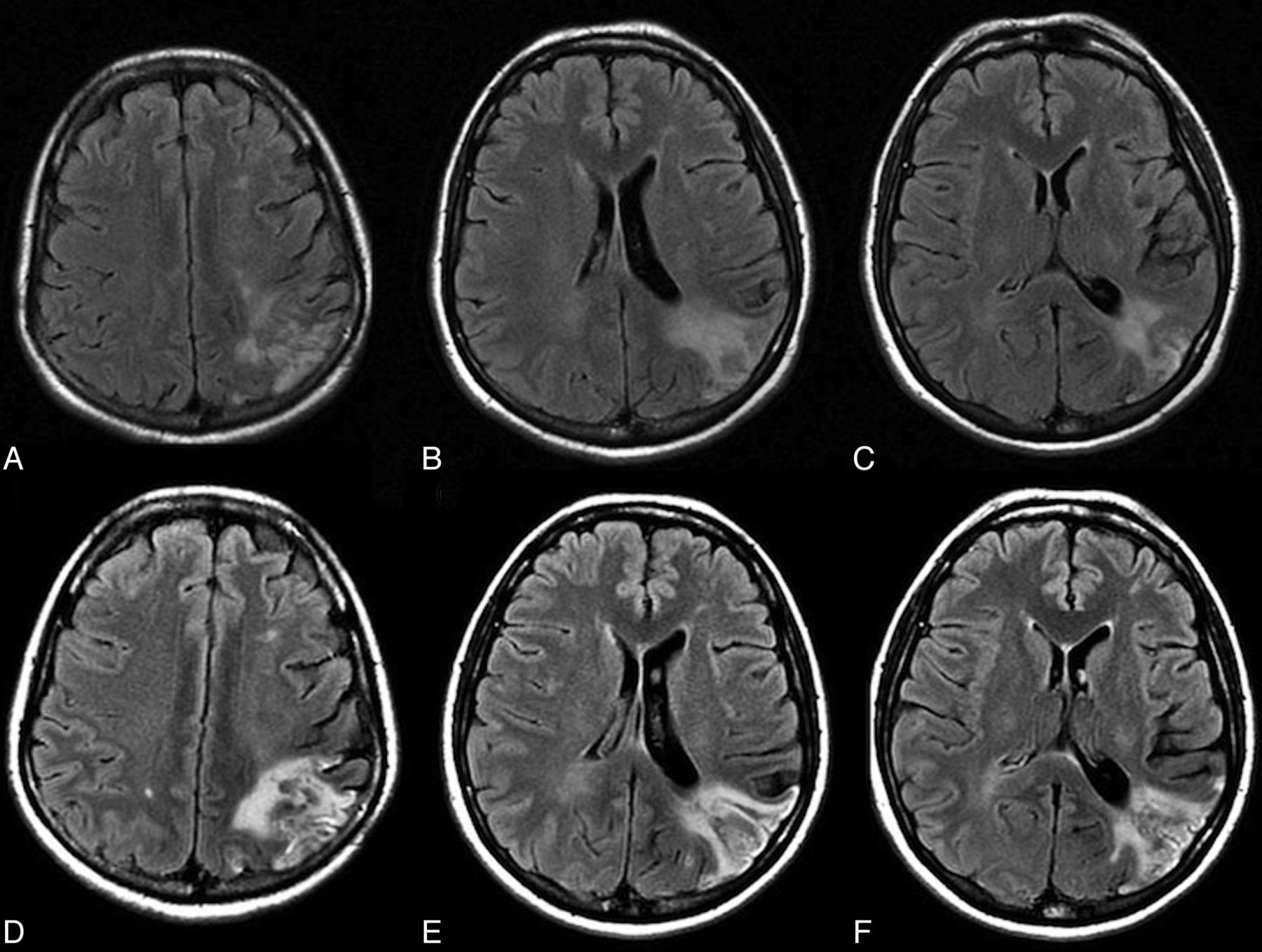
- Fig 3.
A 51-year-old woman with 90% left internal carotid artery stenosis. Color-coded cerebral DSA demonstrated an ipsilateral atretic transverse sinus in Fig 1. A–C, Prestent MR imaging shows an old infarct in the occipital subcortical white matter. She had headache with nausea 8 hours after the procedure. D–F, MR imaging 12 hours after the procedure shows increased signal intensity over the left occipital subcortical white matter.


Tables
- Table 1:
Patient characteristics between those with an ipsilateral stenotic transverse sinus (group A) and those with a normal transverse sinus (group B)
Group A (n = 15) Group B (n = 34) Age (yr) 74.5 + 10.2 73.9 + 10.3 Sex (M/F) 12:3 31:5 Hypertension 5 (33%) 13 (38%) MR imaging evidence of prior stroke 6 (40%) 16 (47%) Stenosis degree 82% 81% Lack of collaterals in MRA 2 (13%) 6 (18%) - Table 2:
Peritherapeutic circulation times and MR imaging findings between those with an ipsilateral stenotic transverse sinus (group A) and those with a normal transverse sinus (group B)
Group A (n = 15) Group B (n = 34) P Value DSA Prestent CCT 6.9 ± 1.2 6.2 ± 1.1 .14 Poststent CCT 6.2 ± 1.5 6.2 ± 1.5 .98 ΔCCT 0.65 ± 1.3 −0.12 ± 1.4 .045a MR imaging High SI in periventricular white matter 3 0 <.001a Gyral effacement 2 0 <.001a Note:—SI indicates signal intensity.
↵a Statically significant.
- Table 3:
Characteristics of 3 patients with high SI in the white matter in FLAIR sequences
Patient Recent Infarct HPS Age (yr) Sex Complications Stenotic Degree Pre-CCT (sec) Post-CCT (sec) ΔCCT (sec) 1 No Headache 51 F No 85% 7.5 5.84 1.66 2 No No 79 F No 77% 5.5 5.03 0.47 3 No No 81 M No 78% 10.0 9.0 1.0 Note:—SI indicates signal intensity.


Jump to section
Related Articles
Cited By...
- Prediction of hyperperfusion phenomenon after carotid artery stenting and carotid angioplasty using quantitative DSA with cerebral circulation time imaging
- Quantifying the Cerebral Hemodynamics of Dural Arteriovenous Fistula in Transverse Sigmoid Sinus Complicated by Sinus Stenosis: A Retrospective Cohort Study
- Peritherapeutic Hemodynamic Changes of Carotid Stenting Evaluated with Quantitative DSA in Patients with Carotid Stenosis
- Changes of Time-Attenuation Curve Blood Flow Parameters in Patients with and without Carotid Stenosis