Advertisement

Main menu

  • Home
  • Content
    • Current Issue
    • Accepted Manuscripts
    • Article Preview
    • Past Issue Archive
    • Video Articles
    • AJNR Case Collection
    • Case of the Week Archive
    • Case of the Month Archive
    • Classic Case Archive
  • Special Collections
    • AJNR Awards
    • Low-Field MRI
    • Alzheimer Disease
    • ASNR Foundation Special Collection
    • Photon-Counting CT
    • View All
  • Multimedia
    • AJNR Podcasts
    • AJNR SCANtastic
    • Trainee Corner
    • MRI Safety Corner
    • Imaging Protocols
  • For Authors
    • Submit a Manuscript
    • Submit a Video Article
    • Submit an eLetter to the Editor/Response
    • Manuscript Submission Guidelines
    • Statistical Tips
    • Fast Publishing of Accepted Manuscripts
    • Graphical Abstract Preparation
    • Imaging Protocol Submission
    • Author Policies
  • About Us
    • About AJNR
    • Editorial Board
    • Editorial Board Alumni
  • More
    • Become a Reviewer/Academy of Reviewers
    • Subscribers
    • Permissions
    • Alerts
    • Feedback
    • Advertisers
    • ASNR Home

User menu

  • Alerts
  • Log in

Search

  • Advanced search
American Journal of Neuroradiology

American Journal of Neuroradiology

ASHNR American Society of Functional Neuroradiology ASHNR American Society of Pediatric Neuroradiology ASSR
  • Alerts
  • Log in

Advanced Search

  • Home
  • Content
    • Current Issue
    • Accepted Manuscripts
    • Article Preview
    • Past Issue Archive
    • Video Articles
    • AJNR Case Collection
    • Case of the Week Archive
    • Case of the Month Archive
    • Classic Case Archive
  • Special Collections
    • AJNR Awards
    • Low-Field MRI
    • Alzheimer Disease
    • ASNR Foundation Special Collection
    • Photon-Counting CT
    • View All
  • Multimedia
    • AJNR Podcasts
    • AJNR SCANtastic
    • Trainee Corner
    • MRI Safety Corner
    • Imaging Protocols
  • For Authors
    • Submit a Manuscript
    • Submit a Video Article
    • Submit an eLetter to the Editor/Response
    • Manuscript Submission Guidelines
    • Statistical Tips
    • Fast Publishing of Accepted Manuscripts
    • Graphical Abstract Preparation
    • Imaging Protocol Submission
    • Author Policies
  • About Us
    • About AJNR
    • Editorial Board
    • Editorial Board Alumni
  • More
    • Become a Reviewer/Academy of Reviewers
    • Subscribers
    • Permissions
    • Alerts
    • Feedback
    • Advertisers
    • ASNR Home
  • Follow AJNR on Twitter
  • Visit AJNR on Facebook
  • Follow AJNR on Instagram
  • Join AJNR on LinkedIn
  • RSS Feeds

AJNR Awards, New Junior Editors, and more. Read the latest AJNR updates

Research ArticlePediatrics

CT and MRI of Pediatric Skull Lesions with Fluid-Fluid Levels

S.A. Nabavizadeh, L.T. Bilaniuk, T. Feygin, K.V. Shekdar, R.A. Zimmerman and A. Vossough
American Journal of Neuroradiology March 2014, 35 (3) 604-608; DOI: https://doi.org/10.3174/ajnr.A3712
S.A. Nabavizadeh
aFrom the Department of Radiology, The Children's Hospital of Philadelphia, University of Pennsylvania, Philadelphia, Pennsylvania.
  • Find this author on Google Scholar
  • Find this author on PubMed
  • Search for this author on this site
L.T. Bilaniuk
aFrom the Department of Radiology, The Children's Hospital of Philadelphia, University of Pennsylvania, Philadelphia, Pennsylvania.
  • Find this author on Google Scholar
  • Find this author on PubMed
  • Search for this author on this site
T. Feygin
aFrom the Department of Radiology, The Children's Hospital of Philadelphia, University of Pennsylvania, Philadelphia, Pennsylvania.
  • Find this author on Google Scholar
  • Find this author on PubMed
  • Search for this author on this site
K.V. Shekdar
aFrom the Department of Radiology, The Children's Hospital of Philadelphia, University of Pennsylvania, Philadelphia, Pennsylvania.
  • Find this author on Google Scholar
  • Find this author on PubMed
  • Search for this author on this site
R.A. Zimmerman
aFrom the Department of Radiology, The Children's Hospital of Philadelphia, University of Pennsylvania, Philadelphia, Pennsylvania.
  • Find this author on Google Scholar
  • Find this author on PubMed
  • Search for this author on this site
A. Vossough
aFrom the Department of Radiology, The Children's Hospital of Philadelphia, University of Pennsylvania, Philadelphia, Pennsylvania.
  • Find this author on Google Scholar
  • Find this author on PubMed
  • Search for this author on this site
  • Article
  • Figures & Data
  • Info & Metrics
  • Responses
  • References
  • PDF
Loading

Article Figures & Data

Figures

  • Tables
  • Fig 1.
    • Download figure
    • Open in new tab
    • Download powerpoint
    Fig 1.

    MR imaging in a patient with Langerhans cell histiocytosis. Axial T1- (A) and T2-weighted (B) images reveal multiple FFLs. An enhancing solid soft-tissue component is seen medially on postcontrast axial (C) and coronal (D) T1-weighted images.

  • Fig 2.
    • Download figure
    • Open in new tab
    • Download powerpoint
    Fig 2.

    CT and MR imaging findings in a patient with an aneurysmal bone cyst. Coronal CT scan (A) reveals marked bone expansion by a well-defined cystic-appearing mass projecting into the left temporal fossa with very thin remnants of the bony cortex seen in the wall (arrows). T2-weighted (B) and precontrast T1-weighted (C) images reveal multiple FFLs. Peripheral and septal enhancement is noted on CT (A) and postcontrast T1-weighted images (D), but no large enhancing solid soft-tissue component is identified.

  • Fig 3.
    • Download figure
    • Open in new tab
    • Download powerpoint
    Fig 3.

    CT and MR imaging findings in a patient with Langerhans cell histiocytosis. Noncontrast CT (A) reveals a lytic lesion with a prominent soft-tissue component and well-defined bone destruction. An FFL is faintly seen (arrow). T2-weighted image (B) and postcontrast T1-weighted image (C) show a few FFLs and enhancement of soft-tissue components, respectively. Axial T2-weighted image (D) following chemotherapy now reveals an increase in the number of FFLs.
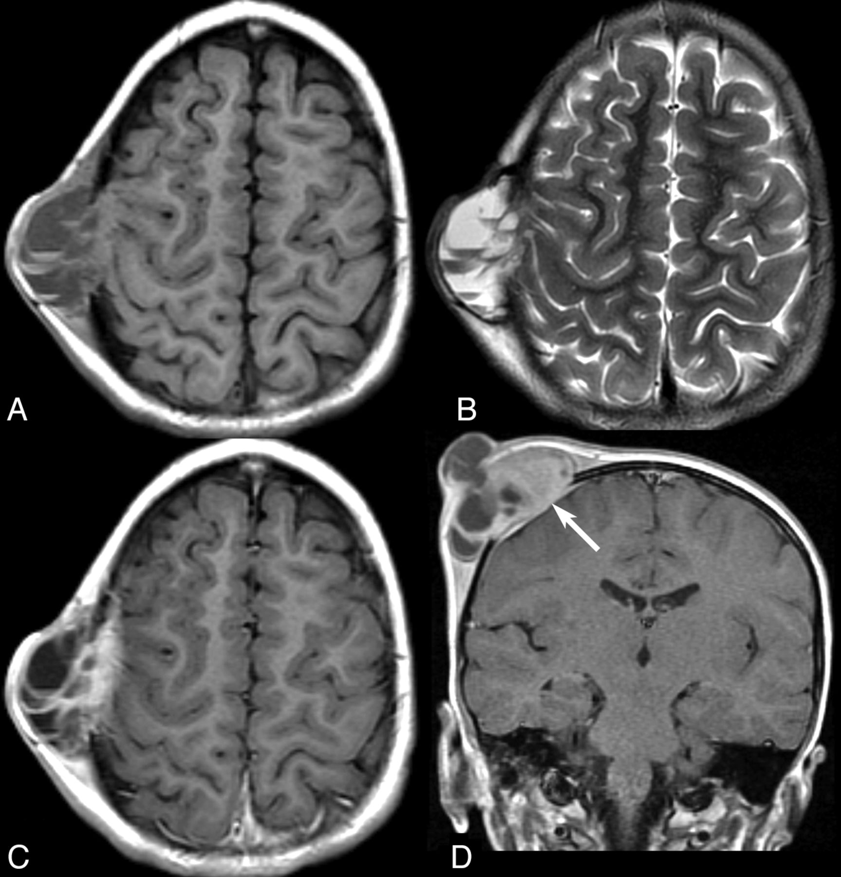
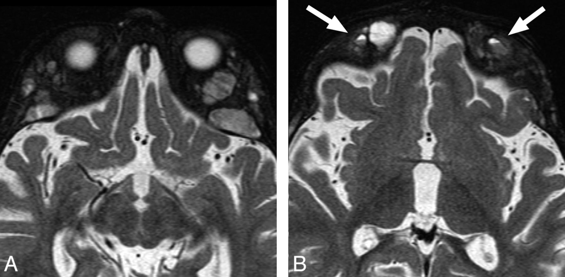
  • Fig 4.
    • Download figure
    • Open in new tab
    • Download powerpoint
    Fig 4.

    MR imaging findings in a patient with neuroblastoma demonstrate typical bone marrow infiltration and expansion by metastases (A). Multiple small FFLs are seen in the lesions (B).

  • Fig 5.
    • Download figure
    • Open in new tab
    • Download powerpoint
    Fig 5.

    MR imaging of a 3-month-old patient found to have a bump on the head on physical examination. T2-weighted axial image shows an FFL within a cephalohematoma (arrow), confirmed to be within the bone on multiple planes and sequences (not shown).

Tables

  • Figures
  • Demographic, clinical, imaging, and histopathologic findings of pediatric patients with FFLs in skull lesions

    PatientSexAge (yr)LocationCTNo. of FFLs on CTMRINo. of FFLs on MRIHistory of TraumaHistopathologyFinal Diagnosis
    1M4Right parietalNCCT9CE MRI17Minor head traumaLCHLCH
    2F0.75Occipital–N/ACE MRI2−LCHLCH
    3F13Left temporalCECT3CE MRI19−ABCABC
    4F10Left parietalNCCT0CE MRI1Forceps deliveryNoneCephalohematoma
    5M0.25Left parietal–N/ACE MRI4−NoneCephalohematoma
    6M1.5Sphenoid–N/ACE MRI2−NoneNeuroblastoma
    7F12Left frontalNCCT3CE MRI5Minor head traumaLCHLCH
    8M11Left temporalCECT2CE MRI6−ABC in the setting of fibrous dysplasiaABC
    9M0.5Left orbitCECT1CE MRI14−LCHLCH
    10M0.02Right parietal–N/ACE MRI3+NoneCephalohematoma
    11F16Right occipital and temporalNCCT7CE MRI29−ABCABC
    • Note:—CECT indicates contrast-enhanced CT; CE, contrast-enhanced; −, none; +, positive for trauma; N/A, not applicable.

PreviousNext
Back to top

In this issue

American Journal of Neuroradiology: 35 (3)
American Journal of Neuroradiology
Vol. 35, Issue 3
1 Mar 2014
  • Table of Contents
  • Index by author
  • Complete Issue (PDF)
Advertisement
Print
Download PDF
Email Article

Thank you for your interest in spreading the word on American Journal of Neuroradiology.

NOTE: We only request your email address so that the person you are recommending the page to knows that you wanted them to see it, and that it is not junk mail. We do not capture any email address.

Enter multiple addresses on separate lines or separate them with commas.
CT and MRI of Pediatric Skull Lesions with Fluid-Fluid Levels
(Your Name) has sent you a message from American Journal of Neuroradiology
(Your Name) thought you would like to see the American Journal of Neuroradiology web site.
CAPTCHA
This question is for testing whether or not you are a human visitor and to prevent automated spam submissions.
Cite this article
S.A. Nabavizadeh, L.T. Bilaniuk, T. Feygin, K.V. Shekdar, R.A. Zimmerman, A. Vossough
CT and MRI of Pediatric Skull Lesions with Fluid-Fluid Levels
American Journal of Neuroradiology Mar 2014, 35 (3) 604-608; DOI: 10.3174/ajnr.A3712

Citation Manager Formats

  • BibTeX
  • Bookends
  • EasyBib
  • EndNote (tagged)
  • EndNote 8 (xml)
  • Medlars
  • Mendeley
  • Papers
  • RefWorks Tagged
  • Ref Manager
  • RIS
  • Zotero
0 Responses
Respond to this article
Share
Bookmark this article
CT and MRI of Pediatric Skull Lesions with Fluid-Fluid Levels
S.A. Nabavizadeh, L.T. Bilaniuk, T. Feygin, K.V. Shekdar, R.A. Zimmerman, A. Vossough
American Journal of Neuroradiology Mar 2014, 35 (3) 604-608; DOI: 10.3174/ajnr.A3712
del.icio.us logo Twitter logo Facebook logo Mendeley logo
  • Tweet Widget
  • Facebook Like
  • Google Plus One
Purchase

Jump to section

  • Article
    • Abstract
    • ABBREVIATIONS:
    • Case Series
    • Discussion
    • Conclusions
    • REFERENCES
  • Figures & Data
  • Info & Metrics
  • Responses
  • References
  • PDF

Related Articles

  • No related articles found.
  • PubMed
  • Google Scholar

Cited By...

  • Point-of-Care Ultrasound Findings Associated With Langerhans Cell Histiocytosis in the Pediatric Emergency Department
  • Crossref (25)
  • Google Scholar

This article has been cited by the following articles in journals that are participating in Crossref Cited-by Linking.

  • Skull Vault Lesions: A Review
    Lucie Colas, Sabine Caron, Anne Cotten
    American Journal of Roentgenology 2015 205 4
  • Pediatric Cranial Fasciitis: Discussion of Cases and Systematic Review of the Literature
    Mohammed Alshareef, Gibson Klapthor, Ahmed Alshareef, Zayed Almadidy, Zachary Wright, Libby Infinger, Ramin Eskandari
    World Neurosurgery 2019 125
  • Craniospinal Polyostotic Fibrous Dysplasia, Aneurysmal Bone Cysts, and Chiari Type 1 Malformation Coexistence in a Patient with McCune-Albright Syndrome
    Kamran Urgun, Baran Yılmaz, Zafer Orkun Toktaş, Akın Akakın, Deniz Konya, Mustafa Kemal Demir, Türker Kılıç
    Pediatric Neurosurgery 2016 51 5
  • Imaging of focal calvarial lesions
    I. Mitra, M. Duraiswamy, J. Benning, H.M. Joy
    Clinical Radiology 2016 71 4
  • Eosinophilic Granuloma of Skull with Fluid Level and Epidural Hematoma: A Case Report and Review of the Literature
    Nishanth Sadashiva, Satyakam Baruah, Shilpa Rao, Anita Mahadevan, Kannepalli Narasinga Rao, Vikas Vazhayil, Sampath Somanna
    Pediatric Neurosurgery 2017 52 1
  • Calcified cephalohematoma as an unusual cause of EEG anomalies: case report
    Vera Vigo, Domenica Immacolata Battaglia, Paolo Frassanito, Gianpiero Tamburrini, Massimo Caldarelli, Luca Massimi
    Journal of Neurosurgery: Pediatrics 2017 19 1
  • Fluid-fluid Levels in Musculoskeletal Tumor Imaging
    Shinji Tsukamoto, Costantino Errani, Fabio Facchini, Panayiotis Papagelopoulos, Andreas F. Mavrogenis
    Current Medical Imaging Formerly Current Medical Imaging Reviews 2021 17 2
  • Langerhans Cell Histiocytosis with Multiple Fluid–fluid Levels in the Parietal Bone
    Kazuki Matsushita, Taro Shimono, Yukio Miki
    Magnetic Resonance in Medical Sciences 2020 19 1
  • A clinical approach to parasellar lesions in the transition age
    Emilia Sbardella, Giulia Puliani, Tiziana Feola, Riccardo Pofi, Rosa Pirchio, Franz Sesti, Federica Verdecchia, Daniele Gianfrilli, Daniel Moffat, Andrea M. Isidori, Ashley B. Grossman
    Journal of Neuroendocrinology 2021 33 6
  • Pediatric focal calvarial lesions: an illustrated review
    Tyler McDermott, Mohammad Amarneh, Yutaka Sato, Pankaj Watal, Vijapura Charmi, Michaelangelo Fuortes, Sami Faruqui, T. Shawn Sato
    Pediatric Radiology 2023 53 13

More in this TOC Section

  • SyMRI & MR Fingerprinting in Brainstem Myelination
  • Comparison of Image Quality and Radiation Dose in Pediatric Temporal Bone CT Using Photon-Counting Detector CT and Energy-Integrating Detector CT
  • Dual-Layer Detector CT for PEDS Image Quality
Show more Pediatrics

Similar Articles

Advertisement

Indexed Content

  • Current Issue
  • Accepted Manuscripts
  • Article Preview
  • Past Issues
  • Editorials
  • Editor's Choice
  • Fellows' Journal Club
  • Letters to the Editor
  • Video Articles

Cases

  • Case Collection
  • Archive - Case of the Week
  • Archive - Case of the Month
  • Archive - Classic Case

More from AJNR

  • Trainee Corner
  • Imaging Protocols
  • MRI Safety Corner
  • Book Reviews

Multimedia

  • AJNR Podcasts
  • AJNR Scantastics

Resources

  • Turnaround Time
  • Submit a Manuscript
  • Submit a Video Article
  • Submit an eLetter to the Editor/Response
  • Manuscript Submission Guidelines
  • Statistical Tips
  • Fast Publishing of Accepted Manuscripts
  • Graphical Abstract Preparation
  • Imaging Protocol Submission
  • Evidence-Based Medicine Level Guide
  • Publishing Checklists
  • Author Policies
  • Become a Reviewer/Academy of Reviewers
  • News and Updates

About Us

  • About AJNR
  • Editorial Board
  • Editorial Board Alumni
  • Alerts
  • Permissions
  • Not an AJNR Subscriber? Join Now
  • Advertise with Us
  • Librarian Resources
  • Feedback
  • Terms and Conditions
  • AJNR Editorial Board Alumni

American Society of Neuroradiology

  • Not an ASNR Member? Join Now

© 2025 by the American Society of Neuroradiology All rights, including for text and data mining, AI training, and similar technologies, are reserved.
Print ISSN: 0195-6108 Online ISSN: 1936-959X

Powered by HighWire