
Article Figures & Data
Figures
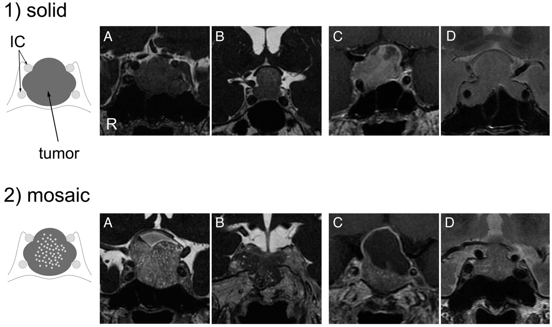
- Fig 1.
Classification of pituitary macroadenomas by use of CE-FIESTA, CE-T1WI, and T2WI. 1, Solid type. Schematic drawing and coronal images of CE-FIESTA (A and B), CE-T1WI (C), and T2WI (D) show homogeneous patterns of SI without intratumoral hyperintense dots. 2, Mosaic type. Schematic drawing and coronal images of CE-FIESTA (A and B), CE-T1WI (C), and T2WI (D) show intratumoral hyperintense dots within the tumor.
- Fig 2.
Macroadenoma with hard consistency in a 77-year-old male patient (case 13). A, Coronal CE-T1WI shows a large heterogeneous enhanced pituitary mass. B, Coronal T2WI shows an isointense mass with respect to normal white matter. C, Coronal CE-FIESTA shows a homogeneous tumor SI pattern without intratumoral hyperintense dots. D, Histologic examination of the resected tumor indicates small size and the formation of multiple nests surrounded by attenuated collagen tissue (Masson trichrome stain; scale bar, 200 μm).
- Fig 3.
Macroadenoma with soft consistency in a 51-year-old female patient (case 28). A, Coronal CE-T1WI shows a large, homogeneous, enhanced pituitary mass. B, Coronal T2WI shows a relatively hyperintense mass with respect to normal white matter. C, Coronal CE-FIESTA shows numerous hyperintense dots within the tumor. D, Histologic examination of the resected tumor indicates small cells with scant collagen in a small, restricted perivascular area (Masson trichrome stain; scale bar, 200 μm).
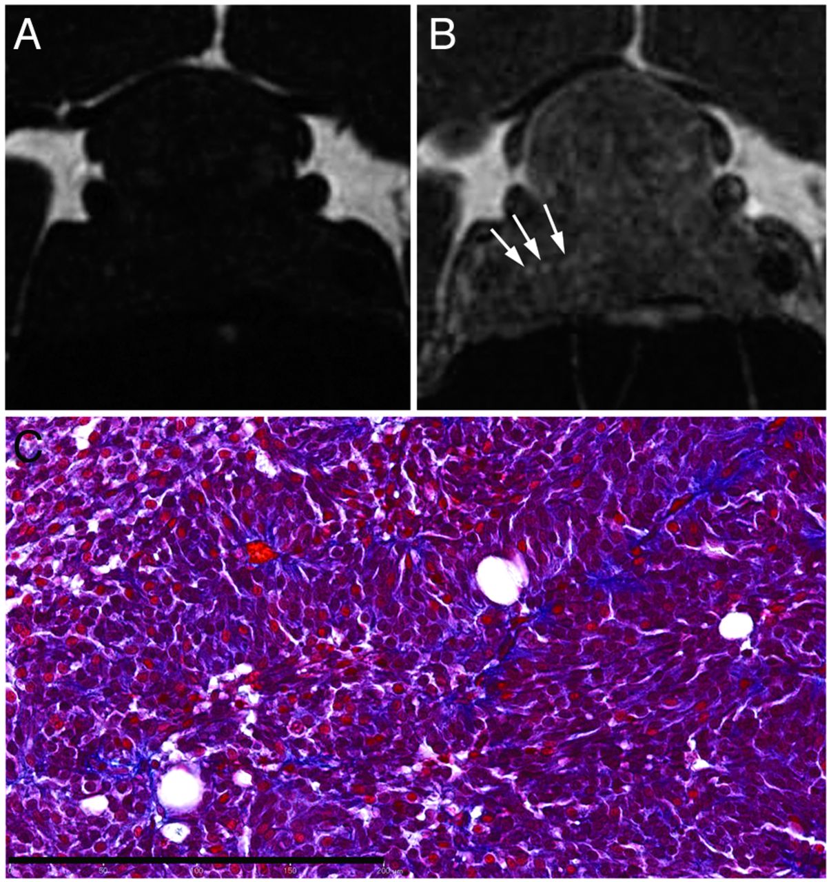
- Fig 4.
Macroadenoma with soft consistency in a 44-year-old female patient (case 29). A, Unenhanced coronal FIESTA image, and B, corresponding coronal CE-FIESTA image. Intratumoral hyperintense dots are shown on CE-FIESTA (arrows in B) but not on unenhanced FIESTA. C, Histologic examination of the resected tumor shows scant collagenous tissues in a perivascular area (Masson trichrome stain; scale bar, 200 μm).


Tables
Hard (5 Cases) Soft (24 Cases) P Value Agea 46.4 ± 20.8 (5) 56.0 ± 16.7 (24) .2687 Sex Male (18) 4 14 .6221 Female (11) 1 10 Preoperative clinical symptoms Visual disturbance (16) 3 13 >.9999 Others (13) 2 11 Headache (6) Hormone abnormality (4) Incidental (3) Clinical endocrine classification Functioning (11) 2 9 >.9999 Nonfunctioning (18) 3 15 Maximum tumor size, mma 27.8 ± 6.4 (5) 29.1 ± 8.9 (24) .7679 Relative postoperative tumor size, %a 72.0 ± 24.4 (4) 28.2 ± 12.1 (23) <.0001 Note:—Numbers in parentheses indicate number of cases in each category.
↵a Data are mean ± SD.
- Table 2:
Effectiveness of CE-FIESTA and CE-T1WI findings for surgery of pituitary macroadenomas
MR Sequences MR Findings Radiologist 1 MR Findings Radiologist 2 Tumor Consistency at Surgery P Value Tumor Consistency at Surgery P Value Hard (n = 5) Soft (n = 24) Hard (n = 5) Soft (n = 24) CE-FIESTA Solid 5 3 .0005 Solid 5 2 .0002 Mosaic 0 21 Mosaic 0 22 CE-T1WI Solid 4 18 >.9999 Solid 4 16 >.9999 Mosaic 1 6 Mosaic 1 8 T2WI Solid 3 15 >.9999 Solid 3 11 .6513 Mosaic 2 9 Mosaic 2 13 Relative Postoperative Tumor Size, % P Value Relative Postoperative Tumor Size, % P Value CE-FIESTA Solid (7) 53.4 ± 29.5 .0040 Solid (6) 58.2 ± 28.8 .0008 Mosaic (20) 28.2 ± 12.6 Mosaic (21) 28.0 ± 12.5 CE-T1WI Solid (22) 36.6 ± 22.7 .3458 Solid (22) 36.9 ± 21.2 .4533 Mosaic (5) 26.5 ± 8.3 Mosaic (5) 30.3 ± 21.4 T2WI Solid (18) 38.4 ± 24.7 .2021 Solid (14) 36.0 ± 27.1 .7565 Mosaic (9) 27.3 ± 7.0 Mosaic (13) 33.4 ± 12.7 Note:—Numbers in parentheses indicate number of cases in each category.
Data are mean ± SD.
Radiologist 1 Radiologist 2 CE-FIESTA Sensitivity 1.00 (0.83–1.00) 1.00 (0.83–1.00) Specificity 0.88 (0.84–0.88) 0.92 (0.88–0.92) Accuracy 0.90 (0.87–0.90) 0.93 (0.9–0.93) Positive predictive value 0.63 (0.56–0.67) 0.71 (0.63–0.75) Negative predictive value 1.00 (0.95–1.00) 1.00 (0.96–1.00) CE-T1WI Sensitivity 0.80 (0.67–0.83) 0.80 (0.67–0.83) Specificity 0.25 (0.24–0.28) 0.33 (0.32–0.36) Accuracy 0.34 (0.33–0.37) 0.41 (0.40–0.43) Positive predictive value 0.18 (0.17–0.28) 0.20 (0.19–0.31) Negative predictive value 0.86 (0.75–0.88) 0.89 (0.80–0.90) T2WI Sensitivity 0.60 (0.38–0.66) 0.60 (0.38–0.66) Specificity 0.38 (0.36–0.40) 0.54 (0.48–0.56) Accuracy 0.41 (0.4–0.43) 0.55 (0.47–0.57) Positive predictive value 0.17 (0.16–0.21) 0.21 (0.20–0.27) Negative predictive value 0.82 (0.75–0.83) 0.87 (0.81–0.88) Note:—Numbers in parentheses indicate number of cases in each category.
- Table 4:
Tumor consistency, SI ratio, and CE-FIESTA findings compared with collagen contents
Tumor Consistency at Surgery P Value Hard (n = 5) Soft (n = 24) Collagen contents, % 46.6 ± 20.2 14.4 ± 14.5 .0002 T2WI SI ratioa 1.20 ± 0.16 1.29 ± 0.19 .3271 CE-FIESTA SI ratioa 1.32 ± 0.35 1.85 ± 0.17 .0106 MR Sequences MR Findings Radiologist 1 Radiologist 2 P Value Collagen Contents, % P Value MR Findings Collagen Contents, % CE-FIESTA Solid (8) 40.9 ± 26.3 <.0001 Solid (7) 35.7 ± 25.2 .012 Mosaic (21) 12.0 ± 7.5 Mosaic (22) 14.9 ± 15.0 CE-T1WI Solid (22) 21.1 ± 21.2 .5733 Solid (20) 22.2 ± 20.4 .3642 Mosaic (7) 16.2 ± 14.5 Mosaic (9) 14.9 ± 18.0 T2WI Solid (18) 20.8 ± 23.5 .7632 Solid (14) 20.6 ± 21.7 .8734 Mosaic (11) 18.5 ± 11.7 Mosaic (15) 19.4 ± 18.2 Note:—Numbers in parentheses indicate number of cases in each category.
Data are mean ± SD.
↵a SI on T2WI and CE-FIESTA of tumor to SI on each of white matter, respectively.


