
Article Figures & Data
Figures
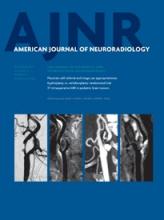
- Fig 1.
Anatomy of the subcallosal artery and RAH and their supplying basal forebrain regions. A, Specimen shows the subcallosal artery. The unpaired subcallosal artery (arrowheads) originates from the posterosuperior aspect of the ACoA, divides into 2 stems, and ascends dorsally into the lamina terminalis (LT) cistern, then curves upward and forward along the LT, paraterminal gyrus (PTG), subcallosal area (SbA), the rostrum (CCr) and genu (CCg) of the corpus callosum, and the anterior cingulate gyrus (CGa), thus exhibiting a characteristic S-shaped curve. OC indicates optic chiasm. B, Illustrative figure of the subcallosal artery (arrowheads) supplying the basal forebrain on the basis of our previous study regarding the microsurgical anatomy of the artery.16 The subcallosal artery originates from the posterosuperior aspect of the ACoA, ascends dorsally into the LT cistern, and supplies the 8 regions of the basal forebrain as follows: preoptic area (POpA), PTG including a part of the septum pellucidum (SP), SbA, anterior commissure (AC), and column of fornix (FxCo), then curves forward and upward to supply the CCr, CCg, and CGa. FM indicates foramen of Monro; MB, mammillary body; A2, A2 segment of the anterior cerebral artery; 3V, third ventricle. C, Specimen of the anterior cerebral and ACoA complex injected with methacrylic resin viewed from the posterior side. The subcallosal artery (arrowheads) is seen arising from the ACoA, A1, right and left A1 segments, and A2, right and left A2 segments, of the anterior cerebral arteries. D, Coronal microangiogram of the RAH on both sides and the unpaired subcallosal artery (reproduced, with permission, from Takahashi S, Goto K, Fukasawa H, et al. Computed tomography of cerebral infarction along the distribution of the basal perforating arteries. Part I. Striate arterial group. Radiology 1985;155:107–18). Both internal carotid arteries have been retracted inferiorly to demonstrate the cisternal course of the RAH (red arrows), which follows a curved or tortuous course along the A1 segment of the anterior cerebral artery (white arrows). The branches of the RAH are distributed to a part of the basal forebrain. The subcallosal artery (arrowhead) is also seen arising from the ACoA (yellow arrow).
- Fig 2.
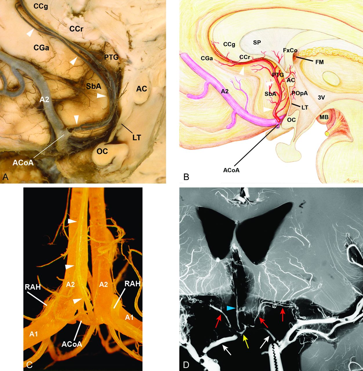
Summary of the infarcted foci on MR images in each region according to the vascular supply. The number on each bar graph represents the number of hemispheres in which infarcted foci of each region were found. Eight regions of the subcallosal artery: column of the fornix (FxCo), anterior commissure (AC), paraterminal gyrus (PTG), subcallosal area (SbA), genu of the corpus callosum (CCg), rostrum of the corpus callosum (CCr), anterior cingulate gyrus (CGa), and preoptic area (POpA). Five regions of the RAH: caudate nucleus (Cd), anterior limb of the internal capsule (ICa), putamen (Pt), nucleus accumbens (NAc), and globus pallidus (GP). Three other regions defined as the regions of unspecified vascular supply: diagonal band of Broca (DBB), bed nucleus of the stria terminalis (BNST), and substantia innominata (SI). The asterisk indicates that metallic artifacts from aneurysmal clips completely obscured the SbA in 2 hemispheres of 2 patients unilaterally and DBB in 1 patient bilaterally. Number sign indicates that “other frontal” represents the frontal lobe other than the orbitofrontal and basal forebrain region.
- Fig 3.
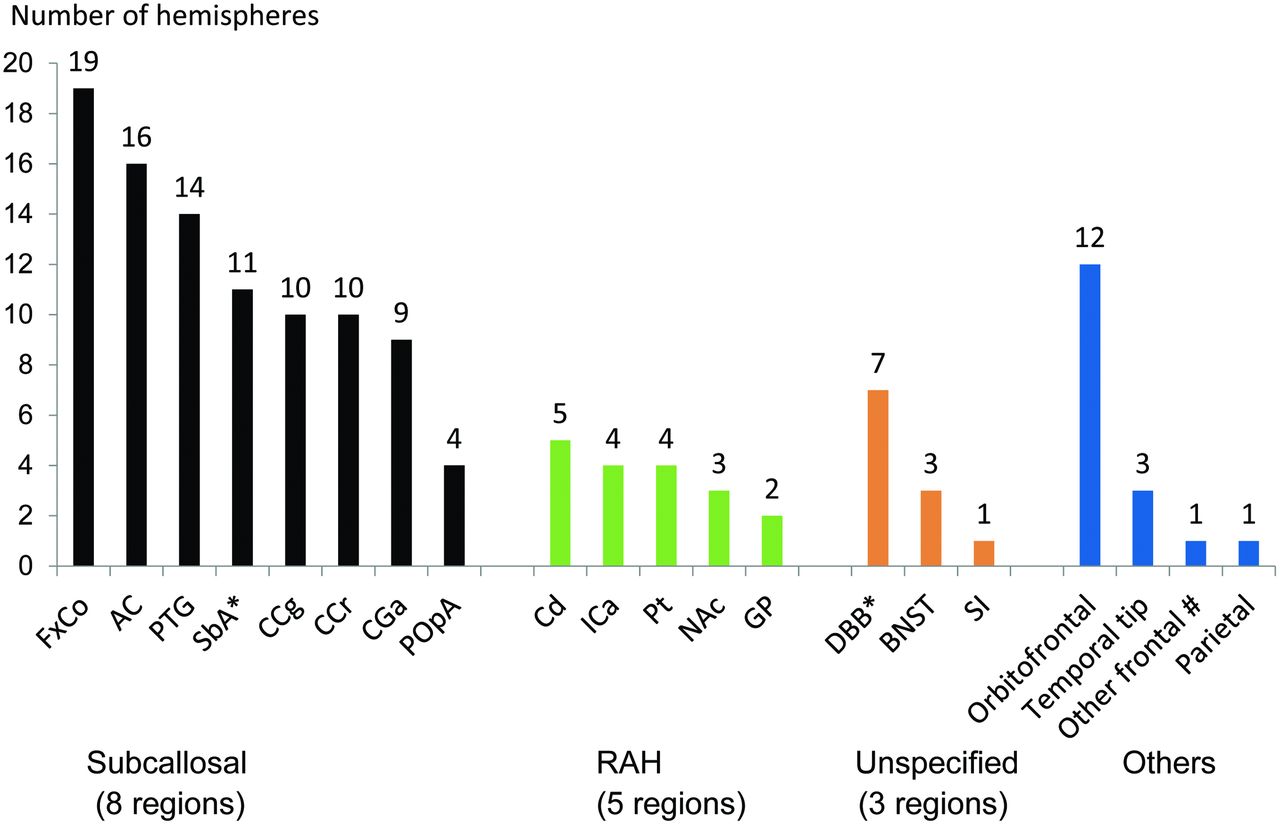
A 45-year-old who man presented with a ruptured aneurysm of the anterior communicating artery. Surgical clipping of the aneurysm was performed the day of onset (patient 4). Neuropsychological examination 3 months after the rupture confirmed amnesia. The imaging anatomy of the basal forebrain is detailed in On-line Fig 2: paramedian sagittal (A), coronal (B), and axial (C) and its next superior section (D), volumetric isotropic turbo spin-echo acquisition of T2WI (T2WI-VISTA) images shows infarcted foci in the midline (light blue arrows) and paramedian parts (dashed light blue arrows) of the anterior commissure along with the pars libera (red arrows) and pars tecta (dashed red arrows, C) of the columns of the fornices. Note that on coronal (B) and axial (C) images, infarcted foci in the bilateral anterior commissure show a characteristic bow-tie-like appearance and are associated with the infarcted foci in the adjoining bilateral pars libera and pars tecta of the column of the fornix. Other than the columns of the fornix and anterior commissure, no other regions are involved in the basal forebrain. The orbitofrontal region and temporal tip on the left were also involved, presumably damaged by the surgical procedure of the aneurysmal clipping (not shown).
- Fig 4.
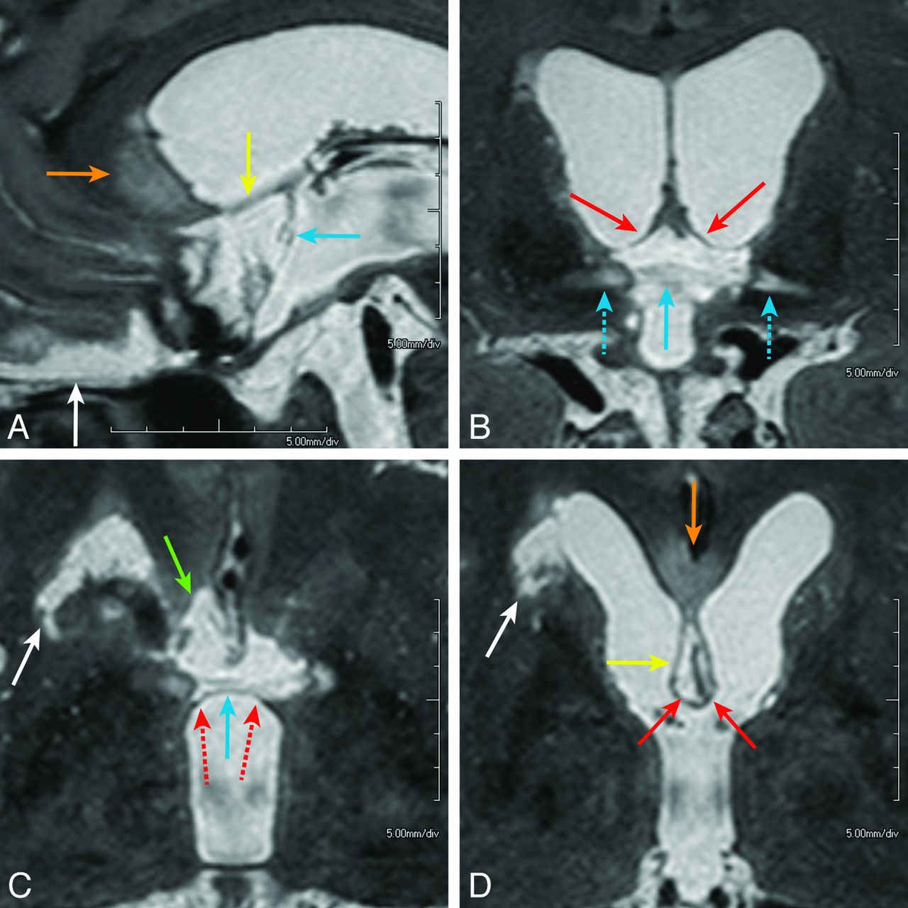
A 52-year-old man presented with a ruptured aneurysm of the anterior communicating artery. Surgical trapping of the ACoA for the ruptured ACoA aneurysm was performed the day of onset (patient 1). Neuropsychological examination 2 months after the rupture confirmed amnesia. Midsagittal (A), coronal (B), and axial (C and D) images of T2WI-VISTA show the infarcted foci involving the pars libera of the column of the fornix (B and D, solid red arrows) and anterior commissure (A–C, light blue arrows), paraterminal gyrus (A and D, yellow arrows), subcallosal area (C, green arrow), and genu or rostrum of the corpus callosum (A and D, orange arrows). The entire extent of the lesions shows sagittally elongated bandlike infarctions along the medial aspect of the brain, which probably represent the distribution of the characteristic S-shaped course of the subcallosal artery. On coronal (B) and axial (C) images, infarction in the bilateral anterior commissure shows a bow-tie-like appearance and is associated with infarcted foci in the adjoining pars libera (solid red arrows) and pars tecta (dashed red arrows) of the bilateral columns of the fornices. Lateral extension of hyperintense lesions along the lateral part of the anterior commissure (B, dashed light blue arrows) may indicate Wallerian degeneration of the midline infarction in the anterior commissure (solid light blue arrow). Infarction in the head of the caudate nucleus on the right is also seen (white arrows, C and D), which presumably represents involvement of the right recurrent artery of Heubner. The orbitofrontal region is also involved (A, white arrow).


Tables
Patient 1 2 3 4 5 6 7 8 9 10 Age (yr) (sex) 52/M 42/M 39/M 45/M 54/M 45/M 69/M 55/M 39/M 59/F Ruptured/unruptured R R R R R R U R R U CT gradea 3 4 3 3 4 3 NA 4 4 NA Treatment Trap Clip Clip 2nd Clip Clip Clip Clip Clip Clip Clip Months from treatmentb 2 4 4b 3 13 3 3 5 3 4 MMSEc 24 25 26 28 25 28 26 24 23 27 IQd 82 97 111 110 120 106 102 83 89 92 MQe 58 59 68 92 92 87 86 64 68 67 IQ minus MQ 24 38 43 18 28 19 16 19 21 25 Attention/concentration 81 80 114 138 131 115 115 91 94 133 Delayed recall <50 <50 <50 <50 64 73 62 <50 <50 <50 Note:—R indicates ruptured; U, unruptured; MMSE, Mini-Mental State Examination; IVH, intraventricular hemorrhage in the bilateral lateral ventricles; Trap, trapping; Clip, clipping; NA, not applied; WAIS-III, Wechsler Adult Intelligence Scale III; WMS-R, Wechsler Memory Scale-Revised.
↵a CT grade, proposed by Claassen et al,39 classifies the severity of aneurysmal subarachnoid hemorrhage on CT scans at onset into 5 grades from 0 to 4, according to the appearance of both of SAH and intraventricular hemorrhage in the bilateral lateral ventricles: grade 0, no SAH or IVH; grade 1, minimal SAH, no IVH; grade 2, minimal SAH, with IVH; grade 3, thick SAH, no IVH; grade 4, thick SAH, with IVH. In this scaling, the definition of “thick” is “completely filling” ≥1 cistern or fissure. In 2 patients with unruptured aneurysm (patients 7 and 10), grading was not applied.
↵b Months after second clipping.
↵c MMSE, used to assess cognitive impairment (full score 30).40
↵d IQ evaluated by the Wechsler Adult Intelligence Scale III.18
↵e MQ, general memory quotient, attention/concentration quotient, and delayed recall quotient, evaluated by Wechsler Memory Scale-Revised.19 Each quotient has a mean of 100 in the normal population and an SD of 15. A substantial difference between IQ (by the WAIS-III) and MQ (by the WMS-R) scores indicates that the person with amnesia has a particular impairment in memory—but not in the “intelligence” per se.20
Patient No. (R/L) 1 2 3 4 5 6 7 8 9 10 R L R L R L R L R L R L R L R L R L R L Vascular territory Subcallosal (n = 8) + + + + + + + + + + + + + + + + + + + RAH (n = 5) + + + + + Unspecified (n = 3) + + + + + + + + Note:—R/L indicates right and left hemisphere; +, infarcted focus present in a vascular territory; blank space, sparing of the territory.
↵a For vascular territories of subcallosal, RAH, and unspecified, refer to Fig. 2.


