Advertisement

Main menu

  • Home
  • Content
    • Current Issue
    • Accepted Manuscripts
    • Article Preview
    • Past Issue Archive
    • Video Articles
    • AJNR Case Collection
    • Case of the Week Archive
    • Case of the Month Archive
    • Classic Case Archive
  • Special Collections
    • AJNR Awards
    • Low-Field MRI
    • Alzheimer Disease
    • ASNR Foundation Special Collection
    • Photon-Counting CT
    • View All
  • Multimedia
    • AJNR Podcasts
    • AJNR SCANtastic
    • Trainee Corner
    • MRI Safety Corner
    • Imaging Protocols
  • For Authors
    • Submit a Manuscript
    • Submit a Video Article
    • Submit an eLetter to the Editor/Response
    • Manuscript Submission Guidelines
    • Statistical Tips
    • Fast Publishing of Accepted Manuscripts
    • Graphical Abstract Preparation
    • Imaging Protocol Submission
    • Author Policies
  • About Us
    • About AJNR
    • Editorial Board
    • Editorial Board Alumni
  • More
    • Become a Reviewer/Academy of Reviewers
    • Subscribers
    • Permissions
    • Alerts
    • Feedback
    • Advertisers
    • ASNR Home

User menu

  • Alerts
  • Log in

Search

  • Advanced search
American Journal of Neuroradiology

American Journal of Neuroradiology

ASHNR American Society of Functional Neuroradiology ASHNR American Society of Pediatric Neuroradiology ASSR
  • Alerts
  • Log in

Advanced Search

  • Home
  • Content
    • Current Issue
    • Accepted Manuscripts
    • Article Preview
    • Past Issue Archive
    • Video Articles
    • AJNR Case Collection
    • Case of the Week Archive
    • Case of the Month Archive
    • Classic Case Archive
  • Special Collections
    • AJNR Awards
    • Low-Field MRI
    • Alzheimer Disease
    • ASNR Foundation Special Collection
    • Photon-Counting CT
    • View All
  • Multimedia
    • AJNR Podcasts
    • AJNR SCANtastic
    • Trainee Corner
    • MRI Safety Corner
    • Imaging Protocols
  • For Authors
    • Submit a Manuscript
    • Submit a Video Article
    • Submit an eLetter to the Editor/Response
    • Manuscript Submission Guidelines
    • Statistical Tips
    • Fast Publishing of Accepted Manuscripts
    • Graphical Abstract Preparation
    • Imaging Protocol Submission
    • Author Policies
  • About Us
    • About AJNR
    • Editorial Board
    • Editorial Board Alumni
  • More
    • Become a Reviewer/Academy of Reviewers
    • Subscribers
    • Permissions
    • Alerts
    • Feedback
    • Advertisers
    • ASNR Home
  • Follow AJNR on Twitter
  • Visit AJNR on Facebook
  • Follow AJNR on Instagram
  • Join AJNR on LinkedIn
  • RSS Feeds

AJNR Awards, New Junior Editors, and more. Read the latest AJNR updates

Research ArticleBrain
Open Access

Long-Term White Matter Changes after Severe Traumatic Brain Injury: A 5-Year Prospective Cohort

J. Dinkel, A. Drier, O. Khalilzadeh, V. Perlbarg, V. Czernecki, R. Gupta, F. Gomas, P. Sanchez, D. Dormont, D. Galanaud, R.D. Stevens, L. Puybasset and for NICER (Neuro Imaging for Coma Emergence and Recovery) Consortium
American Journal of Neuroradiology January 2014, 35 (1) 23-29; DOI: https://doi.org/10.3174/ajnr.A3616
J. Dinkel
aFrom the Department of Radiology (J.D., O.K., R.G.), Massachusetts General Hospital, Harvard Medical School, Boston, Massachusetts
bDepartment of Radiology (J.D.), University Hospital Heidelberg, Heidelberg, Germany
  • Find this author on Google Scholar
  • Find this author on PubMed
  • Search for this author on this site
A. Drier
cDepartments of Neuroradiology (A.D., D.D., D.G.)
  • Find this author on Google Scholar
  • Find this author on PubMed
  • Search for this author on this site
O. Khalilzadeh
aFrom the Department of Radiology (J.D., O.K., R.G.), Massachusetts General Hospital, Harvard Medical School, Boston, Massachusetts
  • Find this author on Google Scholar
  • Find this author on PubMed
  • Search for this author on this site
V. Perlbarg
fINSERM, UMRS 678 (V.P.)
  • Find this author on Google Scholar
  • Find this author on PubMed
  • Search for this author on this site
V. Czernecki
dNeurology (V.C.)
gINSERM, CRICM UMR S975 (V.C.), Université Pierre et Marie Curie-Paris 6, Paris, France
  • Find this author on Google Scholar
  • Find this author on PubMed
  • Search for this author on this site
R. Gupta
aFrom the Department of Radiology (J.D., O.K., R.G.), Massachusetts General Hospital, Harvard Medical School, Boston, Massachusetts
  • Find this author on Google Scholar
  • Find this author on PubMed
  • Search for this author on this site
F. Gomas
hDepartment of Anesthesiology and Intensive Care (F.G.), Louis Mourier Hospital, Colombes, France
  • Find this author on Google Scholar
  • Find this author on PubMed
  • Search for this author on this site
P. Sanchez
eAnesthesiology and Intensive Care (P.S., L.P.), Pitié Salpêtrière Hospital, Assistance Publique–Hôpitaux de Paris, France
  • Find this author on Google Scholar
  • Find this author on PubMed
  • Search for this author on this site
D. Dormont
cDepartments of Neuroradiology (A.D., D.D., D.G.)
  • Find this author on Google Scholar
  • Find this author on PubMed
  • Search for this author on this site
D. Galanaud
cDepartments of Neuroradiology (A.D., D.D., D.G.)
  • Find this author on Google Scholar
  • Find this author on PubMed
  • Search for this author on this site
R.D. Stevens
iDivision of Neuroscience Critical Care (R.D.S.), Department of Anesthesiology Critical Care Medicine, Johns Hopkins University School of Medicine, Baltimore, Maryland.
  • Find this author on Google Scholar
  • Find this author on PubMed
  • Search for this author on this site
L. Puybasset
eAnesthesiology and Intensive Care (P.S., L.P.), Pitié Salpêtrière Hospital, Assistance Publique–Hôpitaux de Paris, France
  • Find this author on Google Scholar
  • Find this author on PubMed
  • Search for this author on this site
  • Article
  • Figures & Data
  • Info & Metrics
  • Responses
  • References
  • PDF
Loading

Article Figures & Data

Figures

  • Tables
  • Fig 1.
    • Download figure
    • Open in new tab
    • Download powerpoint
    Fig 1.

    MR images of a patient with severe TBI at baseline (A and D), year 2 (B and E), and year 5 (C and F). On the conventional FLAIR images (A–C), the hyperintensity of the right frontal lobe (arrow) decreases with time with some minimal atrophic changes. The corresponding FA maps (D–F) are presented. In the acute setting, the normalized FA was slightly reduced in the genu (0.80) and body (0.92) of the corpus callosum and in the bilateral corona radiata (right: 0.96; left: 0.98) compared with control participants (normalized value = 1). At 2 years, most of the normalized FA values in these tracts had further decreased: genu (0.81) and body (0.79) of corpus callosum; corona radiata (right: 0.89; left: 0.96). At 5 years, the values slightly increased: genu (0.82) and body (0.85) of corpus callosum; corona radiata (right: 0.92; left: 0.97).

  • Fig 2.
    • Download figure
    • Open in new tab
    • Download powerpoint
    Fig 2.

    Normalized FA (A), and Lt (B) values of patients and control participants at baseline and follow-up steps. The x-axis represents the ROIs (in posterior fossa: 1- MCP: middle cerebellar peduncle; 2- antBS: anterior brain stem; 3- postBS: posterior brain stem; 4- CP-R: right cerebral peduncle; 5- CP-L: left cerebral peduncle; in deep brain: 6- g-CC: genu of the corpus callosum; 7- b-CC: body of the corpus callosum; 8-second-CC: splenium of the corpus callosum; 9- ALIC-R: right side anterior arm of the internal capsule; 10- ALIC-L: left side anterior arm of the internal capsule; 11- PLIC-R: right side posterior arm of the internal capsule; 12- PLIC-L: left side posterior arm of the internal capsule; in superficial brain regions: 13- SS-R: right stratum sagittale; 14- SS-L: left stratum sagittale; 15- SLF-R: right superior longitudinal fasciculus; 16- SLF-L: left superior longitudinal fasciculus; 17- EC-R: right external capsule; 18- EC-L: left external capsule; 19- CR-R: right corona radiata; 20- CR-L: left corona radiata). There were no significant differences in DTI parameters between baseline and year 2 in the control group and between year 2 and year 5 in the TBI group. ‡: P < .05; for comparison of baseline values between control participants and patients (corrected P). *: P < .05; for repeated-measures ANOVA in the patient group between baseline and follow-up steps. Data are presented as mean and standard error of mean (handles). The connection lines are for clarity.

  • Fig 3.
    • Download figure
    • Open in new tab
    • Download powerpoint
    Fig 3.

    The changes (year 2 vs baseline) in normalized FA (A) and Lt (B) in the presented ROIs were significantly higher in the patients vs healthy control participants. P < .05 for comparison between patients and control participants in all of the presented ROIs.

  • Fig 4.
    • Download figure
    • Open in new tab
    • Download powerpoint
    Fig 4.

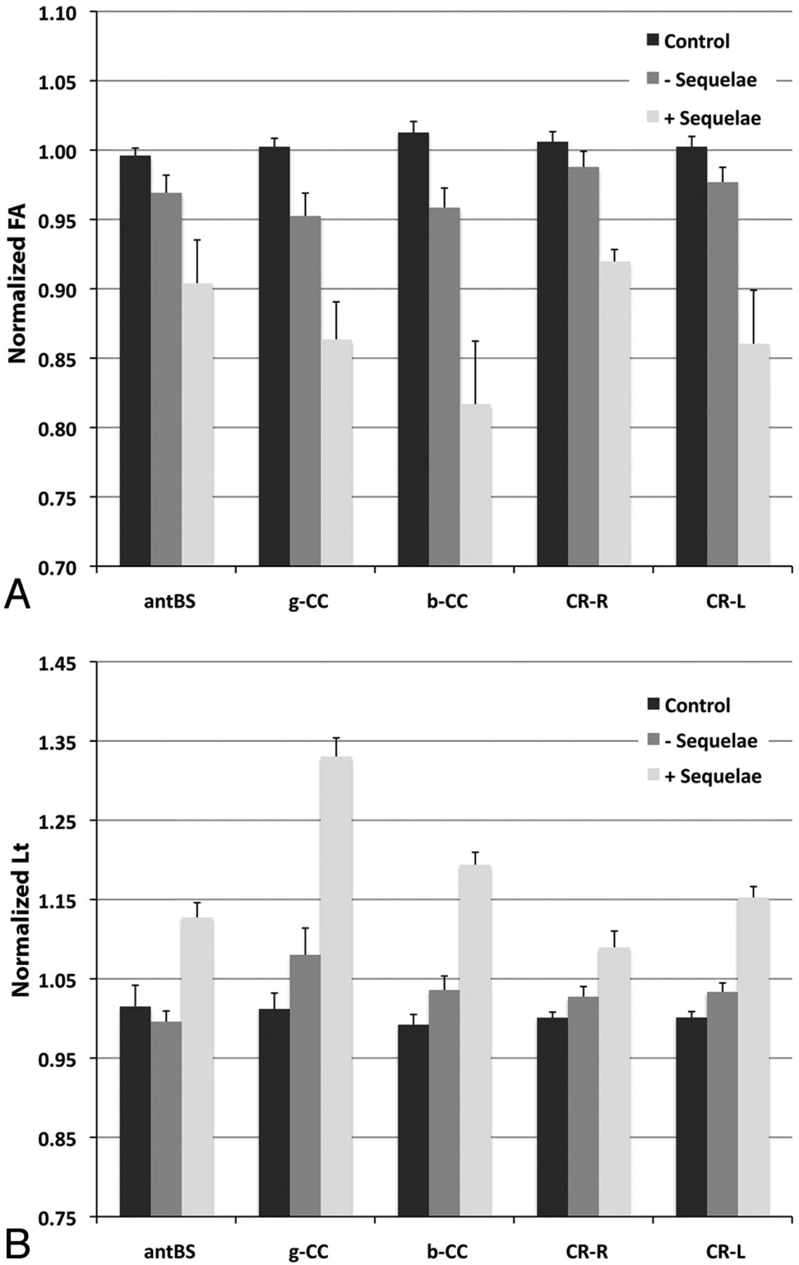
    The normalized FA (A) and Lt (B) values at baseline were significantly different in patients with cognitive sequelae at year 5 (including amnesia, aphasia, and dyspraxia) vs healthy control participants. P < .05 for ANOVA among the groups and for comparison between healthy control participants and patients with sequelae in all of the presented ROIs.

Tables

  • Figures
  • Patient characteristics

    Range
    Age (y)32.4 (9.0)(18–49)
    Men/women, n13/0−
    GCS at admission, n (SD)6 (4)(3–8)
    ICU stay duration (d)47.5 (23.1)(10–97)
    Assisted ventilation duration (d)33.1 (16.9)(9–72)
    Type of accident, n (%)
        MVA10 (77)−
        Assault, fall, other3 (23)−
    Hematoma, n (%)a
        Epidural3 (23)−
        Subdural1 (8)−
        Subarachnoid hemorrhage8 (61)−
    Midline shift3 (23)−
    Compressed third ventricle4 (31)−
    Contusion, n (%)b10 (77)−
    Neurosurgical intervention, n (%)2 (15)−
    Neurologic sequelae, n (%)
        Amnesia2 (15)−
        Aphasia1 (8)−
        Dyspraxia2 (15)−
    • Note:—Variables are presented as mean (standard deviation) or number (percentage).

    • GCS indicates Glasgow Coma Scale; ICU, intensive care unit; SD, standard deviation; MVA, motor-vehicle collision.

    • ↵a Hematoma was defined as an area spontaneously hyperdense on the first CT scan.

    • ↵b Contusion was defined as a focal area appearing hypodense or of mixed density on the CT scan.

PreviousNext
Back to top

In this issue

American Journal of Neuroradiology: 35 (1)
American Journal of Neuroradiology
Vol. 35, Issue 1
1 Jan 2014
  • Table of Contents
  • Index by author
  • Complete Issue (PDF)
Advertisement
Print
Download PDF
Email Article

Thank you for your interest in spreading the word on American Journal of Neuroradiology.

NOTE: We only request your email address so that the person you are recommending the page to knows that you wanted them to see it, and that it is not junk mail. We do not capture any email address.

Enter multiple addresses on separate lines or separate them with commas.
Long-Term White Matter Changes after Severe Traumatic Brain Injury: A 5-Year Prospective Cohort
(Your Name) has sent you a message from American Journal of Neuroradiology
(Your Name) thought you would like to see the American Journal of Neuroradiology web site.
CAPTCHA
This question is for testing whether or not you are a human visitor and to prevent automated spam submissions.
Cite this article
J. Dinkel, A. Drier, O. Khalilzadeh, V. Perlbarg, V. Czernecki, R. Gupta, F. Gomas, P. Sanchez, D. Dormont, D. Galanaud, R.D. Stevens, L. Puybasset, for NICER (Neuro Imaging for Coma Emergence and Recovery) Consortium
Long-Term White Matter Changes after Severe Traumatic Brain Injury: A 5-Year Prospective Cohort
American Journal of Neuroradiology Jan 2014, 35 (1) 23-29; DOI: 10.3174/ajnr.A3616

Citation Manager Formats

  • BibTeX
  • Bookends
  • EasyBib
  • EndNote (tagged)
  • EndNote 8 (xml)
  • Medlars
  • Mendeley
  • Papers
  • RefWorks Tagged
  • Ref Manager
  • RIS
  • Zotero
0 Responses
Respond to this article
Share
Bookmark this article
Long-Term White Matter Changes after Severe Traumatic Brain Injury: A 5-Year Prospective Cohort
J. Dinkel, A. Drier, O. Khalilzadeh, V. Perlbarg, V. Czernecki, R. Gupta, F. Gomas, P. Sanchez, D. Dormont, D. Galanaud, R.D. Stevens, L. Puybasset, for NICER (Neuro Imaging for Coma Emergence and Recovery) Consortium
American Journal of Neuroradiology Jan 2014, 35 (1) 23-29; DOI: 10.3174/ajnr.A3616
del.icio.us logo Twitter logo Facebook logo Mendeley logo
  • Tweet Widget
  • Facebook Like
  • Google Plus One
Purchase

Jump to section

  • Article
    • Abstract
    • ABBREVIATIONS:
    • Materials and Methods
    • Results
    • Discussion
    • Conclusions
    • Footnotes
    • REFERENCES
  • Figures & Data
  • Info & Metrics
  • Responses
  • References
  • PDF

Related Articles

  • No related articles found.
  • PubMed
  • Google Scholar

Cited By...

  • Longitudinal increases in structural connectome segregation and functional connectome integration are associated with better recovery after mild TBI
  • Toward Precision and Reproducibility of Diffusion Tensor Imaging: A Multicenter Diffusion Phantom and Traveling Volunteer Study
  • Crossref (53)
  • Google Scholar

This article has been cited by the following articles in journals that are participating in Crossref Cited-by Linking.

  • In-vivo Dynamics of the Human Hippocampus across the Menstrual Cycle
    Claudia Barth, Christopher J Steele, Karsten Mueller, Vivien P. Rekkas, Katrin Arélin, Andre Pampel, Inga Burmann, Jürgen Kratzsch, Arno Villringer, Julia Sacher
    Scientific Reports 2016 6 1
  • Toward Precision and Reproducibility of Diffusion Tensor Imaging: A Multicenter Diffusion Phantom and Traveling Volunteer Study
    E.M. Palacios, A.J. Martin, M.A. Boss, F. Ezekiel, Y.S. Chang, E.L. Yuh, M.J. Vassar, D.M. Schnyer, C.L. MacDonald, K.L. Crawford, A. Irimia, A.W. Toga, P. Mukherjee
    American Journal of Neuroradiology 2017 38 3
  • Traumatic Brain Injury Incidence, Clinical Overview, and Policies in the US Military Health System Since 2000
    Thomas M. Swanson, Brad M. Isaacson, Cherina M. Cyborski, Louis M. French, Jack W. Tsao, Paul F. Pasquina
    Public Health Reports® 2017 132 2
  • Extended Anatomical Grading in Diffuse Axonal Injury Using MRI: Hemorrhagic Lesions in the Substantia Nigra and Mesencephalic Tegmentum Indicate Poor Long-Term Outcome
    Sami Abu Hamdeh, Niklas Marklund, Marianne Lannsjö, Tim Howells, Raili Raininko, Johan Wikström, Per Enblad
    Journal of Neurotrauma 2017 34 2
  • Traumatic Brain Injury and Risk of Long-Term Brain Changes, Accumulation of Pathological Markers, and Developing Dementia: A Review
    Christian LoBue, Catherine Munro, Jeffrey Schaffert, Nyaz Didehbani, John Hart,, Hunt Batjer, C. Munro Cullum
    Journal of Alzheimer's Disease 2019 70 3
  • White matter disruption in moderate/severe pediatric traumatic brain injury: Advanced tract-based analyses
    Emily L. Dennis, Yan Jin, Julio E. Villalon-Reina, Liang Zhan, Claudia L. Kernan, Talin Babikian, Richard B. Mink, Christopher J. Babbitt, Jeffrey L. Johnson, Christopher C. Giza, Paul M. Thompson, Robert F. Asarnow
    NeuroImage: Clinical 2015 7
  • Dynamic Changes in White Matter Abnormalities Correlate With Late Improvement and Deterioration Following TBI
    Virginia F. J. Newcombe, Marta M. Correia, Christian Ledig, Maria G. Abate, Joanne G. Outtrim, Doris Chatfield, Thomas Geeraerts, Anne E. Manktelow, Eleftherios Garyfallidis, John D. Pickard, Barbara J. Sahakian, Peter J. A. Hutchinson, Daniel Rueckert, Jonathan P. Coles, Guy B. Williams, David K. Menon
    Neurorehabilitation and Neural Repair 2016 30 1
  • Corpus callosum and epilepsies
    Iris Unterberger, Richard Bauer, Gerald Walser, Gerhard Bauer
    Seizure 2016 37
  • Longitudinal increases in structural connectome segregation and functional connectome integration are associated with better recovery after mild TBI
    Amy F. Kuceyeski, Keith W. Jamison, Julia P. Owen, Ashish Raj, Pratik Mukherjee
    Human Brain Mapping 2019 40 15
  • The application of a mathematical model linking structural and functional connectomes in severe brain injury
    A. Kuceyeski, S. Shah, J.P. Dyke, S. Bickel, F. Abdelnour, N.D. Schiff, H.U. Voss, A. Raj
    NeuroImage: Clinical 2016 11

More in this TOC Section

  • Fast Contrast-Enhanced 4D MRA and 4D Flow MRI Using Constrained Reconstruction (HYPRFlow): Potential Applications for Brain Arteriovenous Malformations
  • Multimodal CT Provides Improved Performance for Lacunar Infarct Detection
  • Optimal MRI Sequence for Identifying Occlusion Location in Acute Stroke: Which Value of Time-Resolved Contrast-Enhanced MRA?
Show more BRAIN

Similar Articles

Advertisement

Indexed Content

  • Current Issue
  • Accepted Manuscripts
  • Article Preview
  • Past Issues
  • Editorials
  • Editor's Choice
  • Fellows' Journal Club
  • Letters to the Editor
  • Video Articles

Cases

  • Case Collection
  • Archive - Case of the Week
  • Archive - Case of the Month
  • Archive - Classic Case

More from AJNR

  • Trainee Corner
  • Imaging Protocols
  • MRI Safety Corner
  • Book Reviews

Multimedia

  • AJNR Podcasts
  • AJNR Scantastics

Resources

  • Turnaround Time
  • Submit a Manuscript
  • Submit a Video Article
  • Submit an eLetter to the Editor/Response
  • Manuscript Submission Guidelines
  • Statistical Tips
  • Fast Publishing of Accepted Manuscripts
  • Graphical Abstract Preparation
  • Imaging Protocol Submission
  • Evidence-Based Medicine Level Guide
  • Publishing Checklists
  • Author Policies
  • Become a Reviewer/Academy of Reviewers
  • News and Updates

About Us

  • About AJNR
  • Editorial Board
  • Editorial Board Alumni
  • Alerts
  • Permissions
  • Not an AJNR Subscriber? Join Now
  • Advertise with Us
  • Librarian Resources
  • Feedback
  • Terms and Conditions
  • AJNR Editorial Board Alumni

American Society of Neuroradiology

  • Not an ASNR Member? Join Now

© 2025 by the American Society of Neuroradiology All rights, including for text and data mining, AI training, and similar technologies, are reserved.
Print ISSN: 0195-6108 Online ISSN: 1936-959X

Powered by HighWire