Advertisement
  • Main menu
  • User menu
  • Search
  • English ▼
    • English
    • Afrikaans
    • Albanian
    • Amharic
    • Arabic
    • Armenian
    • Azerbaijani
    • Basque
    • Belarusian
    • Bengali
    • Bosnian
    • Bulgarian
    • Catalan
    • Cebuano
    • Chichewa
    • Chinese (Simplified)
    • Chinese (Traditional)
    • Corsican
    • Croatian
    • Czech
    • Danish
    • Dutch
    • Esperanto
    • Estonian
    • Filipino
    • Finnish
    • French
    • Frisian
    • Galician
    • Georgian
    • German
    • Greek
    • Gujarati
    • Haitian Creole
    • Hausa
    • Hawaiian
    • Hebrew
    • Hindi
    • Hmong
    • Hungarian
    • Icelandic
    • Igbo
    • Indonesian
    • Irish
    • Italian
    • Japanese
    • Javanese
    • Kannada
    • Kazakh
    • Khmer
    • Korean
    • Kurdish (Kurmanji)
    • Kyrgyz
    • Lao
    • Latin
    • Latvian
    • Lithuanian
    • Luxembourgish
    • Macedonian
    • Malagasy
    • Malay
    • Malayalam
    • Maltese
    • Maori
    • Marathi
    • Mongolian
    • Myanmar (Burmese)
    • Nepali
    • Norwegian
    • Pashto
    • Persian
    • Polish
    • Portuguese
    • Punjabi
    • Romanian
    • Russian
    • Samoan
    • Scottish Gaelic
    • Serbian
    • Sesotho
    • Shona
    • Sindhi
    • Sinhala
    • Slovak
    • Slovenian
    • Somali
    • Spanish
    • Sudanese
    • Swahili
    • Swedish
    • Tajik
    • Tamil
    • Telugu
    • Thai
    • Turkish
    • Ukrainian
    • Urdu
    • Uzbek
    • Vietnamese
    • Welsh
    • Xhosa
    • Yiddish
    • Yoruba
    • Zulu

Main menu

  • Home
  • Content
    • Current Issue
    • Accepted Manuscripts
    • Article Preview
    • Past Issue Archive
    • Video Articles
    • AJNR Case Collection
    • Case of the Week Archive
    • Case of the Month Archive
    • Classic Case Archive
  • Special Collections
    • Advancing NeuroMRI with High-Relaxivity Contrast Agents
    • AJNR Awards
    • Low-Field MRI
    • Alzheimer Disease
    • ASNR Foundation Special Collection
    • Photon-Counting CT
    • View All
  • Multimedia
    • AJNR Podcasts
    • AJNR SCANtastic
    • Trainee Corner
    • MRI Safety Corner
    • Imaging Protocols
  • For Authors
    • Submit a Manuscript
    • Submit a Video Article
    • Submit an eLetter to the Editor/Response
    • Manuscript Submission Guidelines
    • Statistical Tips
    • Fast Publishing of Accepted Manuscripts
    • Graphical Abstract Preparation
    • Imaging Protocol Submission
    • Author Policies
  • About Us
    • About AJNR
    • Editorial Board
    • Editorial Board Alumni
  • More
    • Become a Reviewer/Academy of Reviewers
    • Subscribers
    • Permissions
    • Alerts
    • Feedback
    • Advertisers
    • ASNR Home

User menu

  • Alerts
  • Log in

Search

  • Advanced search
American Journal of Neuroradiology

American Journal of Neuroradiology

ASHNR American Society of Functional Neuroradiology ASHNR American Society of Pediatric Neuroradiology ASSR
  • Alerts
  • Log in

Advanced Search

English ▼
  • English
  • Afrikaans
  • Albanian
  • Amharic
  • Arabic
  • Armenian
  • Azerbaijani
  • Basque
  • Belarusian
  • Bengali
  • Bosnian
  • Bulgarian
  • Catalan
  • Cebuano
  • Chichewa
  • Chinese (Simplified)
  • Chinese (Traditional)
  • Corsican
  • Croatian
  • Czech
  • Danish
  • Dutch
  • Esperanto
  • Estonian
  • Filipino
  • Finnish
  • French
  • Frisian
  • Galician
  • Georgian
  • German
  • Greek
  • Gujarati
  • Haitian Creole
  • Hausa
  • Hawaiian
  • Hebrew
  • Hindi
  • Hmong
  • Hungarian
  • Icelandic
  • Igbo
  • Indonesian
  • Irish
  • Italian
  • Japanese
  • Javanese
  • Kannada
  • Kazakh
  • Khmer
  • Korean
  • Kurdish (Kurmanji)
  • Kyrgyz
  • Lao
  • Latin
  • Latvian
  • Lithuanian
  • Luxembourgish
  • Macedonian
  • Malagasy
  • Malay
  • Malayalam
  • Maltese
  • Maori
  • Marathi
  • Mongolian
  • Myanmar (Burmese)
  • Nepali
  • Norwegian
  • Pashto
  • Persian
  • Polish
  • Portuguese
  • Punjabi
  • Romanian
  • Russian
  • Samoan
  • Scottish Gaelic
  • Serbian
  • Sesotho
  • Shona
  • Sindhi
  • Sinhala
  • Slovak
  • Slovenian
  • Somali
  • Spanish
  • Sudanese
  • Swahili
  • Swedish
  • Tajik
  • Tamil
  • Telugu
  • Thai
  • Turkish
  • Ukrainian
  • Urdu
  • Uzbek
  • Vietnamese
  • Welsh
  • Xhosa
  • Yiddish
  • Yoruba
  • Zulu
  • Home
  • Content
    • Current Issue
    • Accepted Manuscripts
    • Article Preview
    • Past Issue Archive
    • Video Articles
    • AJNR Case Collection
    • Case of the Week Archive
    • Case of the Month Archive
    • Classic Case Archive
  • Special Collections
    • Advancing NeuroMRI with High-Relaxivity Contrast Agents
    • AJNR Awards
    • Low-Field MRI
    • Alzheimer Disease
    • ASNR Foundation Special Collection
    • Photon-Counting CT
    • View All
  • Multimedia
    • AJNR Podcasts
    • AJNR SCANtastic
    • Trainee Corner
    • MRI Safety Corner
    • Imaging Protocols
  • For Authors
    • Submit a Manuscript
    • Submit a Video Article
    • Submit an eLetter to the Editor/Response
    • Manuscript Submission Guidelines
    • Statistical Tips
    • Fast Publishing of Accepted Manuscripts
    • Graphical Abstract Preparation
    • Imaging Protocol Submission
    • Author Policies
  • About Us
    • About AJNR
    • Editorial Board
    • Editorial Board Alumni
  • More
    • Become a Reviewer/Academy of Reviewers
    • Subscribers
    • Permissions
    • Alerts
    • Feedback
    • Advertisers
    • ASNR Home
  • Follow AJNR on Twitter
  • Visit AJNR on Facebook
  • Follow AJNR on Instagram
  • Join AJNR on LinkedIn
  • RSS Feeds

AJNR Awards, New Junior Editors, and more. Read the latest AJNR updates


Improved Turnaround Times | Median time to first decision: 12 days

Research ArticleSpine Imaging and Spine Image-Guided Interventions

Optimal Contrast Concentration for CT-Guided Epidural Steroid Injections

P.G. Kranz, M. Abbott, D. Abbott and J.K. Hoang
American Journal of Neuroradiology January 2014, 35 (1) 191-195; DOI: https://doi.org/10.3174/ajnr.A3626
P.G. Kranz
aFrom the Departments of Radiology (P.G.K., M.A., J.K.H.)
  • Find this author on Google Scholar
  • Find this author on PubMed
  • Search for this author on this site
M. Abbott
aFrom the Departments of Radiology (P.G.K., M.A., J.K.H.)
  • Find this author on Google Scholar
  • Find this author on PubMed
  • Search for this author on this site
D. Abbott
bBiostatistics and Bioinformatics (D.A.), Duke University Medical Center, Durham, North Carolina.
  • Find this author on Google Scholar
  • Find this author on PubMed
  • Search for this author on this site
J.K. Hoang
aFrom the Departments of Radiology (P.G.K., M.A., J.K.H.)
  • Find this author on Google Scholar
  • Find this author on PubMed
  • Search for this author on this site
  • Article
  • Figures & Data
  • Info & Metrics
  • Responses
  • References
  • PDF
Loading

Article Figures & Data

Figures

  • Tables
  • Fig 1.
    • Download figure
    • Open in new tab
    • Download powerpoint
    Fig 1.

    Phantom study assessing needle visibility. A, Scout image from CT demonstrating the configuration of the phantom. The central chamber houses contrast and a 22-gauge needle. Axial cross-sectional images of the chamber are shown with various contrast dilutions: 50 mg/mL (B), 66 mg/mL (C), 100 mg/mL (D), 133 mg/mL (E), and 150 mg/mL (F).

  • Fig 2.
    • Download figure
    • Open in new tab
    • Download powerpoint
    Fig 2.

    Axial CTF images show representative examples of visibility scores on the basis of our 5-point scoring system. A, Score of +2. The contrast is too dense to visualize the location of the needle tip. B, Score of 0. The needle tip can be distinguished from the injected contrast; adequate soft tissue contrast is also provided. C, Score of −2. The injected contrast is too dilute to provide adequate soft tissue contrast.
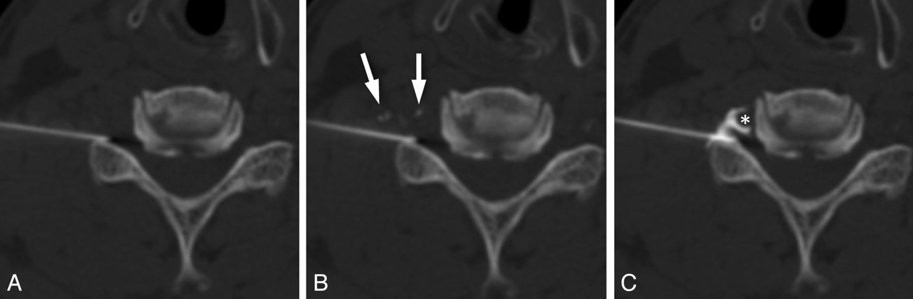
  • Fig 3.
    • Download figure
    • Open in new tab
    • Download powerpoint
    Fig 3.

    Importance of precise needle placement in cervical epidural steroid injections. A, Axial CTF image obtained before contrast injection. B, Image obtained immediately after contrast injection shows transient filling of small vessels (arrows). No epidural spread is seen. C, Image obtained after advancing the needle <2 mm demonstrates foraminal epidural contrast spread, with no vascular filling. Note the close proximity of the needle tip to the vertebral artery (*). This case highlights the importance of accurate localization of the needle tip and the need to be able to make small but precise adjustments in needle position during epidural injections.

Tables

  • Figures
    • View popup
    Table 1:

    Five-point scoring system for the clinical study

    Too ConcentratedOptimalToo Dilute
    Visibility score+2+10−1−2
    Needle tipNot visibleBorderlineVisibleVisibleVisible
    Soft tissue contrastAdequateAdequateAdequateBorderlineNot acceptable
    • View popup
    Table 2:

    Visibility scores for the clinical study

    Visibility Score
    −2−10+1+2
    Total scores (n = 120)n = 9n = 10n = 34n = 25n = 42
    (2 readers)(7.5%)(8.3%)(28.3%)(20.8%)(35.0%)
    Iodine concentration
        66 mg/mL (n = 34)n = 7n = 5n = 15n = 6n = 1
    (20.6%)(14.7%)(44.1%)(17.6%)(2.9%)
        100 mg/mL (n = 32)n = 2n = 5n = 9n = 10n = 6
    (6.3%)(15.6%)(28.1%)(31.3%)(18.8%)
        133 mg/mL (n = 26)n = 0n = 0n = 2n = 3n = 21
    (0%)(0%)(7.7%)(11.5%)(80.8%)
        150 mg/mL (n = 28)n = 0n = 0n = 8n = 6n = 14
    (0%)(0%)(28.6%)(21.4%)(50.0%)
    Injection technique
        ILESI (n = 64)n = 0n = 3n = 14n = 17n = 30
    (0%)(4.7%)(21.9%)(26.6%)(46.9%)
        TFESI (n = 56)n = 9n = 7n = 20n = 8n = 12
    (16.1%)(12.5%)(35.7%)(14.3%)(21.4%)
    • View popup
    Table 3:

    Comparison of visibility scores on the basis of combined iodine concentration and injection technique

    Iodine Concentration + Injection TechniqueP Valuea
    (66 mg/mL and TFESI) versus (100 mg/mL and TFESI).011
    (66 mg/mL and ILESI) versus (100 mg/mL and ILESI).016
    (66 mg/mL and ILESI) versus (66 mg/mL and TFESI).051
    (100 mg/mL and ILESI) versus (100 mg/mL and TFESI).021
    • ↵a P values are based on results of generalized estimating equations test of difference contrast statements with the use of a Bonferroni-corrected significance threshold of 0.0083.

PreviousNext
Back to top

In this issue

American Journal of Neuroradiology: 35 (1)
American Journal of Neuroradiology
Vol. 35, Issue 1
1 Jan 2014
  • Table of Contents
  • Index by author
  • Complete Issue (PDF)
Advertisement
Print
Download PDF
Email Article
Cite this article
0 Responses
Respond to this article
Share
Bookmark this article
Optimal Contrast Concentration for CT-Guided Epidural Steroid Injections
P.G. Kranz, M. Abbott, D. Abbott, J.K. Hoang
American Journal of Neuroradiology Jan 2014, 35 (1) 191-195; DOI: 10.3174/ajnr.A3626
del.icio.us logo Twitter logo Facebook logo Mendeley logo
  • Tweet Widget
Purchase

Jump to section

  • Article
    • Abstract
    • ABBREVIATIONS:
    • Materials and Methods
    • Results
    • Discussion
    • Conclusions
    • Footnotes
    • REFERENCES
  • Figures & Data
  • Info & Metrics
  • Responses
  • References
  • PDF

Related Articles

  • No related articles found.
  • PubMed
  • Google Scholar

Cited By...

  • Intrathecal gadolinium: a fatal flaw
  • Incidence of Inadvertent Intravascular Injection during CT Fluoroscopy-Guided Epidural Steroid Injections
  • Crossref
  • Google Scholar

This article has not yet been cited by articles in journals that are participating in Crossref Cited-by Linking.

More in this TOC Section

  • Dynamic CT Myelography: Patient Positioning
  • Evaluation of SIH MR Scoring Systems in Normals
  • Management Outcomes For VO Spine Biopsy
Show more SPINE IMAGING AND SPINE IMAGE-GUIDED INTERVENTIONS

Similar Articles

Advertisement

Indexed Content

  • Current Issue
  • Accepted Manuscripts
  • Article Preview
  • Past Issues
  • Editorials
  • Editor's Choice
  • Fellows' Journal Club
  • Letters to the Editor
  • Video Articles

Cases

  • Case Collection
  • Archive - Case of the Week
  • Archive - Case of the Month
  • Archive - Classic Case

More from AJNR

  • Trainee Corner
  • Imaging Protocols
  • MRI Safety Corner
  • Book Reviews

Multimedia

  • AJNR Podcasts
  • AJNR Scantastics

Resources

  • Turnaround Time
  • Submit a Manuscript
  • Submit a Video Article
  • Submit an eLetter to the Editor/Response
  • Manuscript Submission Guidelines
  • Statistical Tips
  • Fast Publishing of Accepted Manuscripts
  • Graphical Abstract Preparation
  • Imaging Protocol Submission
  • Evidence-Based Medicine Level Guide
  • Publishing Checklists
  • Author Policies
  • Become a Reviewer/Academy of Reviewers
  • News and Updates

About Us

  • About AJNR
  • Editorial Board
  • Editorial Board Alumni
  • Alerts
  • Permissions
  • Not an AJNR Subscriber? Join Now
  • Advertise with Us
  • Librarian Resources
  • Feedback
  • Terms and Conditions
  • AJNR Editorial Board Alumni

American Society of Neuroradiology

  • Not an ASNR Member? Join Now

© 2025 by the American Society of Neuroradiology All rights, including for text and data mining, AI training, and similar technologies, are reserved.
Print ISSN: 0195-6108 Online ISSN: 1936-959X

Powered by HighWire
Email this Article

Thank you for your interest in spreading the word on American Journal of Neuroradiology.

NOTE: We only request your email address so that the person you are recommending the page to knows that you wanted them to see it, and that it is not junk mail. We do not capture any email address.

Enter multiple addresses on separate lines or separate them with commas.
Optimal Contrast Concentration for CT-Guided Epidural Steroid Injections
(Your Name) has sent you a message from American Journal of Neuroradiology
(Your Name) thought you would like to see the American Journal of Neuroradiology web site.
CAPTCHA
This question is for testing whether or not you are a human visitor and to prevent automated spam submissions.
Cite this article
P.G. Kranz, M. Abbott, D. Abbott, J.K. Hoang
Optimal Contrast Concentration for CT-Guided Epidural Steroid Injections
American Journal of Neuroradiology Jan 2014, 35 (1) 191-195; DOI: 10.3174/ajnr.A3626

Citation Manager Formats

  • BibTeX
  • Bookends
  • EasyBib
  • EndNote (tagged)
  • EndNote 8 (xml)
  • Medlars
  • Mendeley
  • Papers
  • RefWorks Tagged
  • Ref Manager
  • RIS
  • Zotero

We use cookies on this site to enhance your user experience. By clicking any link on this page you are giving your consent for us to set cookies.