
Article Figures & Data
Figures
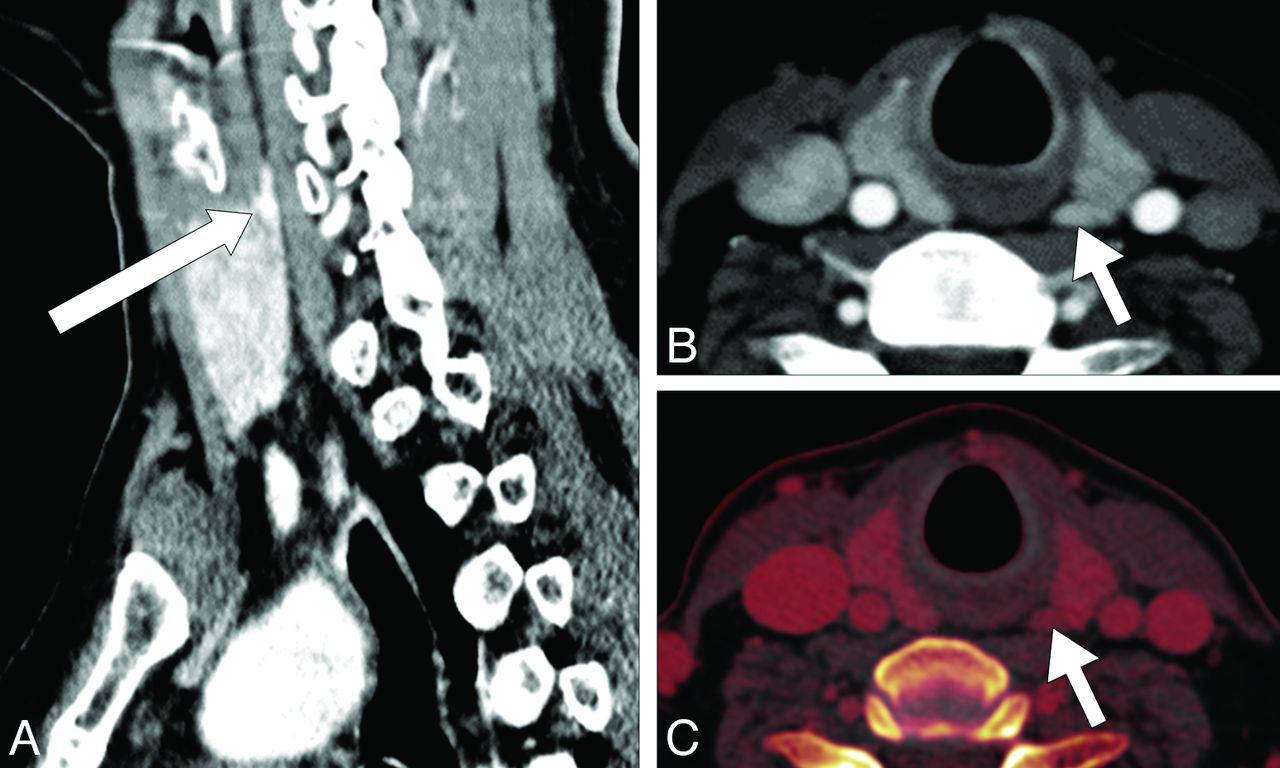
- Fig 1.
CT examination of the patient by using our modified 4D CT protocol shows a 6 × 3 mm nodular parathyroid adenoma (arrows) lying in the superomedial aspect, posterior to the left thyroid lobe in both sagittal (A) and axial (B) sections. The iodine overlay image (acquired by using dual-energy CT in the venous phase), also obtained by using this protocol, allows the measurement of iodine concentration in the tissues, to differentiate between the parathyroid adenoma and surrounding thyroid tissues (C). Directed parathyroidectomy and histopathologic examination, thereafter, confirmed the presence of a parathyroid adenoma.
- Fig 2.
Modified 4D CT by using our suggested protocol demonstrates the enhancement characteristics of a hyperplastic parathyroid adenoma (region of interest 1) and an adjacent soft-tissue structure (ie, a normal-functioning thyroid gland) (region of interest 2). Contrast-enhancement analysis on the parathyroid adenoma shows an attenuation value of 36.1 HU on the virtual noncontrast scan (A), which rapidly enhanced to 175.5 HU in the arterial phase (B), and immediately decreased to 100.3 HU in the dual-energy venous phase (C) and 75.1 HU in the delayed (D) image. The parathyroid adenoma can be easily distinguished from the surrounding soft tissues on the basis of its characteristic “rapid contrast uptake and washout” feature.
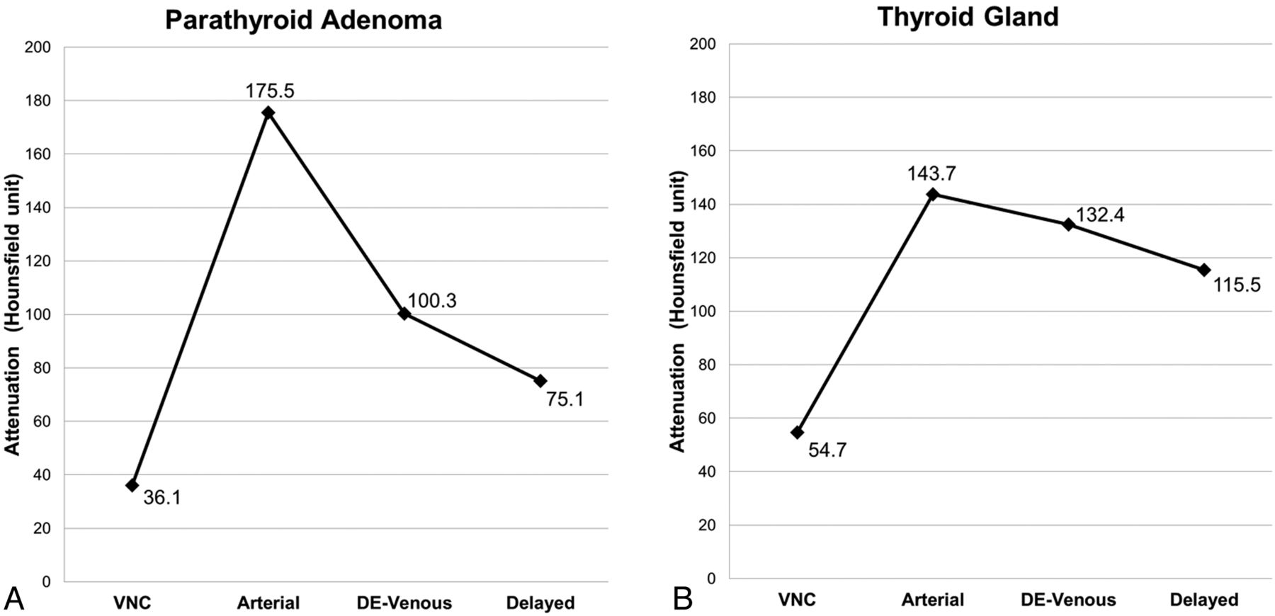
- Fig 3.
Attenuation plots show the characteristic dynamic contrast-enhancement pattern of the parathyroid adenoma (ie, rapid contrast uptake and washout, (A) and a normal-functioning right thyroid gland (B) measured from the same scans obtained from the patient diagnosed with PHPT by using our modified 4D CT protocol. The mean Hounsfield unit attenuations of the imaged structures in the VNC (reconstructed from the dual-energy venous [DE-venous]), arterial (by using bolus tracking), DE-venous (55 seconds), and delayed (85 seconds) phases were measured by using region-of-interest analysis as shown in Fig 2.


Tables
Phase Measurement CTDIvol (mGy) DLP (mGy.cm) Precontrast 27.16 452 Arterial 27.32 475 Venous 27.08 471 Delayed 27.32 475 Total 108.88 1873 Note:—CTDIvol indicates CT dose index volume; DLP, dose-length product.
↵a These measurements obtained during our modified 4D CT scan on another weight- and sex-matched patient were compared with values obtained during standard 4D CT (including the precontrast scan) of our patient. They showed an estimated 20% reduction in the radiation exposure.
Phase Measurement CTDIvol (mGy) DLP (mGy.cm) Arterial 26.92 475 DE-venousb 31.78 583 Delayed 26.68 471 Total 85.38 1529 % Reduction from standard protocol 21.6 18.4 Note:—CTDIvol indicates CT dose index volume; DLP, dose-length product; DE, dual-energy.
↵a These measurements obtained during our modified 4D CT scan on the current patient were compared with values obtained during standard 4D CT (including the precontrast scan) of another weight- and sex-matched patient. Both CTDIvol and DLP measurements were recorded during modified 4D CT (excluding the precontrast scan) for the current patient (61.6-kg female patient) illustrated in this case presentation. They showed an estimated 20% reduction in the radiation exposure.
↵b The venous phase scan was acquired using dual-energy CT (DE-venous) to obtain the VNC image.
In this issue


Related Articles
Cited By...
- No citing articles found.