Advertisement

Main menu

  • Home
  • Content
    • Current Issue
    • Accepted Manuscripts
    • Article Preview
    • Past Issue Archive
    • Video Articles
    • AJNR Case Collection
    • Case of the Week Archive
    • Case of the Month Archive
    • Classic Case Archive
  • Special Collections
    • AJNR Awards
    • Low-Field MRI
    • Alzheimer Disease
    • ASNR Foundation Special Collection
    • Photon-Counting CT
    • View All
  • Multimedia
    • AJNR Podcasts
    • AJNR SCANtastic
    • Trainee Corner
    • MRI Safety Corner
    • Imaging Protocols
  • For Authors
    • Submit a Manuscript
    • Submit a Video Article
    • Submit an eLetter to the Editor/Response
    • Manuscript Submission Guidelines
    • Statistical Tips
    • Fast Publishing of Accepted Manuscripts
    • Graphical Abstract Preparation
    • Imaging Protocol Submission
    • Author Policies
  • About Us
    • About AJNR
    • Editorial Board
    • Editorial Board Alumni
  • More
    • Become a Reviewer/Academy of Reviewers
    • Subscribers
    • Permissions
    • Alerts
    • Feedback
    • Advertisers
    • ASNR Home

User menu

  • Alerts
  • Log in

Search

  • Advanced search
American Journal of Neuroradiology

American Journal of Neuroradiology

ASHNR American Society of Functional Neuroradiology ASHNR American Society of Pediatric Neuroradiology ASSR
  • Alerts
  • Log in

Advanced Search

  • Home
  • Content
    • Current Issue
    • Accepted Manuscripts
    • Article Preview
    • Past Issue Archive
    • Video Articles
    • AJNR Case Collection
    • Case of the Week Archive
    • Case of the Month Archive
    • Classic Case Archive
  • Special Collections
    • AJNR Awards
    • Low-Field MRI
    • Alzheimer Disease
    • ASNR Foundation Special Collection
    • Photon-Counting CT
    • View All
  • Multimedia
    • AJNR Podcasts
    • AJNR SCANtastic
    • Trainee Corner
    • MRI Safety Corner
    • Imaging Protocols
  • For Authors
    • Submit a Manuscript
    • Submit a Video Article
    • Submit an eLetter to the Editor/Response
    • Manuscript Submission Guidelines
    • Statistical Tips
    • Fast Publishing of Accepted Manuscripts
    • Graphical Abstract Preparation
    • Imaging Protocol Submission
    • Author Policies
  • About Us
    • About AJNR
    • Editorial Board
    • Editorial Board Alumni
  • More
    • Become a Reviewer/Academy of Reviewers
    • Subscribers
    • Permissions
    • Alerts
    • Feedback
    • Advertisers
    • ASNR Home
  • Follow AJNR on Twitter
  • Visit AJNR on Facebook
  • Follow AJNR on Instagram
  • Join AJNR on LinkedIn
  • RSS Feeds

AJNR Awards, New Junior Editors, and more. Read the latest AJNR updates

LetterLetter

Dual-Energy 4-Phase CT Scan in Primary Hyperparathyroidism

D. Lau, H. Yang and P.L. Kei
American Journal of Neuroradiology August 2013, 34 (8) E91-E93; DOI: https://doi.org/10.3174/ajnr.A3657
D. Lau
aDepartment of Diagnostic Radiology Singapore General Hospital Singapore
  • Find this author on Google Scholar
  • Find this author on PubMed
  • Search for this author on this site
H. Yang
aDepartment of Diagnostic Radiology Singapore General Hospital Singapore
  • Find this author on Google Scholar
  • Find this author on PubMed
  • Search for this author on this site
P.L. Kei
aDepartment of Diagnostic Radiology Singapore General Hospital Singapore
  • Find this author on Google Scholar
  • Find this author on PubMed
  • Search for this author on this site
  • Article
  • Figures & Data
  • Info & Metrics
  • Responses
  • References
  • PDF
Loading

We read with interest the article by Gafton et al1 published in 2012. The authors illustrated the use of a dual-phase CT protocol for the enhancement characterization of parathyroid lesions and suggested the elimination of precontrast and delayed scan phases from 4D CT to reduce radiation exposure. In this letter, we describe the alternative approach of our institution to 4D CT. Our modified 4D CT protocol uses virtual noncontrast (VNC) images obtained from dual-energy CT-reconstructed venous-enhanced images to replace true precontrast scans. This process allows a complete examination of the contrast enhancement and washout characteristics of parathyroid adenomas on 4D CT, without reducing the number of scan phases.

Here we present a 57-year-old female patient who was suspected of having primary hyperparathyroidism (PHPT) and was scanned by using our modified 4D CT protocol. The institutional review board approval was waived for this dose-saving study. The patient was referred for neck imaging using our modified protocol after negative technetium Tc99m-sestamibi scintigraphy findings, despite the presence of elevated total serum calcium and parathyroid hormone levels. A modified 4-phase CT scan was obtained, with a dual-energy scan acquired during the venous phase.

Image Acquisition and Processing

All scans were obtained with the patient in the supine position by using a 64-section dual-source CT scanner (Somatom Definition; Siemens, Erlangen, Germany) to acquire contiguous axial images from the lower margin of the mandible to the level of the inferior aortic arch. Intravenous injection of 50 mL of iodinated contrast material (iohexol, Omnipaque, 350 mg/mL; GE Healthcare, Milwaukee, Wisconsin), followed by 50 mL of saline chaser bolus, was administered via the right antecubital vein at a rate of 3 mL/s by using a 20-gauge catheter. Scan acquisition in the arterial, dual-energy venous, and delayed phases was performed at 25, 55, and 85 seconds, respectively. Single-energy CT in the arterial and delayed phases was performed by using a collimation of 16 × 0.75 mm and a pitch value of 1 at 120 kV(peak)/62 mAs, whereas dual-energy CT was performed in the venous phase by using a 32 × 0.6 mm collimation, a pitch value of 1, and tube current settings of 80 kVp/210 mAs (tube A) and 140 kVp/50 mAs (tube B). Both single- and dual-energy CT scans were obtained with a gantry rotation speed of 0.8 seconds, with on-line tube current modulation (Care Dose 4D; Siemens). The estimated CT dose index and dose-length product were recorded for the scan session.

All images were subsequently reconstructed into axial and sagittal views with a section thickness of 3 mm and an increment of 3 mm, with an FOV of 20 cm and a 512 × 512 matrix, by using a B30f kernel for single-energy CT and a D30f kernel for dual-energy CT on a syngo MultiModality Workplace (MMWP), Version VE 31A (Siemens). The dual-energy venous raw data from both detectors were reconstructed into 80 kV(peak), 140 kVp, and weighted average 120 kVp images (30% of the image information was derived from the 80 kVp image and 70% from the 140 kVp image). Further postprocessing to the dual-energy venous dataset by using the Liver Virtual Unenhanced application on the syngo MMWP was performed to generate the iodine map from the individual CT voxels, as well as the subtraction of the iodine signal from the dataset to produce a VNC-enhanced image.

Image Analysis

All images were analyzed on syngo MMWP. Circular regions of interest were defined and positioned within the center of the parathyroid adenoma to measure the contrast attenuation values. The size was chosen as approximately 5.0 mm2 to cover almost the entire lesion but avoiding beam-hardening and partial volume artifacts at the borders of the lesion. The mean ± SD values in Hounsfield units measured from the respective images acquired from each scan phase were recorded. Results from the modified 4D CT scan are shown in Figs 1⇓–3. A subsequent directed parathyroidectomy revealed the presence of a parathyroid adenoma (Figs 1⇓–3).

Fig 1.
  • Download figure
  • Open in new tab
  • Download powerpoint
Fig 1.

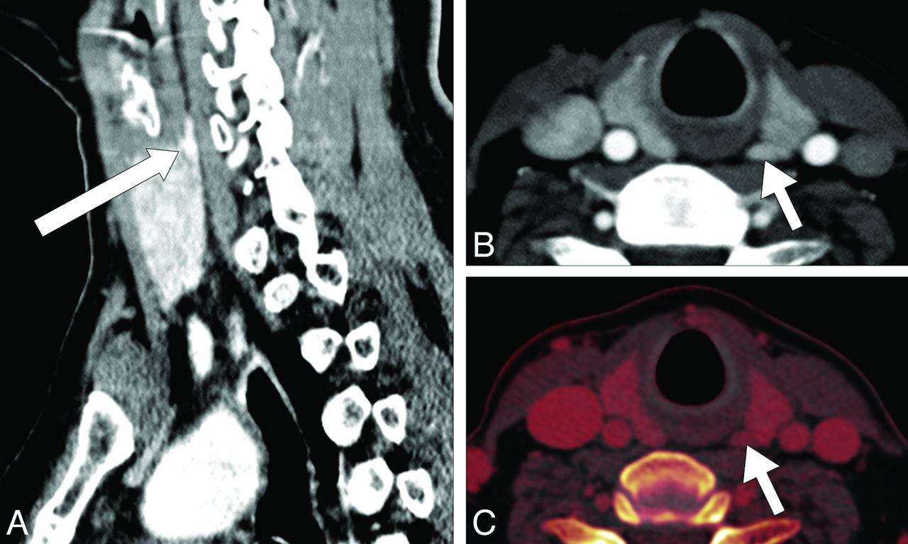
CT examination of the patient by using our modified 4D CT protocol shows a 6 × 3 mm nodular parathyroid adenoma (arrows) lying in the superomedial aspect, posterior to the left thyroid lobe in both sagittal (A) and axial (B) sections. The iodine overlay image (acquired by using dual-energy CT in the venous phase), also obtained by using this protocol, allows the measurement of iodine concentration in the tissues, to differentiate between the parathyroid adenoma and surrounding thyroid tissues (C). Directed parathyroidectomy and histopathologic examination, thereafter, confirmed the presence of a parathyroid adenoma.

Fig 2.
  • Download figure
  • Open in new tab
  • Download powerpoint
Fig 2.

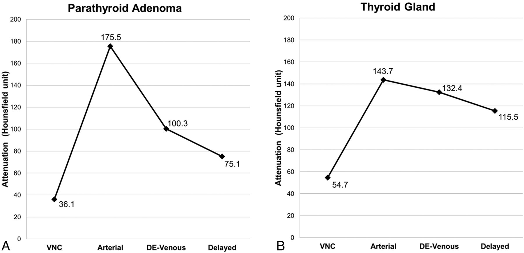
Modified 4D CT by using our suggested protocol demonstrates the enhancement characteristics of a hyperplastic parathyroid adenoma (region of interest 1) and an adjacent soft-tissue structure (ie, a normal-functioning thyroid gland) (region of interest 2). Contrast-enhancement analysis on the parathyroid adenoma shows an attenuation value of 36.1 HU on the virtual noncontrast scan (A), which rapidly enhanced to 175.5 HU in the arterial phase (B), and immediately decreased to 100.3 HU in the dual-energy venous phase (C) and 75.1 HU in the delayed (D) image. The parathyroid adenoma can be easily distinguished from the surrounding soft tissues on the basis of its characteristic “rapid contrast uptake and washout” feature.

Fig 3.
  • Download figure
  • Open in new tab
  • Download powerpoint
Fig 3.

Attenuation plots show the characteristic dynamic contrast-enhancement pattern of the parathyroid adenoma (ie, rapid contrast uptake and washout, (A) and a normal-functioning right thyroid gland (B) measured from the same scans obtained from the patient diagnosed with PHPT by using our modified 4D CT protocol. The mean Hounsfield unit attenuations of the imaged structures in the VNC (reconstructed from the dual-energy venous [DE-venous]), arterial (by using bolus tracking), DE-venous (55 seconds), and delayed (85 seconds) phases were measured by using region-of-interest analysis as shown in Fig 2.

Our findings suggest that VNC images could potentially replace true precontrast images to provide an accurate differentiation of the parathyroid adenoma from surrounding normal tissues, by characterizing its contrast enhancement with time. It has been reported in the literature that VNC images can provide a good approximation of the attenuation values similar to that in true precontrast images and, accordingly, lower radiation exposure in patients by omitting the need for precontrast scanning.2 The resulting images obtained by using our modified protocol provided a reasonably good diagnostic anatomic localization of the parathyroid adenoma (Fig 1). A complete 4-phase examination of the enhancement characteristics of the parathyroid adenoma (rapid contrast uptake and washout) was feasible (Figs 2 and 3), with an estimated reduction in the overall radiation exposure by approximately 20% (Tables 1 and 2). In addition, with appropriate software (which we do not have), measurement of the tissue iodine concentration (in milligrams/deciliter) is also possible on the iodine overlay image. This has potentially more useful implications in head and neck imaging than conventional 4D CT because it allows the radiologist to determine whether the lesion is arising from thyroid tissues or is extrathyroidal in nature.

View this table:
  • View inline
  • View popup
Table 1:

Standard 4D CT protocol in another patienta

View this table:
  • View inline
  • View popup
Table 2:

Modified 4D CT protocol in our current patienta

In our experience, provision of images from all 4 phases, including the precontrast and delayed scans, is helpful in determining the baseline attenuation value and dynamic changes with time. Various authors have proposed modified 4D CT methods, mostly by eliminating 1 or 2 phases from the scan technique.1,3 These may reduce the diagnostic confidence of the reporter. We feel that the provision of 4 phases is crucial for the localization of small or ectopic parathyroid adenoma or lesions situated in crowded structures in the neck or other distal locations such as the mediastinum.4,5

However, one drawback in using this modified technique is the subtraction of iodine attenuation from the thyroid glands on VNC images, thereby possibly negating the potential benefit of this modified 4D CT study. Admittedly, a true precontrast scan does confer additional information of soft-tissue contrast between the parathyroid adenoma and surrounding soft tissue (thyroid tissue is inherently denser compared with adenomatous tissue, due to its high iodine concentration); this, however, comes with added radiation burden. In the authors' experience, careful analysis of the VNC, arterial, venous, and delay phases can help in separating parathyroid adenoma tissues from normal surrounding soft tissues (such as the thyroid gland). In addition, for patients with PHPT and concurrent thyroiditis, in which the attenuation of the thyroid gland is low to begin with, having an iodine overlay map allows the quantification of iodine concentration of a particular lesion, enabling accurate lesion characterization.

In conclusion, we believe our modified dual-energy protocol can allow reduction in radiation exposure during 4D CT parathyroid scanning without compromising the need for studying the enhancement characteristics of these lesions. Nevertheless, further studies should be conducted to validate the real clinical impact of our suggested protocol for diagnosing PHPT in a larger group of patients.

Acknowledgments

We thank the Singapore General Hospital Department of Radiology for support of this study and the technologists Muhammad Illyyas and Joanne Chia for performing the scan and image reconstruction.

REFERENCES

  1. 1.↵
    1. Gafton AR,
    2. Glastonbury CM,
    3. Eastwood JD,
    4. et al
    . Parathyroid lesions: characterization with dual-phase arterial and venous enhanced CT of the neck. AJNR Am J Neuroradiol 2012;33:949–52
    Abstract/FREE Full Text
  2. 2.↵
    1. Tawfik AM,
    2. Kerl JM,
    3. Razek AA,
    4. et al
    . Image quality and radiation dose of dual-energy CT of the head and neck compared with a standard 120-kVp acquisition. AJNR Am J Neuroradiol 2011;32:1994–99
    Abstract/FREE Full Text
  3. 3.↵
    1. Kutler DI,
    2. Moquete R,
    3. Kazam E,
    4. et al
    . Parathyroid localization with modified 4D-computed tomography and ultrasonography for patients with primary hyperparathyroidism. Laryngoscope 2011;121:1219–24
    CrossRefPubMed
  4. 4.↵
    1. Hunter GJ,
    2. Schellingerhout D,
    3. Vu TH,
    4. et al
    . Accuracy of four-dimensional CT for the localization of abnormal parathyroid glands in patients with primary hyperparathyroidism. Radiology 2012;264:789–95
    CrossRefPubMed
  5. 5.↵
    1. Chazen JL,
    2. Gupta A,
    3. Dunning A,
    4. et al
    . Diagnostic accuracy of 4D-CT parathyroid adenomas and hyperplasia. AJNR Am J Neuroradiol 2012;33:429–33
    Abstract/FREE Full Text
  • © 2013 by American Journal of Neuroradiology
PreviousNext
Back to top

In this issue

American Journal of Neuroradiology: 34 (8)
American Journal of Neuroradiology
Vol. 34, Issue 8
1 Aug 2013
  • Table of Contents
  • Index by author
Advertisement
Print
Download PDF
Email Article

Thank you for your interest in spreading the word on American Journal of Neuroradiology.

NOTE: We only request your email address so that the person you are recommending the page to knows that you wanted them to see it, and that it is not junk mail. We do not capture any email address.

Enter multiple addresses on separate lines or separate them with commas.
Dual-Energy 4-Phase CT Scan in Primary Hyperparathyroidism
(Your Name) has sent you a message from American Journal of Neuroradiology
(Your Name) thought you would like to see the American Journal of Neuroradiology web site.
CAPTCHA
This question is for testing whether or not you are a human visitor and to prevent automated spam submissions.
Cite this article
D. Lau, H. Yang, P.L. Kei
Dual-Energy 4-Phase CT Scan in Primary Hyperparathyroidism
American Journal of Neuroradiology Aug 2013, 34 (8) E91-E93; DOI: 10.3174/ajnr.A3657

Citation Manager Formats

  • BibTeX
  • Bookends
  • EasyBib
  • EndNote (tagged)
  • EndNote 8 (xml)
  • Medlars
  • Mendeley
  • Papers
  • RefWorks Tagged
  • Ref Manager
  • RIS
  • Zotero
0 Responses
Respond to this article
Share
Bookmark this article
Dual-Energy 4-Phase CT Scan in Primary Hyperparathyroidism
D. Lau, H. Yang, P.L. Kei
American Journal of Neuroradiology Aug 2013, 34 (8) E91-E93; DOI: 10.3174/ajnr.A3657
del.icio.us logo Twitter logo Facebook logo Mendeley logo
  • Tweet Widget
  • Facebook Like
  • Google Plus One
Purchase

Jump to section

  • Article
    • Acknowledgments
    • REFERENCES
  • Figures & Data
  • Info & Metrics
  • Responses
  • References
  • PDF

Related Articles

  • No related articles found.
  • PubMed
  • Google Scholar

Cited By...

  • No citing articles found.
  • Crossref (8)
  • Google Scholar

This article has been cited by the following articles in journals that are participating in Crossref Cited-by Linking.

  • Material Separation Using Dual-Energy CT: Current and Emerging Applications
    Manuel Patino, Andrea Prochowski, Mukta D. Agrawal, Frank J. Simeone, Rajiv Gupta, Peter F. Hahn, Dushyant V. Sahani
    RadioGraphics 2016 36 4
  • Dual-Energy CT in Head and Neck Imaging
    Elise D. Roele, Veronique C. M. L. Timmer, Lauretta A. A. Vaassen, Anna M. J. L. van Kroonenburgh, A. A. Postma
    Current Radiology Reports 2017 5 5
  • Dual-Energy CT Characteristics of Parathyroid Adenomas on 25-and 55-Second 4D-CT Acquisitions
    Reza Forghani, Michael Roskies, Xiaoyang Liu, Xianming Tan, Alex Mlynarek, Richard J. Payne, Jaykumar R. Nair, Michael P. Hier, Mark Levental
    Journal of Computer Assisted Tomography 2016 40 5
  • Clinical applications of dual-energy CT in head and neck imaging
    Daniel Thomas Ginat, Michael Mayich, Laleh Daftari-Besheli, Rajiv Gupta
    European Archives of Oto-Rhino-Laryngology 2016 273 3
  • Dual energy 4D-CT of parathyroid adenomas not clearly localized by sestamibi scintigraphy and ultrasonography – a retrospective study
    M. Woisetschläger, O. Gimm, K. Johansson, G. Wallin, I. Albert-Garcia, A. Spångeus
    European Journal of Radiology 2020 124
  • Dual energy computed tomography should be a first line preoperative localization imaging test for primary hyperparathyroidism patients
    Jake Hiebert, Cameron Hague, Shangmei Hou, Sam M. Wiseman
    The American Journal of Surgery 2018 215 5
  • Renal Adiposity Does not Preclude Quantitative Assessment of Renal Function Using Dual-Energy Multidetector CT in Mildly Obese Human Subjects
    Christopher M. Ferguson, Alfonso Eirin, Gregory J. Michalak, Ahmad F. Hedayat, Abdelrhman M. Abumoawad, Ahmed Saad, Xiangyang Zhu, Stephen C. Textor, Cynthia H. McCollough, Lilach O. Lerman
    Academic Radiology 2019 26 11
  • Dual-layer spectral CT virtual-non-contrast images aid in parathyroid adenoma analysis and radiation dose reduction: confirmation of findings from dual-energy CT
    Danielle Maraia, Steven Hemmerdinger, Patrick Chiarolanzio, Hasit Mehta, Sana Ali, William Gomes, Javin Schefflein, Maynard High, Edwin Gulko
    Clinical Imaging 2022 84

More in this TOC Section

  • Letter to the Editor regarding “Automated Volumetric Software in Dementia: Help or Hindrance to the Neuroradiologist?”
  • Reply:
  • Brain AVM’s Nidus: What if We Hadn’t Understood Anything?
Show more LETTERS

Similar Articles

Advertisement

Indexed Content

  • Current Issue
  • Accepted Manuscripts
  • Article Preview
  • Past Issues
  • Editorials
  • Editor's Choice
  • Fellows' Journal Club
  • Letters to the Editor
  • Video Articles

Cases

  • Case Collection
  • Archive - Case of the Week
  • Archive - Case of the Month
  • Archive - Classic Case

More from AJNR

  • Trainee Corner
  • Imaging Protocols
  • MRI Safety Corner
  • Book Reviews

Multimedia

  • AJNR Podcasts
  • AJNR Scantastics

Resources

  • Turnaround Time
  • Submit a Manuscript
  • Submit a Video Article
  • Submit an eLetter to the Editor/Response
  • Manuscript Submission Guidelines
  • Statistical Tips
  • Fast Publishing of Accepted Manuscripts
  • Graphical Abstract Preparation
  • Imaging Protocol Submission
  • Evidence-Based Medicine Level Guide
  • Publishing Checklists
  • Author Policies
  • Become a Reviewer/Academy of Reviewers
  • News and Updates

About Us

  • About AJNR
  • Editorial Board
  • Editorial Board Alumni
  • Alerts
  • Permissions
  • Not an AJNR Subscriber? Join Now
  • Advertise with Us
  • Librarian Resources
  • Feedback
  • Terms and Conditions
  • AJNR Editorial Board Alumni

American Society of Neuroradiology

  • Not an ASNR Member? Join Now

© 2025 by the American Society of Neuroradiology All rights, including for text and data mining, AI training, and similar technologies, are reserved.
Print ISSN: 0195-6108 Online ISSN: 1936-959X

Powered by HighWire