
Article Figures & Data
Figures
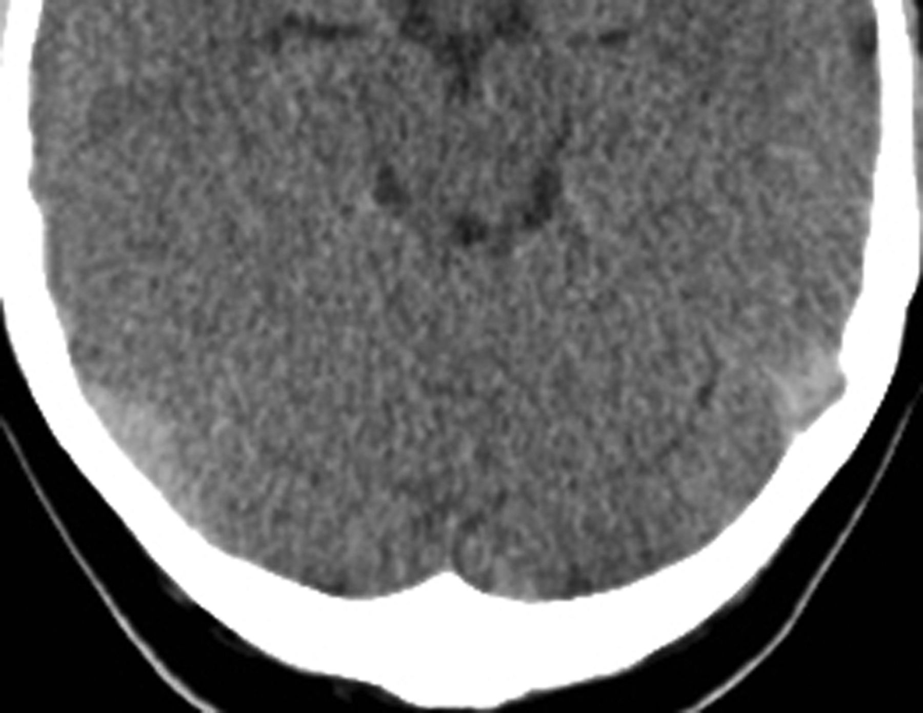
- Fig 1.
Axial brain CT scan in a patient with false-negative results (left sigmoid sinus according to the measurements of 2 readers [mean HU, 55; HCT, 39.6; H:H ratio, 1.39]).
- Fig 2.
Axial brain CT scan in a control participant with false-positive results in the transverse sinuses according to the measurements of 1 reader (mean HU, 66.3; HCT 43; H:H ratio, 1.54).
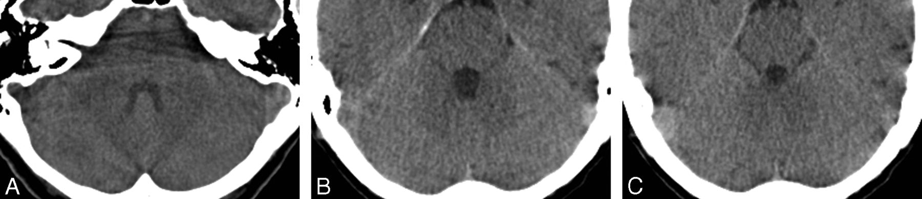
- Fig 3.
Axial brain CT scan in a patient with a thrombosis in the superior sagittal sinus (A) and right transverse sinus (B) (mean HU, 76.6; HCT, 40.2; H:H ratio, 1.86).
- Fig 4.
The average attenuation values among the 3 readers was significantly different in patients with acute CVST (73.9 ± 9.2 HU) compared with control participants (52.8 ± 6.7 HU) (A). The H:H ratio showed values of 1.33 ± 0.12 in patients without CVST, and 1.91 ± 0.32 in patients with CVST (P < .0001) (B).
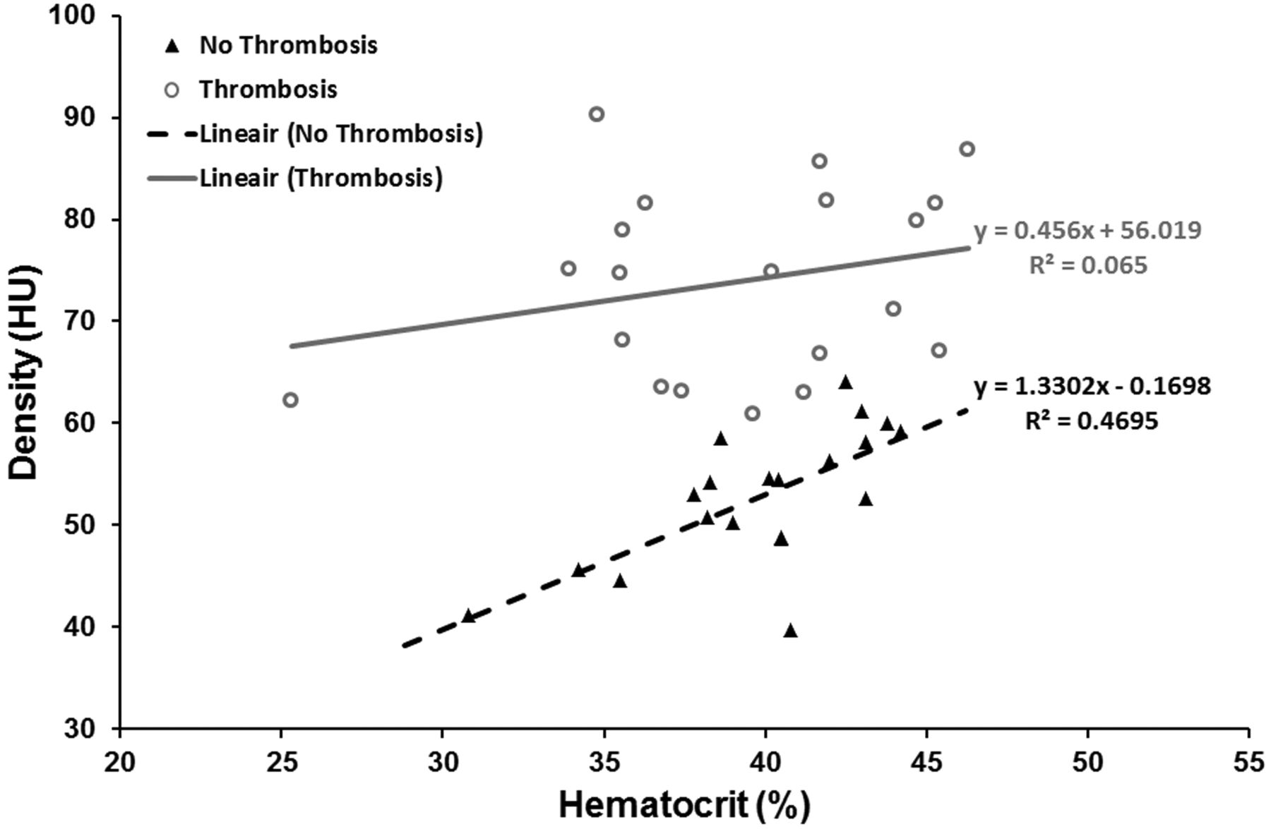
- Fig 5.
Scatterplot of venous sinus attenuation as a function of HCT showing a significant positive correlation between HCT and attenuation values in patients without acute CVST (R2 = 0.47; P = .0009), but not in patients with CVST (R2 = 0.07; P = .28).


Tables
Location of thrombus in 20 patients with acute CVST
Patient No. Location of Thrombus 1 Left TS 2 SSS, SR, bilateral TS 3 Left TS and SS 4 SSS and SR 5 SSS, left TS 6 SSS, left TS 7 SSS, right TS 8 Right TS and SS 9 Left TS and SS 10 Right TS and SS 11 Left TS and SS 12 Right TS and SS 13 SSS, left TS and SS 14 Left TS and SS 15 SSS, SR, right TS and SS 16 SR, right TS and SS 17 Right TS and SS 18 SSS, bilateral TS and SS 19 SSS, right TS and SS 20 SSS, right TS and SS Note:—SR indicates sinus rectus; SS, sigmoid sinus; SSS, superior sagittal sinus; TS, transverse sinus.


