
Article Figures & Data
Figures
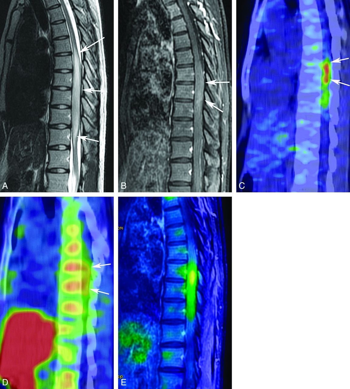
- Fig 1.
A 23-year-old woman with an anaplastic astrocytoma. T2-weighted MR image (A) reveals a tumor of the spinal cord from the eighth thoracic vertebra to the first lumbar vertebra (white arrows). A contrast-enhanced T1-weighted MR image with fat saturation (B) shows mild contrast enhancement of the tumor (white arrows). FDG-PET (C) and MET-PET (D) reveal increased activity of both FDG (SUVmax = 7.4) and MET (SUVmax = 3.2) of the tumor (white arrows). A fusion image of FDG-PET and contrast-enhanced T1-weighted MR imaging with fat saturation (E) clearly shows increased uptake of FDG in the tumor.
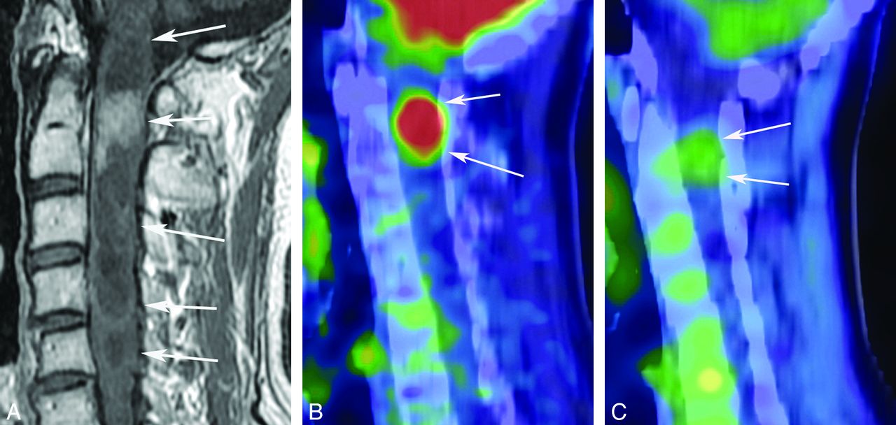
- Fig 2.
A 56-year-old man with an ependymoma. A contrast-enhanced T1-weighted image (A) shows a tumor from the first to fifth cervical vertebra with a cystic and solid component (white arrows). Contrast enhancement of the solid component at the second cervical vertebra is seen (white arrows). FDG-PET (B) and MET-PET (C) show accumulation of both FDG (SUVmax = 11.2) and MET (SUVmax = 2.2) (white arrows).
- Fig 3.
A 12-year-old girl with a cavernous angioma. A T2-weighted MR image (A) shows a mainly hypointense tumor (white arrows) at the twelfth thoracic vertebra. Hypointensity may be due to hemosiderin deposition. A hyperintense area surrounding the hypointense lesion seems to be edema, but its histologic evidence was not acquired. Although accumulation of MET is not evident (B), FDG-PET (C) shows slight activity of the tumor (SUVmax = 5.2) (black arrow).


Tables
SUVmax of each tumor
Patient Age (yr) Sex Pathology Location Cystic Component SUVmax (FDG) SUVmax (MET) 1 23 F Anaplastic astrocytoma Th – 7.4 3.2 2 47 M Hemangioblastoma L + 2.5 2.4 3 18 F Ependymoma C + 10 2.9 4 56 M Ependymoma C + 11.2 2.2 5 30 M Ependymoma (myxopapillary) L + 2.4 2.2 6 41 M Ependymoma (tanycytic) Th – 5.4 2.4 7 12 F Cavernous angioma Th – 5.2 2.0 8 75 F Ependymoma (cellular) C + 7.1 3.5 9 29 F Ependymoma (myxopapillary) L + 3.5 3.2 Note:—Th indicates thoracic cord; L, lumbar cord; C, cervical cord; –, absence of cystic component on MRI; +, existence of cystic component on MRI.
In this issue


Jump to section
Related Articles
Cited By...
- [18F]FDG-PET Evaluation of Spinal Pathology in Patients in Oncology: Pearls and Pitfalls for the Neuroradiologist
- Differentiation between Treatment-Induced Necrosis and Recurrent Tumors in Patients with Metastatic Brain Tumors: Comparison among 11C-Methionine-PET, FDG-PET, MR Permeability Imaging, and MRI-ADC--Preliminary Results
- Genetics of Von Hippel-Lindau Disease
- Intramedullary Spinal Cord Metastases: Visibility on PET and Correlation with MRI Features