
Article Figures & Data
Figures
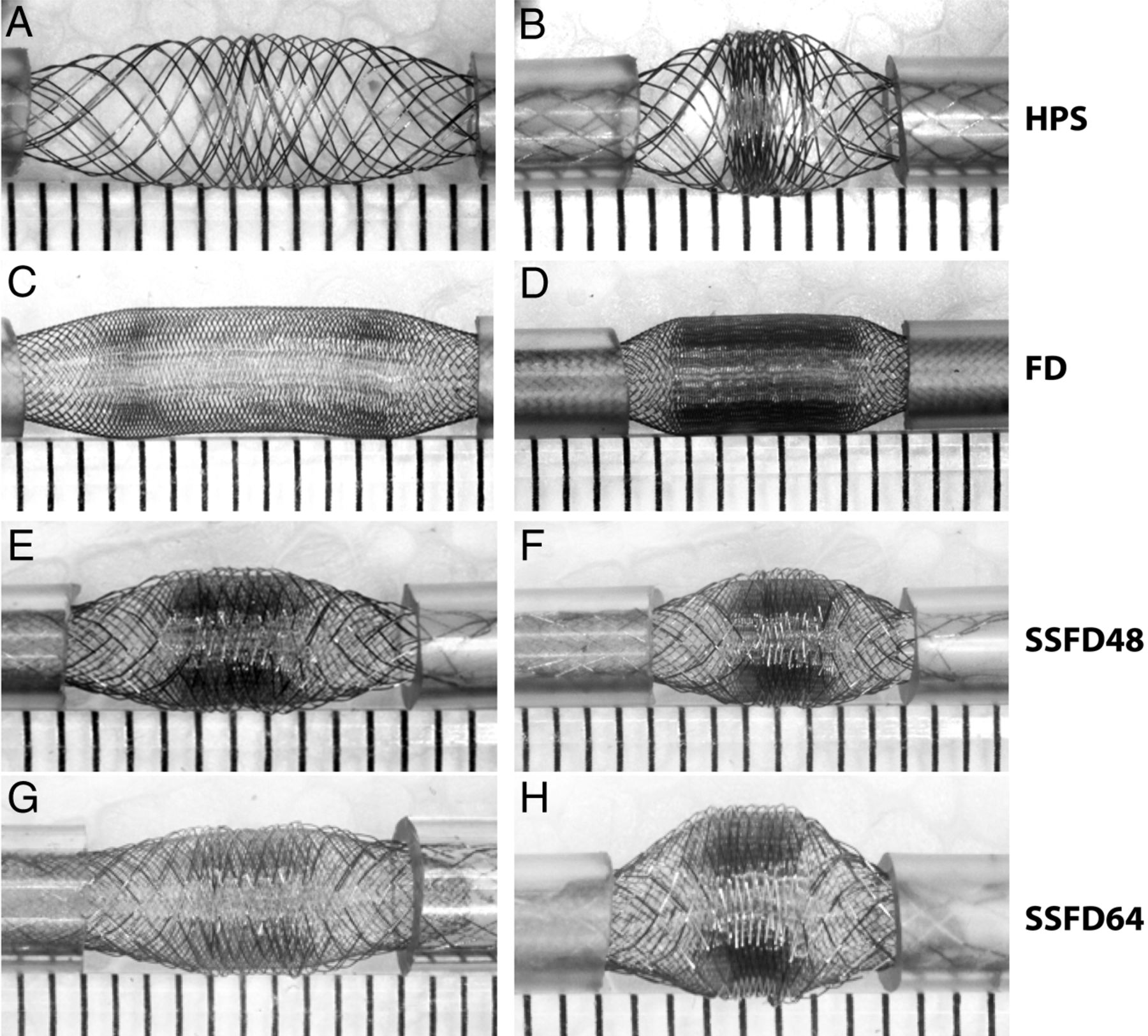
- Fig. 1.
Devices. The first set of experiments is shown, in which various devices were used. Note the similar pattern shared by all braided devices, with a concentration of stent struts in the midportion of the device, compacted between relatively porous segments. The phenomenon, more marked with increasing compaction (as shown on the right side), leads to heterogeneities in porosity along the length of the same device.
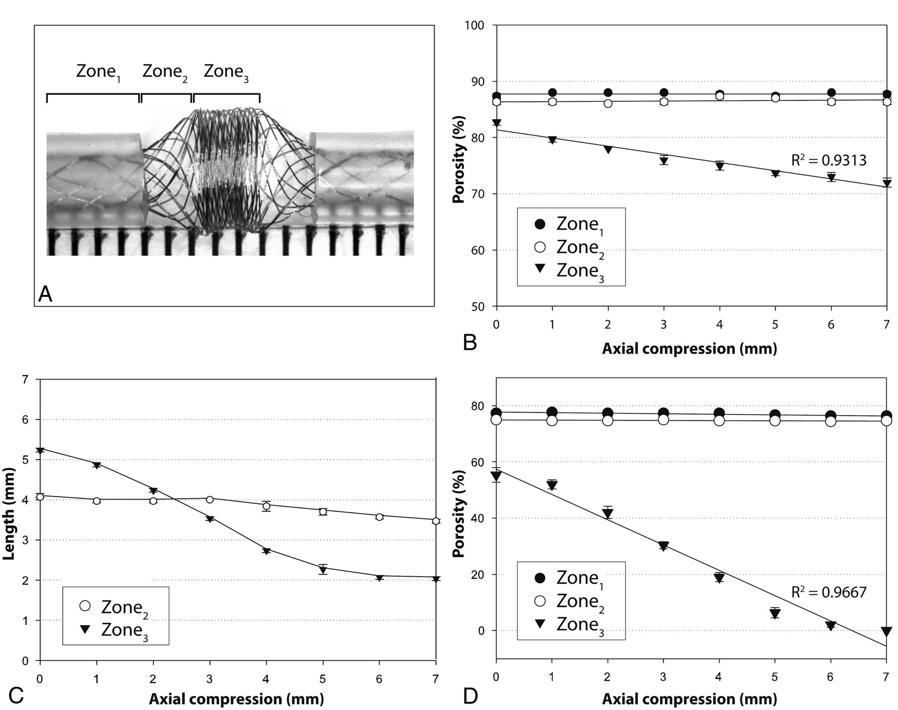
- Fig. 2.
Zones. The 3 characteristic zones are illustrated in a severely compacted HPS (A). Progressive approximation of the tubes (axial compression) leads to decreasing porosities of the compaction Zone3, but Zones1and2 remain relatively unchanged, as shown in B (HPS in 2-mm tubes). Axial compression leads directly to a shortening of Zone3, but the transition Zone2 length remains relatively unchanged (C). The same phenomenon was reproduced with the FD48 (compare B and D), with porosities reaching 0 with severe compaction.
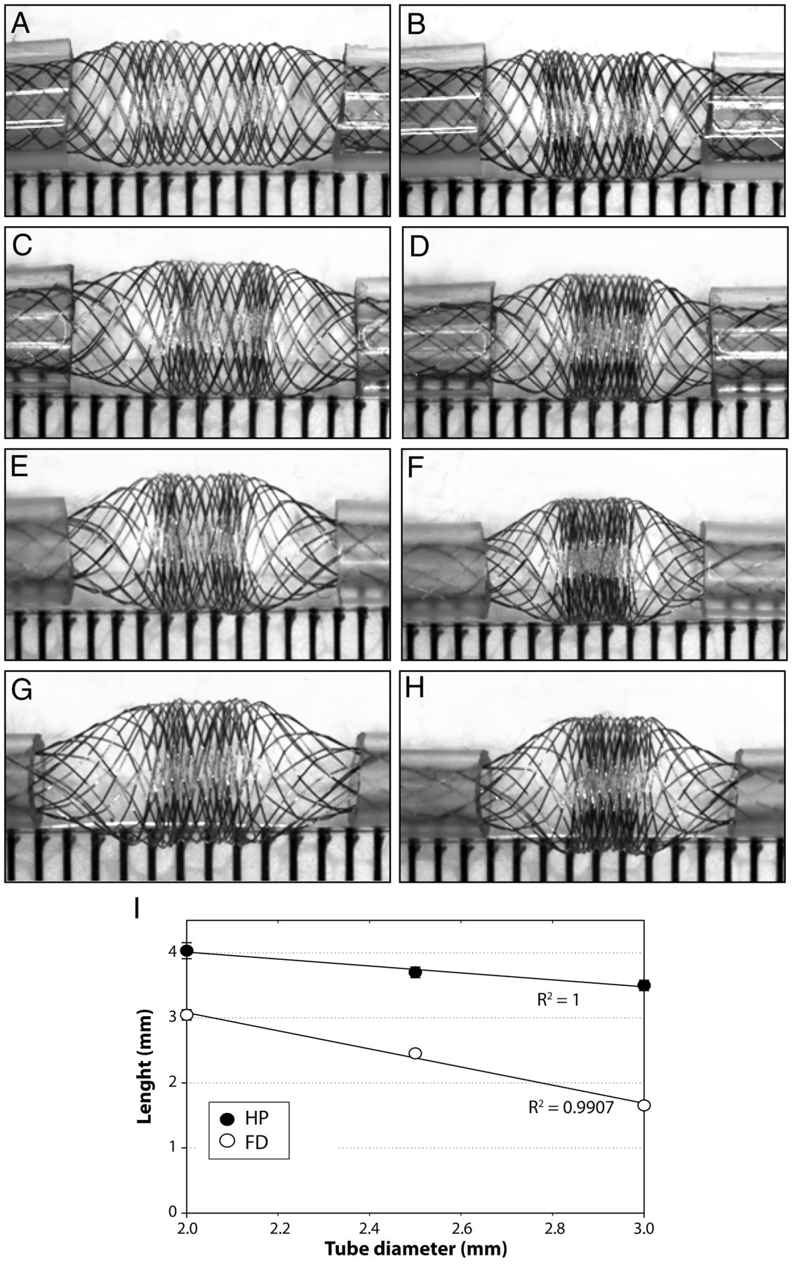
- Fig. 3.
Compaction and diameters. Effects of 2-mm (left column) and 4-mm compaction (right column) and of tube diameters (2–3.5 mm) on the compaction Zone3 and the transition Zone2 of the HPS are shown (A–H). Note that tubes of decreasing diameters lead to increased compaction of Zone3 because the transition Zone2 increases in length as tube diameter decreases. The relationship between the length of the transition Zone2 and the size of tubes is illustrated in I for both the HPS and the FD48.
- Fig. 4.
Curvatures and FDs. Curvatures affect porosities of the convexity and concavity of FD48. The effects are limited to the compaction Zone3, the transition Zone2 being relatively spared.
- Fig. 5.
Concavity at 45°(A, C) and convexity at 135°(B, D) of expansion Zone3 before (A, B), or after (C, D) the stent is compacted. Curvatures affect the porosity of the convexity and concavity sides of the compaction Zone3 (E), whereas the transition Zone2 remains virtually unchanged by comparison (F, showing the porosity of the transition Zone2 with different tubes and different curvatures).
- Fig. 6.
Stenoses can occur at the level of device extremities (A–D); the stenosis is more severe when the length introduced inside the tube is insufficient (compare A and B), and eccentric when the device is curved (D). The relationship between stenoses and length of device introduced inside tubes is shown for FD48 (E) as well as for the 48-wire stent-in-stent endoluminal flow-diverting devices (F).
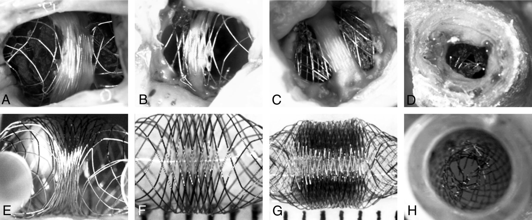
- Fig. 7.
In vivo and in vitro observations. Photographs of 4 autopsy specimens of aneurysms treated with HPSs (A, B) or FDs (C, D) are compared with 4 photographs of benchtop studies designed to mimic in vivo findings (E–H). Note how neointima formation (A–C) tends to be limited to the less porous compaction Zone3, and how the transition Zone2, lacking neointima, may be responsible for failures. The in vivo stent stenosis (D) is reproduced inside a tube (H).


Tables
Device characteristics
HPS FD48 SSFD36 SSFD48 SSFD64 Nominal stent diameter × length (mm) 4.5 × 30 3.5 × 21 3.75 × 32 3.75 × 32 3.75 × 32 P 89 ± 1.5% 77.2 ± 2.9% 72.4 ± 3.1% 70.3 ± 2.8% 66.0 ± 2.6% PD (pores/mm2) 0.6 ± 0.5 6.3 ± 2.5 5.6 ± 1.4 8.2 ± 2.2 10.2 ± 2.9 Number of wires 16 struts 48 braided wires Outer: 16 struts; inner: 36 braided wires Outer: 16 struts; inner: 48 braided wires Outer: 16 struts; inner: 64 braided wires Note:—SSFD indicates stent-in-stent endoluminal flow diverting device.
In this issue


Jump to section
Related Articles
Cited By...
- Subacute Stent Deformities as an Underlying Reason for Vessel Stenosis after Flow Diversion with the p64 Stent: Review and Discussion of Biologic Mechanisms and Consequences
- Braid stability after flow diverter treatment of intracranial aneurysms: a systematic review and meta-analysis
- Comprehensive Analysis of Post-Pipeline Endothelialization and Remodeling
- Predictors of the Effects of Flow Diversion in Very Large and Giant Aneurysms
- Predictive score for complete occlusion of intracranial aneurysms treated by flow-diverter stents using machine learning
- A realistic way to investigate the design, and mechanical properties of flow diverter stents
- Comparison of Pipeline Embolization Device Sizing Based on Conventional 2D Measurements and Virtual Simulation Using the Sim&Size Software: An Agreement Study
- Ostium Ratio and Neck Ratio Could Predict the Outcome of Sidewall Intracranial Aneurysms Treated with Flow Diverters
- Toward Better Understanding of Flow Diversion in Bifurcation Aneurysms
- Flow-Diversion Effect of LEO Stents: Aneurysm Occlusion and Flow Remodeling of Covered Side Branches and Perforators
- Flow Diversion with Low-Profile Braided Stents for the Treatment of Very Small or Uncoilable Intracranial Aneurysms at or Distal to the Circle of Willis
- Compacting a Single Flow Diverter versus Overlapping Flow Diverters for Intracranial Aneurysms: A Computational Study
- Virtual-versus-Real Implantation of Flow Diverters: Clinical Potential and Influence of Vascular Geometry
- Compaction of flow diverters improves occlusion of experimental wide-necked aneurysms
- The Added Value of Volume-of-Interest C-Arm CT Imaging during Endovascular Treatment of Intracranial Aneurysms
- Pipeline endovascular device for the treatment of intracranial aneurysms at the level of the circle of Willis and beyond: multicenter experience
- Preliminary experience with the Pipeline Flex Embolization Device: technical note
- Flow diverters: inter and intra-rater reliability of porosity and pore density measurements
- Visual Outcomes with Flow-Diverter Stents Covering the Ophthalmic Artery for Treatment of Internal Carotid Artery Aneurysms
- The Success of Flow Diversion in Large and Giant Sidewall Aneurysms May Depend on the Size of the Defect in the Parent Artery
- Enhanced Aneurysmal Flow Diversion Using a Dynamic Push-Pull Technique: An Experimental and Modeling Study
- Building Multidevice Pipeline Constructs of Favorable Metal Coverage: A Practical Guide
- Variable Porosity of the Pipeline Embolization Device in Straight and Curved Vessels: A Guide for Optimal Deployment Strategy