Advertisement

Main menu

  • Home
  • Content
    • Current Issue
    • Accepted Manuscripts
    • Article Preview
    • Past Issue Archive
    • Video Articles
    • AJNR Case Collection
    • Case of the Week Archive
    • Case of the Month Archive
    • Classic Case Archive
  • Special Collections
    • AJNR Awards
    • Low-Field MRI
    • Alzheimer Disease
    • ASNR Foundation Special Collection
    • Photon-Counting CT
    • View All
  • Multimedia
    • AJNR Podcasts
    • AJNR SCANtastic
    • Trainee Corner
    • MRI Safety Corner
    • Imaging Protocols
  • For Authors
    • Submit a Manuscript
    • Submit a Video Article
    • Submit an eLetter to the Editor/Response
    • Manuscript Submission Guidelines
    • Statistical Tips
    • Fast Publishing of Accepted Manuscripts
    • Graphical Abstract Preparation
    • Imaging Protocol Submission
    • Author Policies
  • About Us
    • About AJNR
    • Editorial Board
    • Editorial Board Alumni
  • More
    • Become a Reviewer/Academy of Reviewers
    • Subscribers
    • Permissions
    • Alerts
    • Feedback
    • Advertisers
    • ASNR Home

User menu

  • Alerts
  • Log in

Search

  • Advanced search
American Journal of Neuroradiology

American Journal of Neuroradiology

ASHNR American Society of Functional Neuroradiology ASHNR American Society of Pediatric Neuroradiology ASSR
  • Alerts
  • Log in

Advanced Search

  • Home
  • Content
    • Current Issue
    • Accepted Manuscripts
    • Article Preview
    • Past Issue Archive
    • Video Articles
    • AJNR Case Collection
    • Case of the Week Archive
    • Case of the Month Archive
    • Classic Case Archive
  • Special Collections
    • AJNR Awards
    • Low-Field MRI
    • Alzheimer Disease
    • ASNR Foundation Special Collection
    • Photon-Counting CT
    • View All
  • Multimedia
    • AJNR Podcasts
    • AJNR SCANtastic
    • Trainee Corner
    • MRI Safety Corner
    • Imaging Protocols
  • For Authors
    • Submit a Manuscript
    • Submit a Video Article
    • Submit an eLetter to the Editor/Response
    • Manuscript Submission Guidelines
    • Statistical Tips
    • Fast Publishing of Accepted Manuscripts
    • Graphical Abstract Preparation
    • Imaging Protocol Submission
    • Author Policies
  • About Us
    • About AJNR
    • Editorial Board
    • Editorial Board Alumni
  • More
    • Become a Reviewer/Academy of Reviewers
    • Subscribers
    • Permissions
    • Alerts
    • Feedback
    • Advertisers
    • ASNR Home
  • Follow AJNR on Twitter
  • Visit AJNR on Facebook
  • Follow AJNR on Instagram
  • Join AJNR on LinkedIn
  • RSS Feeds

AJNR Awards, New Junior Editors, and more. Read the latest AJNR updates

Article CommentaryBrain
Open Access

MR Imaging of Cerebral Extraventricular Neurocytoma: A Report of 9 Cases

K. Liu, G. Wen, X.-F. Lv, Yan-Jia Deng, Yong-Jian Deng, G.-Q. Hou, X.-L. Zhang, L.-J. Han and J.-L. Ding
American Journal of Neuroradiology March 2013, 34 (3) 541-546; DOI: https://doi.org/10.3174/ajnr.A3264
K. Liu
aFrom the Medical Imaging Center (K.L., G.W., X.-L.Z.)
  • Find this author on Google Scholar
  • Find this author on PubMed
  • Search for this author on this site
G. Wen
aFrom the Medical Imaging Center (K.L., G.W., X.-L.Z.)
  • Find this author on Google Scholar
  • Find this author on PubMed
  • Search for this author on this site
X.-F. Lv
cDepartment of Medical Imaging and Interventional Radiology (X.-F.L., L.-J.H.), Cancer Center, Sun Yat-Sen University, Guangzhou, P.R. China
  • Find this author on Google Scholar
  • Find this author on PubMed
  • Search for this author on this site
Yan-Jia Deng
dDepartment of Radiology (Yan-Jia D.), The 5th Affiliated Hospital of Sun Yat-Sen University, Zhuhai, P.R. China
  • Find this author on Google Scholar
  • Find this author on PubMed
  • Search for this author on this site
Yong-Jian Deng
bDepartment of Pathology (Yong-Jian D.), Nanfang Hospital, Southern Medical University, Guangzhou, P.R. China
  • Find this author on Google Scholar
  • Find this author on PubMed
  • Search for this author on this site
G.-Q. Hou
eDepartment of Medical Image Center (G.-Q.H.), Nanshan Hospital, Shenzhen, P.R. China
  • Find this author on Google Scholar
  • Find this author on PubMed
  • Search for this author on this site
X.-L. Zhang
aFrom the Medical Imaging Center (K.L., G.W., X.-L.Z.)
fDepartment of Radiology (X.-L.Z.), Dongguan Kanghua Hospital, Dongguan, P.R. China
  • Find this author on Google Scholar
  • Find this author on PubMed
  • Search for this author on this site
L.-J. Han
cDepartment of Medical Imaging and Interventional Radiology (X.-F.L., L.-J.H.), Cancer Center, Sun Yat-Sen University, Guangzhou, P.R. China
  • Find this author on Google Scholar
  • Find this author on PubMed
  • Search for this author on this site
J.-L. Ding
gDepartment of Radiology (J.-L.D.), The People's Hospital of Futian Shenzhen, Shenzhen, P.R. China.
  • Find this author on Google Scholar
  • Find this author on PubMed
  • Search for this author on this site
  • Article
  • Figures & Data
  • Info & Metrics
  • Responses
  • References
  • PDF
Loading

Article Figures & Data

Figures

  • Fig. 2.
    • Download figure
    • Open in new tab
    • Download powerpoint
    Fig. 2.

    MR images of a 43-year-old woman with EVN of the right parietal lobe. A, Axial T1WI shows a hypointense well-demarcated large mass (small arrows) with hyperintensity of its central area due to hemorrhage (large arrow). B, Axial FLAIR image shows a heterogeneous hyperintense mass with only slight peritumoral edema. C and D, Coronal and sagittal postcontrast T1WI show mild inhomogeneous “zebra-like” enhancement of the solid portion. E, Hematoxylin and eosin staining shows extensive microcystic change.
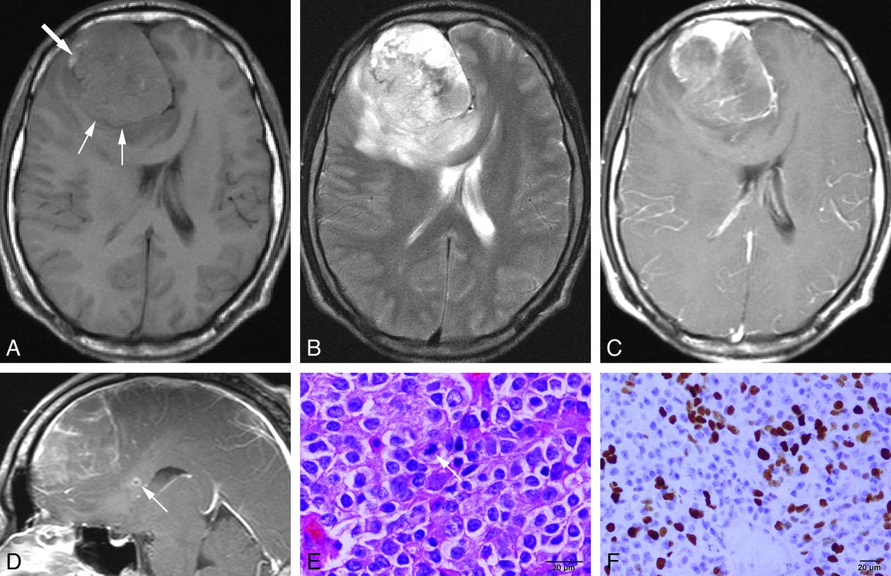
  • Fig. 1.
    • Download figure
    • Open in new tab
    • Download powerpoint
    Fig. 1.

    MR images of a 33-year-old man with atypical EVN of the right frontal lobe. A, Axial precontrast T1WI shows a well-demarcated large mass in the right frontal lobe (small arrows), isointense to gray matter dotted with small foci of hyperintensity representing hemorrhage (large arrow). B, Axial T2WI reveals mixed hyperintense to hypointense components with moderate peritumoral edema. C and D, Axial and sagittal postcontrast T1WI demonstrate a mass with uneven and patchy enhancement. A small focus of infiltration adjacent to the mass is noted in corpus callosum (arrow, D). E, Hematoxylin and eosin staining shows the composition of monotonous small tumor cells with regular round nuclei, and mitosis (arrow). F, Results of immunohistochemical assays demonstrate Ki-67 > 30%.
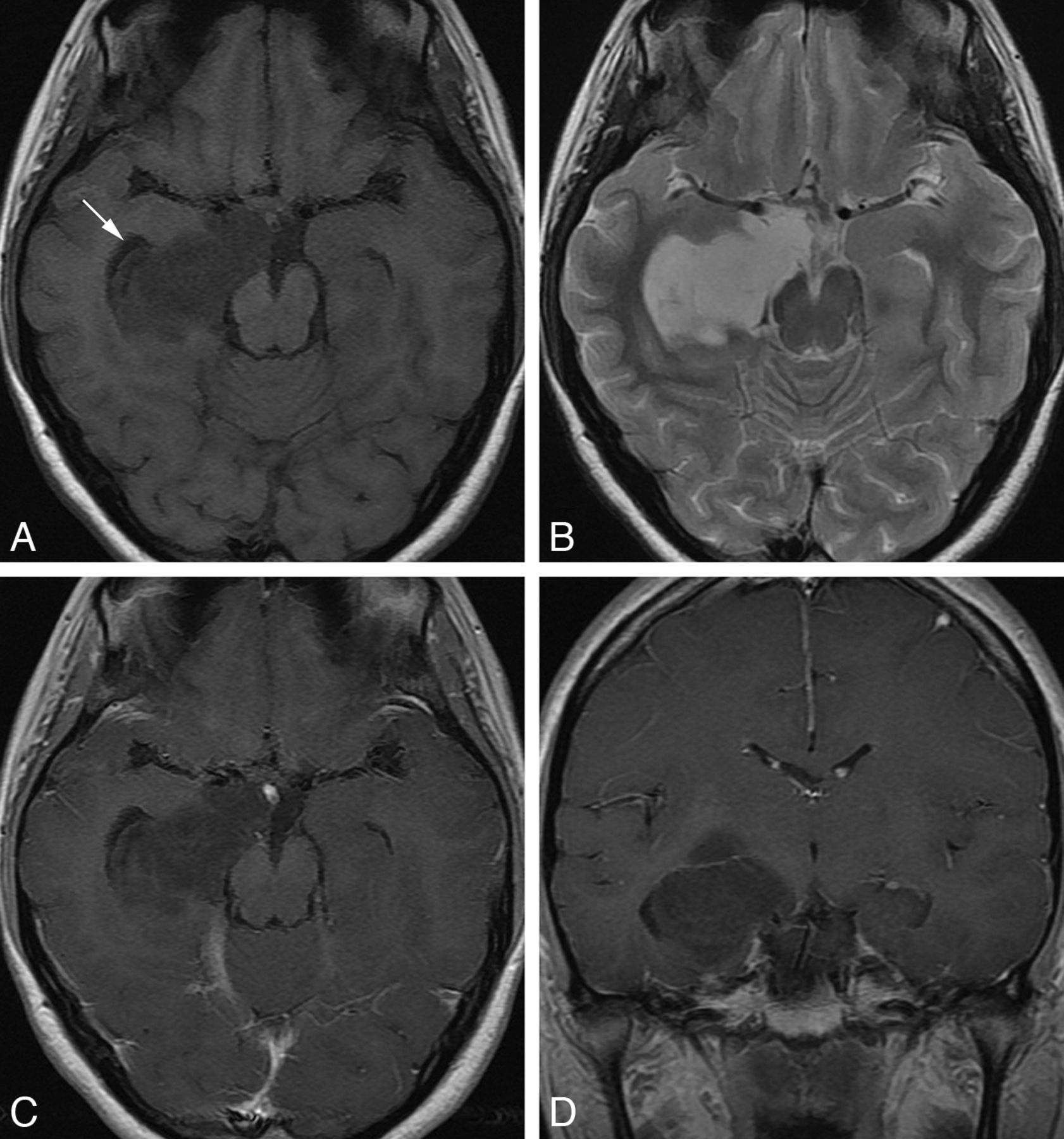
  • Fig. 3.
    • Download figure
    • Open in new tab
    • Download powerpoint
    Fig. 3.

    MR images of a 20-year-old woman with EVN of the right temporal lobe. A, Axial T1WI shows a homogeneous hypointense lesion located deep in the right temporal lobe. The temporal horn of right lateral ventricle is displaced (arrow). B, Axial T2WI image shows markedly hyperintense signal within the lesion approaching that of the CSF. No obvious perilesional edema is observed. C and D, Axial and coronal postcontrast T1WI show no obvious enhancement.

  • Fig. 4.
    • Download figure
    • Open in new tab
    • Download powerpoint
    Fig. 4.

    MR and CT images of a 40-year-old man with EVN of the sellar and suprasellar region. A, Axial T1WI shows a lesion with primarily isointense signal. B, Axial T2WI shows a mixed isointense to hyperintense lesion. C, Coronal postcontrast T1WI demonstrates the heterogeneously strong enhancement of the lesion and the transverse infiltration toward vascular structures on both sides (arrows). D, Axial precontrast CT image shows a lesion with slightly increased attenuation and small foci of decreased attenuation of cystic degeneration.

PreviousNext
Back to top

In this issue

American Journal of Neuroradiology: 34 (3)
American Journal of Neuroradiology
Vol. 34, Issue 3
1 Mar 2013
  • Table of Contents
  • Index by author
Advertisement
Print
Download PDF
Email Article

Thank you for your interest in spreading the word on American Journal of Neuroradiology.

NOTE: We only request your email address so that the person you are recommending the page to knows that you wanted them to see it, and that it is not junk mail. We do not capture any email address.

Enter multiple addresses on separate lines or separate them with commas.
MR Imaging of Cerebral Extraventricular Neurocytoma: A Report of 9 Cases
(Your Name) has sent you a message from American Journal of Neuroradiology
(Your Name) thought you would like to see the American Journal of Neuroradiology web site.
CAPTCHA
This question is for testing whether or not you are a human visitor and to prevent automated spam submissions.
Cite this article
K. Liu, G. Wen, X.-F. Lv, Yan-Jia Deng, Yong-Jian Deng, G.-Q. Hou, X.-L. Zhang, L.-J. Han, J.-L. Ding
MR Imaging of Cerebral Extraventricular Neurocytoma: A Report of 9 Cases
American Journal of Neuroradiology Mar 2013, 34 (3) 541-546; DOI: 10.3174/ajnr.A3264

Citation Manager Formats

  • BibTeX
  • Bookends
  • EasyBib
  • EndNote (tagged)
  • EndNote 8 (xml)
  • Medlars
  • Mendeley
  • Papers
  • RefWorks Tagged
  • Ref Manager
  • RIS
  • Zotero
0 Responses
Respond to this article
Share
Bookmark this article
MR Imaging of Cerebral Extraventricular Neurocytoma: A Report of 9 Cases
K. Liu, G. Wen, X.-F. Lv, Yan-Jia Deng, Yong-Jian Deng, G.-Q. Hou, X.-L. Zhang, L.-J. Han, J.-L. Ding
American Journal of Neuroradiology Mar 2013, 34 (3) 541-546; DOI: 10.3174/ajnr.A3264
del.icio.us logo Twitter logo Facebook logo Mendeley logo
  • Tweet Widget
  • Facebook Like
  • Google Plus One
Purchase

Jump to section

  • Article
    • Abstract
    • ABBREVIATIONS:
    • Materials and Methods
    • Results
    • Discussion
    • Conclusions
    • Acknowledgments
    • Footnotes
    • References
  • Figures & Data
  • Info & Metrics
  • Responses
  • References
  • PDF

Related Articles

  • No related articles found.
  • PubMed
  • Google Scholar

Cited By...

  • Awake craniotomy in an adolescent patient with an extraventricular neurocytoma
  • Crossref (30)
  • Google Scholar

This article has been cited by the following articles in journals that are participating in Crossref Cited-by Linking.

  • Extraventricular neurocytoma, a comprehensive review
    Akshay S. Patil, G. Menon, H. V. Easwer, S. Nair
    Acta Neurochirurgica 2014 156 2
  • Clinical Manifestations of Central Neurocytoma
    Isaac Yang, Nolan Ung, Lawrance K. Chung, Daniel T. Nagasawa, Kimberly Thill, Junmook Park, Stephen Tenn
    Neurosurgery Clinics of North America 2015 26 1
  • Pharmacodynamic Analysis of Magnetic Resonance Imaging-Monitored Focused Ultrasound-Induced Blood-Brain Barrier Opening for Drug Delivery to Brain Tumors
    Po-Chun Chu, Wen-Yen Chai, Han-Yi Hsieh, Jiun-Jie Wang, Shiaw-Pyng Wey, Chiung-Yin Huang, Kuo-Chen Wei, Hao-Li Liu
    BioMed Research International 2013 2013
  • Extraventricular Neurocytomas
    Fadi B. Sweiss, Michaela Lee, Jonathan H. Sherman
    Neurosurgery Clinics of North America 2015 26 1
  • Imaging of Neuronal and Mixed Glioneuronal Tumors
    Ahmed Abdel Khalek Abdel Razek, Nermeen A. Elsebaie, Carlos Zamora, Mauricio Castillo
    Journal of Computer Assisted Tomography 2020 44 3
  • Imaging of extraventricular neurocytoma: a systematic literature review
    Nicola Romano, Margherita Federici, Antonio Castaldi
    La radiologia medica 2020 125 10
  • Central Versus Extraventricular Neurocytoma in Children: A Clinicopathologic Comparison and Review of the Literature
    Stergios Zacharoulis, Andres Morales La Madrid, Pratiti Bandopadhayay, Susan N. Chi, Peter Manley, Nicole N.J. Ullrich, Karen Marcus, Liliana Goumnerova, Robert M. Scott, Mark Proctor, Tina Young-Poussaint, Umberto De Girolami, Mark W. Kieran
    Journal of Pediatric Hematology/Oncology 2016 38 6
  • The 2021 WHO Classification for Gliomas and Implications on Imaging Diagnosis: Part 3—Summary of Imaging Findings on Glioneuronal and Neuronal Tumors
    Yae Won Park, Philipp Vollmuth, Martha Foltyn‐Dumitru, Felix Sahm, Kyu Sung Choi, Ji Eun Park, Sung Soo Ahn, Jong Hee Chang, Se Hoon Kim
    Journal of Magnetic Resonance Imaging 2023 58 6
  • The typical and atypical MR imaging findings of central neurocytomas: Report on eighteen cases and review of the literature
    Zhanlong Ma, Hailang Yan, Haibin Shi, Yan Li, Jiacheng Song, Junwen Huang, Xiongning Hong
    Clinical Neurology and Neurosurgery 2016 146
  • Extraventricular neurocytoma in the left temporal lobe: A case report and review of the literature
    YU-CHEN JI, JING-XIA HU, YAN LI, PENG-XIANG YAN, HUAN-CONG ZUO
    Oncology Letters 2016 11 6

More in this TOC Section

  • Evaluating the Effects of White Matter Multiple Sclerosis Lesions on the Volume Estimation of 6 Brain Tissue Segmentation Methods
  • Quiet PROPELLER MRI Techniques Match the Quality of Conventional PROPELLER Brain Imaging Techniques
  • Predictors of Reperfusion in Patients with Acute Ischemic Stroke
Show more BRAIN

Similar Articles

Advertisement

Indexed Content

  • Current Issue
  • Accepted Manuscripts
  • Article Preview
  • Past Issues
  • Editorials
  • Editor's Choice
  • Fellows' Journal Club
  • Letters to the Editor
  • Video Articles

Cases

  • Case Collection
  • Archive - Case of the Week
  • Archive - Case of the Month
  • Archive - Classic Case

More from AJNR

  • Trainee Corner
  • Imaging Protocols
  • MRI Safety Corner

Multimedia

  • AJNR Podcasts
  • AJNR Scantastics

Resources

  • Turnaround Time
  • Submit a Manuscript
  • Submit a Video Article
  • Submit an eLetter to the Editor/Response
  • Manuscript Submission Guidelines
  • Statistical Tips
  • Fast Publishing of Accepted Manuscripts
  • Graphical Abstract Preparation
  • Imaging Protocol Submission
  • Evidence-Based Medicine Level Guide
  • Publishing Checklists
  • Author Policies
  • Become a Reviewer/Academy of Reviewers
  • News and Updates

About Us

  • About AJNR
  • Editorial Board
  • Editorial Board Alumni
  • Alerts
  • Permissions
  • Not an AJNR Subscriber? Join Now
  • Advertise with Us
  • Librarian Resources
  • Feedback
  • Terms and Conditions
  • AJNR Editorial Board Alumni

American Society of Neuroradiology

  • Not an ASNR Member? Join Now

© 2025 by the American Society of Neuroradiology All rights, including for text and data mining, AI training, and similar technologies, are reserved.
Print ISSN: 0195-6108 Online ISSN: 1936-959X

Powered by HighWire