Advertisement

Main menu

  • Home
  • Content
    • Current Issue
    • Accepted Manuscripts
    • Article Preview
    • Past Issue Archive
    • Video Articles
    • AJNR Case Collection
    • Case of the Week Archive
    • Case of the Month Archive
    • Classic Case Archive
  • Special Collections
    • AJNR Awards
    • Low-Field MRI
    • Alzheimer Disease
    • ASNR Foundation Special Collection
    • Photon-Counting CT
    • View All
  • Multimedia
    • AJNR Podcasts
    • AJNR SCANtastic
    • Trainee Corner
    • MRI Safety Corner
    • Imaging Protocols
  • For Authors
    • Submit a Manuscript
    • Submit a Video Article
    • Submit an eLetter to the Editor/Response
    • Manuscript Submission Guidelines
    • Statistical Tips
    • Fast Publishing of Accepted Manuscripts
    • Graphical Abstract Preparation
    • Imaging Protocol Submission
    • Author Policies
  • About Us
    • About AJNR
    • Editorial Board
    • Editorial Board Alumni
  • More
    • Become a Reviewer/Academy of Reviewers
    • Subscribers
    • Permissions
    • Alerts
    • Feedback
    • Advertisers
    • ASNR Home

User menu

  • Alerts
  • Log in

Search

  • Advanced search
American Journal of Neuroradiology

American Journal of Neuroradiology

ASHNR American Society of Functional Neuroradiology ASHNR American Society of Pediatric Neuroradiology ASSR
  • Alerts
  • Log in

Advanced Search

  • Home
  • Content
    • Current Issue
    • Accepted Manuscripts
    • Article Preview
    • Past Issue Archive
    • Video Articles
    • AJNR Case Collection
    • Case of the Week Archive
    • Case of the Month Archive
    • Classic Case Archive
  • Special Collections
    • AJNR Awards
    • Low-Field MRI
    • Alzheimer Disease
    • ASNR Foundation Special Collection
    • Photon-Counting CT
    • View All
  • Multimedia
    • AJNR Podcasts
    • AJNR SCANtastic
    • Trainee Corner
    • MRI Safety Corner
    • Imaging Protocols
  • For Authors
    • Submit a Manuscript
    • Submit a Video Article
    • Submit an eLetter to the Editor/Response
    • Manuscript Submission Guidelines
    • Statistical Tips
    • Fast Publishing of Accepted Manuscripts
    • Graphical Abstract Preparation
    • Imaging Protocol Submission
    • Author Policies
  • About Us
    • About AJNR
    • Editorial Board
    • Editorial Board Alumni
  • More
    • Become a Reviewer/Academy of Reviewers
    • Subscribers
    • Permissions
    • Alerts
    • Feedback
    • Advertisers
    • ASNR Home
  • Follow AJNR on Twitter
  • Visit AJNR on Facebook
  • Follow AJNR on Instagram
  • Join AJNR on LinkedIn
  • RSS Feeds

AJNR Awards, New Junior Editors, and more. Read the latest AJNR updates

Research ArticlePediatrics

Imaging Changes in Very Young Children with Brain Tumors Treated with Proton Therapy and Chemotherapy

N.D. Sabin, T.E. Merchant, J.H. Harreld, Z. Patay, P. Klimo, I. Qaddoumi, G.T. Armstrong, K. Wright, J. Gray, D.J. Indelicato and A. Gajjar
American Journal of Neuroradiology February 2013, 34 (2) 446-450; DOI: https://doi.org/10.3174/ajnr.A3219
N.D. Sabin
aFrom the Departments of Radiological Sciences (N.D.S., T.E.M., J.H.H., Z.P., J.G.)
  • Find this author on Google Scholar
  • Find this author on PubMed
  • Search for this author on this site
T.E. Merchant
aFrom the Departments of Radiological Sciences (N.D.S., T.E.M., J.H.H., Z.P., J.G.)
  • Find this author on Google Scholar
  • Find this author on PubMed
  • Search for this author on this site
J.H. Harreld
aFrom the Departments of Radiological Sciences (N.D.S., T.E.M., J.H.H., Z.P., J.G.)
  • Find this author on Google Scholar
  • Find this author on PubMed
  • Search for this author on this site
Z. Patay
aFrom the Departments of Radiological Sciences (N.D.S., T.E.M., J.H.H., Z.P., J.G.)
  • Find this author on Google Scholar
  • Find this author on PubMed
  • Search for this author on this site
P. Klimo Jr
bSurgery (P.K.)
eSemmes-Murphey Neurologic and Spine Institute (P.K.), Memphis, Tennessee
fDepartment of Neurosurgery (P.K.), University of Tennessee Health Science Center, Memphis, Tennessee
  • Find this author on Google Scholar
  • Find this author on PubMed
  • Search for this author on this site
I. Qaddoumi
cOncology (I.Q., K.W., A.G.)
  • Find this author on Google Scholar
  • Find this author on PubMed
  • Search for this author on this site
G.T. Armstrong
dEpidemiology and Cancer Control (G.T.A.), St. Jude Children's Research Hospital, Memphis, Tennessee
  • Find this author on Google Scholar
  • Find this author on PubMed
  • Search for this author on this site
K. Wright
cOncology (I.Q., K.W., A.G.)
  • Find this author on Google Scholar
  • Find this author on PubMed
  • Search for this author on this site
J. Gray
cOncology (I.Q., K.W., A.G.)
  • Find this author on Google Scholar
  • Find this author on PubMed
  • Search for this author on this site
D.J. Indelicato
gDepartment of Radiation Oncology (D.J.I.), University of Florida College of Medicine, Gainesville, Florida
hUniversity of Florida Proton Therapy Institute (D.J.I.), Jacksonville, Florida.
  • Find this author on Google Scholar
  • Find this author on PubMed
  • Search for this author on this site
A. Gajjar
aFrom the Departments of Radiological Sciences (N.D.S., T.E.M., J.H.H., Z.P., J.G.)
  • Find this author on Google Scholar
  • Find this author on PubMed
  • Search for this author on this site
  • Article
  • Figures & Data
  • Info & Metrics
  • Responses
  • References
  • PDF
Loading

Article Figures & Data

Figures

  • Tables
  • Fig 1.
    • Download figure
    • Open in new tab
    • Download powerpoint
    Fig 1.

    Axial T2-weighted (left) and axial postgadolinium T1-weighted (right) images through the posterior fossa of patient 2, acquired 3.6 months after completion of PT, reveal prominent T2 prolongation and heterogeneous enhancement within the pons. This portion of the pons was in the high-dose volume of irradiation.

  • Fig 2.
    • Download figure
    • Open in new tab
    • Download powerpoint
    Fig 2.

    Axial FLAIR (A) and postgadolinium coronal T1-weighted (B) images for patient 6, obtained 4.2 months after completion of PT, demonstrate patchy hyperintensity (FLAIR) and focal irregular enhancement (arrows in B) within the pons. Diffusion-weighted (C) and apparent diffusion coefficient map (D) images reveal small areas of restricted diffusion in the areas of abnormal signal and enhancement. This portion of the pons was in the high-dose volume of irradiation. The abnormalities improved on an MR imaging study obtained 1.5 months later and were no longer visible 4 months after they first appeared (E and F).

  • Fig 3.
    • Download figure
    • Open in new tab
    • Download powerpoint
    Fig 3.

    Axial T2 (left) and axial postgadolinium T1-weighted (right) images through the level of the thalami for patient 4 demonstrate enlargement of the right thalamus with hyperintensity on the T2-weighted image without enhancement (arrows).

  • Fig 4.
    • Download figure
    • Open in new tab
    • Download powerpoint
    Fig 4.

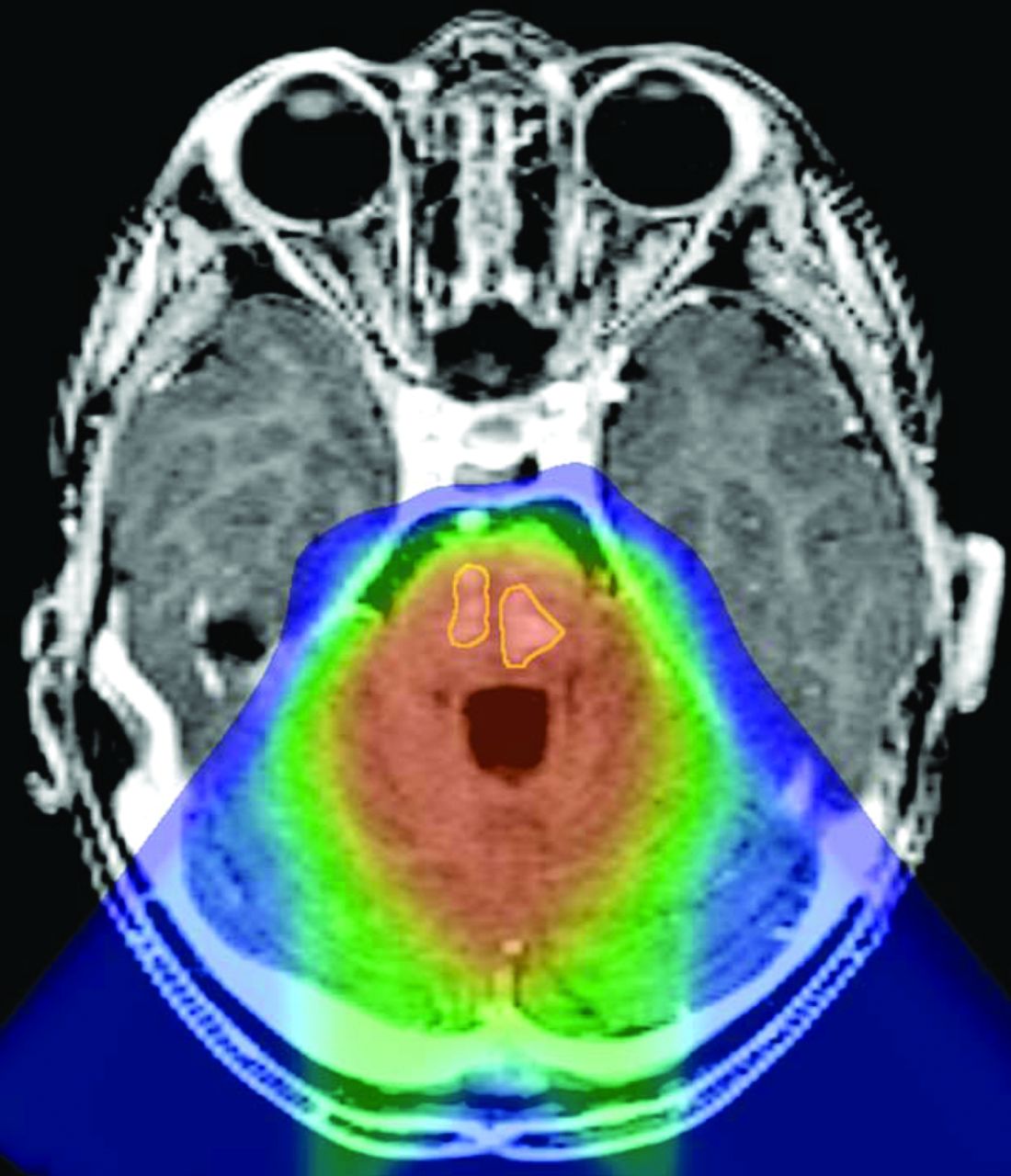
    Reformatted axial postgadolinium T1-weighted magnetization-prepared rapid acquisition of gradient echo image through the posterior fossa of patient 8, coregistered to the proton therapy treatment plan, demonstrates 2 areas of enhancement within the pons (outlined in yellow) located within the high-dose volume of irradiation (orange wash).

Tables

  • Figures
    • View popup
    Table 1:

    Clinical data for 8 patients with imaging changes after proton therapy

    PatientSexDiagnosisLocationAge at Diagnosis (yr)Age at Proton Therapy (yr)
        1FEPFourth ventricle1.72.2
        2MEPFourth ventricle1.92.4
        3FCPCLeft lateral ventricle1.22.6
        4FPNETLeft parietal lobe2.73.2
        5FMBFourth ventricle11.4
        6FEPFourth ventricle33.5
        7FAT/RTPosterior fossa0.61
        8MEPFourth ventricle2.22.6
    Median1.82.5
    Mean1.82.4
    • Note:—EP indicates ependymoma; MB, medulloblastoma.

    • View popup
    Table 2:

    MRI findings observed in 8 patients after proton therapy

    Time from Completion of Proton Therapy to First MRI with Changes (mo)Time from Appearance of Changes to Start of Resolution of Changes (mo)Location of Abnormal Signal/EnhancementPhysical Examination Findings
    Patient
        15.32.2Brain stem, cerebellum, upper cervical spinal cordNone
        23.62.3Brain stemUnilateral weakness
        35.63CerebellumNone
        43.43.1Right thalamus (no enhancement)Unilateral weakness
        56.32.2Brain stemNone
        64.21.5Brain stem and cerebellumUnilateral weakness
        73Patient died of disease before follow-upBrain stemNone
        83.23.5Brain stemAtaxia
    Median3.92.3 (of 7 with follow-up MRI)
    Mean4.32.5 (of 7 with follow-up MRI)
PreviousNext
Back to top

In this issue

American Journal of Neuroradiology: 34 (2)
American Journal of Neuroradiology
Vol. 34, Issue 2
1 Feb 2013
  • Table of Contents
  • Index by author
Advertisement
Print
Download PDF
Email Article

Thank you for your interest in spreading the word on American Journal of Neuroradiology.

NOTE: We only request your email address so that the person you are recommending the page to knows that you wanted them to see it, and that it is not junk mail. We do not capture any email address.

Enter multiple addresses on separate lines or separate them with commas.
Imaging Changes in Very Young Children with Brain Tumors Treated with Proton Therapy and Chemotherapy
(Your Name) has sent you a message from American Journal of Neuroradiology
(Your Name) thought you would like to see the American Journal of Neuroradiology web site.
CAPTCHA
This question is for testing whether or not you are a human visitor and to prevent automated spam submissions.
Cite this article
N.D. Sabin, T.E. Merchant, J.H. Harreld, Z. Patay, P. Klimo, I. Qaddoumi, G.T. Armstrong, K. Wright, J. Gray, D.J. Indelicato, A. Gajjar
Imaging Changes in Very Young Children with Brain Tumors Treated with Proton Therapy and Chemotherapy
American Journal of Neuroradiology Feb 2013, 34 (2) 446-450; DOI: 10.3174/ajnr.A3219

Citation Manager Formats

  • BibTeX
  • Bookends
  • EasyBib
  • EndNote (tagged)
  • EndNote 8 (xml)
  • Medlars
  • Mendeley
  • Papers
  • RefWorks Tagged
  • Ref Manager
  • RIS
  • Zotero
0 Responses
Respond to this article
Share
Bookmark this article
Imaging Changes in Very Young Children with Brain Tumors Treated with Proton Therapy and Chemotherapy
N.D. Sabin, T.E. Merchant, J.H. Harreld, Z. Patay, P. Klimo, I. Qaddoumi, G.T. Armstrong, K. Wright, J. Gray, D.J. Indelicato, A. Gajjar
American Journal of Neuroradiology Feb 2013, 34 (2) 446-450; DOI: 10.3174/ajnr.A3219
del.icio.us logo Twitter logo Facebook logo Mendeley logo
  • Tweet Widget
  • Facebook Like
  • Google Plus One
Purchase

Jump to section

  • Article
    • Abstract
    • ABBREVIATIONS:
    • Case Series
    • Discussion
    • Acknowledgments
    • Footnotes
    • References
  • Figures & Data
  • Info & Metrics
  • Responses
  • References
  • PDF

Related Articles

  • No related articles found.
  • PubMed
  • Google Scholar

Cited By...

  • Radiation Necrosis in Pediatric Patients with Brain Tumors Treated with Proton Radiotherapy
  • Crossref (36)
  • Google Scholar

This article has been cited by the following articles in journals that are participating in Crossref Cited-by Linking.

  • Medulloblastoma
    Paul A. Northcott, Giles W. Robinson, Christian P. Kratz, Donald J. Mabbott, Scott L. Pomeroy, Steven C. Clifford, Stefan Rutkowski, David W. Ellison, David Malkin, Michael D. Taylor, Amar Gajjar, Stefan M. Pfister
    Nature Reviews Disease Primers 2019 5 1
  • Charged-particle therapy in cancer: clinical uses and future perspectives
    Marco Durante, Roberto Orecchia, Jay S. Loeffler
    Nature Reviews Clinical Oncology 2017 14 8
  • Clinical evidence of variable proton biological effectiveness in pediatric patients treated for ependymoma
    Christopher R. Peeler, Dragan Mirkovic, Uwe Titt, Pierre Blanchard, Jillian R. Gunther, Anita Mahajan, Radhe Mohan, David R. Grosshans
    Radiotherapy and Oncology 2016 121 3
  • National Cancer Institute Workshop on Proton Therapy for Children: Considerations Regarding Brainstem Injury
    Daphne Haas-Kogan, Daniel Indelicato, Harald Paganetti, Natia Esiashvili, Anita Mahajan, Torunn Yock, Stella Flampouri, Shannon MacDonald, Maryam Fouladi, Kry Stephen, John Kalapurakal, Stephanie Terezakis, Hanne Kooy, David Grosshans, Mike Makrigiorgos, Kavita Mishra, Tina Young Poussaint, Kenneth Cohen, Thomas Fitzgerald, Vinai Gondi, Arthur Liu, Jeff Michalski, Dragan Mirkovic, Radhe Mohan, Stephanie Perkins, Kenneth Wong, Bhadrasain Vikram, Jeff Buchsbaum, Larry Kun
    International Journal of Radiation Oncology*Biology*Physics 2018 101 1
  • Imaging Changes in Pediatric Intracranial Ependymoma Patients Treated With Proton Beam Radiation Therapy Compared to Intensity Modulated Radiation Therapy
    Jillian R. Gunther, Mariko Sato, Murali Chintagumpala, Leena Ketonen, Jeremy Y. Jones, Pamela K. Allen, Arnold C. Paulino, M. Fatih Okcu, Jack M. Su, Jeffrey Weinberg, Nicholas S. Boehling, Soumen Khatua, Adekunle Adesina, Robert Dauser, William E. Whitehead, Anita Mahajan
    International Journal of Radiation Oncology*Biology*Physics 2015 93 1
  • Radiation Necrosis in Pediatric Patients with Brain Tumors Treated with Proton Radiotherapy
    S.F. Kralik, C.Y. Ho, W. Finke, J.C. Buchsbaum, C.P. Haskins, C.-S. Shih
    American Journal of Neuroradiology 2015 36 8
  • Outcomes and Acute Toxicities of Proton Therapy for Pediatric Atypical Teratoid/Rhabdoid Tumor of the Central Nervous System
    Susan L. McGovern, M. Fatih Okcu, Mark F. Munsell, Nancy Kumbalasseriyil, David R. Grosshans, Mary F. McAleer, Murali Chintagumpala, Soumen Khatua, Anita Mahajan
    International Journal of Radiation Oncology*Biology*Physics 2014 90 5
  • Linear energy transfer incorporated intensity modulated proton therapy optimization
    Wenhua Cao, Azin Khabazian, Pablo P Yepes, Gino Lim, Falk Poenisch, David R Grosshans, Radhe Mohan
    Physics in Medicine & Biology 2017 63 1
  • Brainstem Injury in Pediatric Patients With Posterior Fossa Tumors Treated With Proton Beam Therapy and Associated Dosimetric Factors
    Michelle S. Gentile, Beow Y. Yeap, Harald Paganetti, Claire P. Goebel, Dillon E. Gaudet, Sara L. Gallotto, Elizabeth A. Weyman, Michael L. Morgan, Shannon M. MacDonald, Drosoula Giantsoudi, Judith Adams, Nancy J. Tarbell, Hanne Kooy, Torunn I. Yock
    International Journal of Radiation Oncology*Biology*Physics 2018 100 3
  • Brain Necrosis in Adult Patients After Proton Therapy: Is There Evidence for Dependency on Linear Energy Transfer?
    Andrzej Niemierko, Jan Schuemann, Maximilian Niyazi, Drosoula Giantsoudi, Genevieve Maquilan, Helen A. Shih, Harald Paganetti
    International Journal of Radiation Oncology*Biology*Physics 2021 109 1

More in this TOC Section

  • SyMRI & MR Fingerprinting in Brainstem Myelination
  • Comparison of Image Quality and Radiation Dose in Pediatric Temporal Bone CT Using Photon-Counting Detector CT and Energy-Integrating Detector CT
  • Dual-Layer Detector CT for PEDS Image Quality
Show more Pediatrics

Similar Articles

Advertisement

Indexed Content

  • Current Issue
  • Accepted Manuscripts
  • Article Preview
  • Past Issues
  • Editorials
  • Editor's Choice
  • Fellows' Journal Club
  • Letters to the Editor
  • Video Articles

Cases

  • Case Collection
  • Archive - Case of the Week
  • Archive - Case of the Month
  • Archive - Classic Case

More from AJNR

  • Trainee Corner
  • Imaging Protocols
  • MRI Safety Corner
  • Book Reviews

Multimedia

  • AJNR Podcasts
  • AJNR Scantastics

Resources

  • Turnaround Time
  • Submit a Manuscript
  • Submit a Video Article
  • Submit an eLetter to the Editor/Response
  • Manuscript Submission Guidelines
  • Statistical Tips
  • Fast Publishing of Accepted Manuscripts
  • Graphical Abstract Preparation
  • Imaging Protocol Submission
  • Evidence-Based Medicine Level Guide
  • Publishing Checklists
  • Author Policies
  • Become a Reviewer/Academy of Reviewers
  • News and Updates

About Us

  • About AJNR
  • Editorial Board
  • Editorial Board Alumni
  • Alerts
  • Permissions
  • Not an AJNR Subscriber? Join Now
  • Advertise with Us
  • Librarian Resources
  • Feedback
  • Terms and Conditions
  • AJNR Editorial Board Alumni

American Society of Neuroradiology

  • Not an ASNR Member? Join Now

© 2025 by the American Society of Neuroradiology All rights, including for text and data mining, AI training, and similar technologies, are reserved.
Print ISSN: 0195-6108 Online ISSN: 1936-959X

Powered by HighWire