Advertisement

Main menu

  • Home
  • Content
    • Current Issue
    • Accepted Manuscripts
    • Article Preview
    • Past Issue Archive
    • Video Articles
    • AJNR Case Collection
    • Case of the Week Archive
    • Case of the Month Archive
    • Classic Case Archive
  • Special Collections
    • Advancing NeuroMRI with High-Relaxivity Contrast Agents
    • AJNR Awards
    • Low-Field MRI
    • Alzheimer Disease
    • ASNR Foundation Special Collection
    • Photon-Counting CT
    • View All
  • Multimedia
    • AJNR Podcasts
    • AJNR SCANtastic
    • Trainee Corner
    • MRI Safety Corner
    • Imaging Protocols
  • For Authors
    • Submit a Manuscript
    • Submit a Video Article
    • Submit an eLetter to the Editor/Response
    • Manuscript Submission Guidelines
    • Statistical Tips
    • Fast Publishing of Accepted Manuscripts
    • Graphical Abstract Preparation
    • Imaging Protocol Submission
    • Author Policies
  • About Us
    • About AJNR
    • Editorial Board
    • Editorial Board Alumni
  • More
    • Become a Reviewer/Academy of Reviewers
    • Subscribers
    • Permissions
    • Alerts
    • Feedback
    • Advertisers
    • ASNR Home

User menu

  • Alerts
  • Log in

Search

  • Advanced search
American Journal of Neuroradiology

American Journal of Neuroradiology

ASHNR American Society of Functional Neuroradiology ASHNR American Society of Pediatric Neuroradiology ASSR
  • Alerts
  • Log in

Advanced Search

  • Home
  • Content
    • Current Issue
    • Accepted Manuscripts
    • Article Preview
    • Past Issue Archive
    • Video Articles
    • AJNR Case Collection
    • Case of the Week Archive
    • Case of the Month Archive
    • Classic Case Archive
  • Special Collections
    • Advancing NeuroMRI with High-Relaxivity Contrast Agents
    • AJNR Awards
    • Low-Field MRI
    • Alzheimer Disease
    • ASNR Foundation Special Collection
    • Photon-Counting CT
    • View All
  • Multimedia
    • AJNR Podcasts
    • AJNR SCANtastic
    • Trainee Corner
    • MRI Safety Corner
    • Imaging Protocols
  • For Authors
    • Submit a Manuscript
    • Submit a Video Article
    • Submit an eLetter to the Editor/Response
    • Manuscript Submission Guidelines
    • Statistical Tips
    • Fast Publishing of Accepted Manuscripts
    • Graphical Abstract Preparation
    • Imaging Protocol Submission
    • Author Policies
  • About Us
    • About AJNR
    • Editorial Board
    • Editorial Board Alumni
  • More
    • Become a Reviewer/Academy of Reviewers
    • Subscribers
    • Permissions
    • Alerts
    • Feedback
    • Advertisers
    • ASNR Home
  • Follow AJNR on Twitter
  • Visit AJNR on Facebook
  • Follow AJNR on Instagram
  • Join AJNR on LinkedIn
  • RSS Feeds

AJNR Awards, New Junior Editors, and more. Read the latest AJNR updates


Improved Turnaround Times | Median time to first decision: 12 days

Research ArticleBrain

Intracranial-Derived Atherosclerosis Assessment: An In Vitro Comparison between Virtual Histology by Intravascular Ultrasonography, 7T MRI, and Histopathologic Findings

S. Majidi, J. Sein, M. Watanabe, A.E. Hassan, P.-F. Van de Moortele, M.F.K. Suri, H.B. Clark and A.I. Qureshi
American Journal of Neuroradiology December 2013, 34 (12) 2259-2264; DOI: https://doi.org/10.3174/ajnr.A3631
S. Majidi
aFrom the Zeenat Qureshi Stroke Research Center (S.M., M.W., A.E.H., M.F.K.S., A.I.Q.)
  • Find this author on Google Scholar
  • Find this author on PubMed
  • Search for this author on this site
J. Sein
bCenter for Magnetic Resonance Research (J.S., P.-F.V.d.M.)
  • Find this author on Google Scholar
  • Find this author on PubMed
  • Search for this author on this site
M. Watanabe
aFrom the Zeenat Qureshi Stroke Research Center (S.M., M.W., A.E.H., M.F.K.S., A.I.Q.)
  • Find this author on Google Scholar
  • Find this author on PubMed
  • Search for this author on this site
A.E. Hassan
aFrom the Zeenat Qureshi Stroke Research Center (S.M., M.W., A.E.H., M.F.K.S., A.I.Q.)
  • Find this author on Google Scholar
  • Find this author on PubMed
  • Search for this author on this site
P.-F. Van de Moortele
bCenter for Magnetic Resonance Research (J.S., P.-F.V.d.M.)
  • Find this author on Google Scholar
  • Find this author on PubMed
  • Search for this author on this site
M.F.K. Suri
aFrom the Zeenat Qureshi Stroke Research Center (S.M., M.W., A.E.H., M.F.K.S., A.I.Q.)
  • Find this author on Google Scholar
  • Find this author on PubMed
  • Search for this author on this site
H.B. Clark
cDepartment of Laboratory Medicine and Pathology (H.B.C.), University of Minnesota, Minneapolis, Minnesota.
  • Find this author on Google Scholar
  • Find this author on PubMed
  • Search for this author on this site
A.I. Qureshi
aFrom the Zeenat Qureshi Stroke Research Center (S.M., M.W., A.E.H., M.F.K.S., A.I.Q.)
  • Find this author on Google Scholar
  • Find this author on PubMed
  • Search for this author on this site
  • Article
  • Figures & Data
  • Info & Metrics
  • Responses
  • References
  • PDF
Loading

Article Figures & Data

Figures

  • Tables
  • Fig 1.
    • Download figure
    • Open in new tab
    • Download powerpoint
    Fig 1.

    Characteristics of Eagle Eye Gold IVUS imaging catheter.
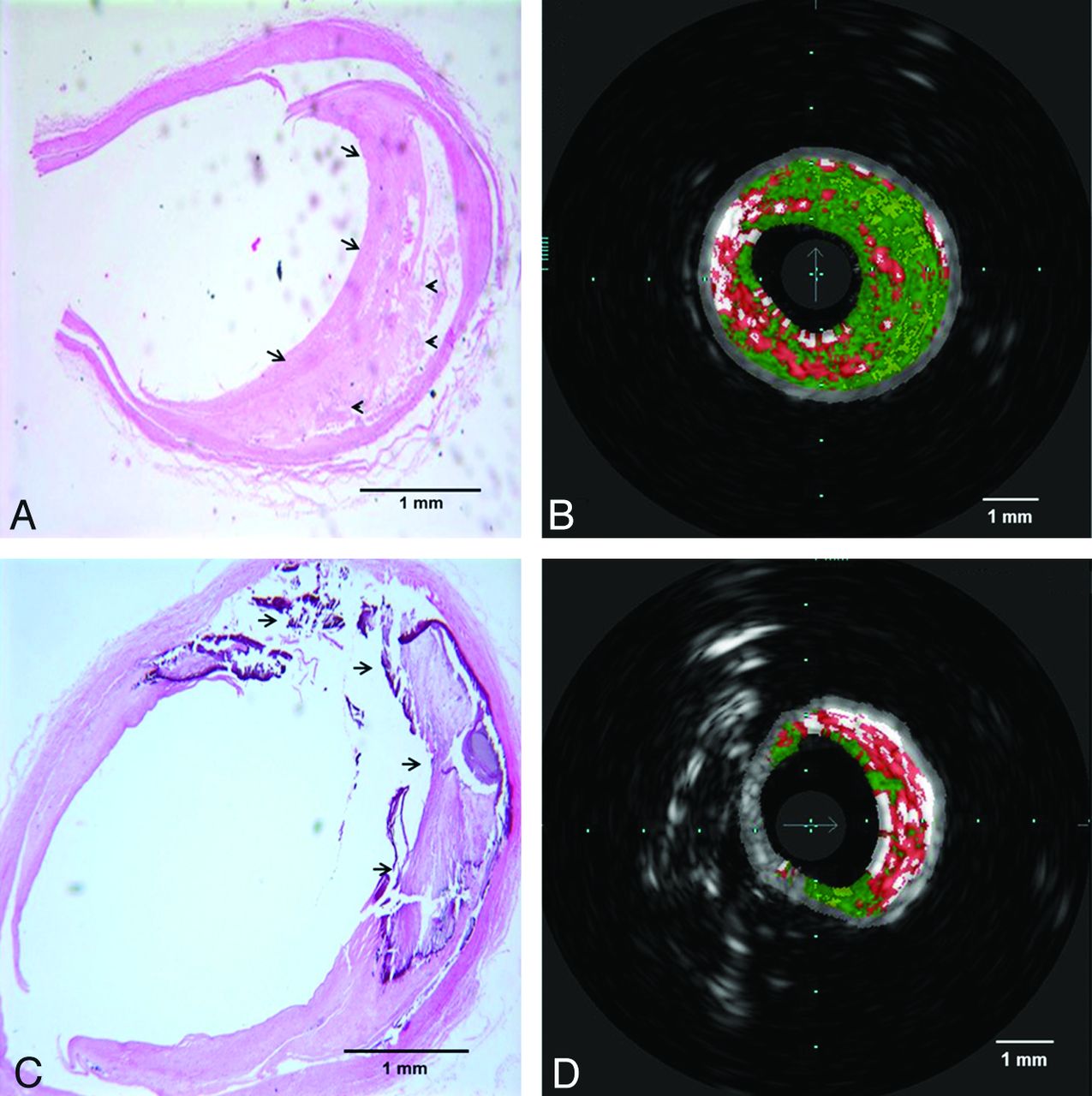
  • Fig 2.
    • Download figure
    • Open in new tab
    • Download powerpoint
    Fig 2.

    Histopathologic sections with corresponding VH-IVUS images. A and B, Fibrous (arrows in A) and fibrofatty (arrowheads in A) tissues in the histologic section are correlated with dark green and light green areas in VH-IVUS, respectively. C and D, attenuated calcium and necrotic area (arrows in C) and its corresponding area in VH-IVUS red (necrosis) and white (attenuated calcium) areas.

  • Fig 3.
    • Download figure
    • Open in new tab
    • Download powerpoint
    Fig 3.

    Four different histopathologic sections of intracranial vessels with atherosclerotic plaque and their corresponding 3D SPACE MR imaging (in the middle) and VH-IVUS (on the right) images. Fibrous areas (arrows) and attenuated calcium (arrowheads) consistently visualized as areas with hyperintense and hypointense signals in MR imaging, respectively.

Tables

  • Figures
    • View popup
    Table 1:

    Comparison of different types of atherosclerotic plaques between VH-IVUS and histologic sections (as reference standard)

    Category of PlaquePlaque Classification by Histological AnalysisPlaque Classification by VH-IVUS Analysis
    True-PositiveTrue-NegativeFalse-PositiveFalse-NegativeSensitivitySpecificityPPVNPV
    No plaque223200100100100100
    Pathologic intima thickening6328035010010090.3
    Fibroatheroma1915114478.973.378.973.3
    Calcified fibroatheromaa0033100970100
    Thin-cap fibroatheroma32301166.696.766.696.7
    Calcified thin-cap fibroatheroma2131115096.85096.8
    Fibrocalcific atheroma223200100100100100
    Total34251977973.596.678.195.6
    • PPV indicates positive predictive value; NPV, negative predictive value.

    • ↵a Number is small and requires cautious interpretation of false-positive or false-negative values.

    • View popup
    Table 2:

    Area of 4 different components of plaque types in histologic sections and VH-IVUS analyses

    Histopathology, % (SD)VH-IVUS, % (SD)Pearson Correlation
    Fibrous45.6 (18.7)50.8 (17.1)0.66
    Fibrofatty35.6 (14.1)12.5 (12.1)0.34
    Dense calcium7.1 (7.6)9.1 (5.2)0.64
    Necrosis5.1 (4.6)20.9 (10.9)0.23
PreviousNext
Back to top

In this issue

American Journal of Neuroradiology: 34 (12)
American Journal of Neuroradiology
Vol. 34, Issue 12
1 Dec 2013
  • Table of Contents
  • Index by author
  • Complete Issue (PDF)
Advertisement
Print
Download PDF
Email Article

Thank you for your interest in spreading the word on American Journal of Neuroradiology.

NOTE: We only request your email address so that the person you are recommending the page to knows that you wanted them to see it, and that it is not junk mail. We do not capture any email address.

Enter multiple addresses on separate lines or separate them with commas.
Intracranial-Derived Atherosclerosis Assessment: An In Vitro Comparison between Virtual Histology by Intravascular Ultrasonography, 7T MRI, and Histopathologic Findings
(Your Name) has sent you a message from American Journal of Neuroradiology
(Your Name) thought you would like to see the American Journal of Neuroradiology web site.
CAPTCHA
This question is for testing whether or not you are a human visitor and to prevent automated spam submissions.
Cite this article
S. Majidi, J. Sein, M. Watanabe, A.E. Hassan, P.-F. Van de Moortele, M.F.K. Suri, H.B. Clark, A.I. Qureshi
Intracranial-Derived Atherosclerosis Assessment: An In Vitro Comparison between Virtual Histology by Intravascular Ultrasonography, 7T MRI, and Histopathologic Findings
American Journal of Neuroradiology Dec 2013, 34 (12) 2259-2264; DOI: 10.3174/ajnr.A3631

Citation Manager Formats

  • BibTeX
  • Bookends
  • EasyBib
  • EndNote (tagged)
  • EndNote 8 (xml)
  • Medlars
  • Mendeley
  • Papers
  • RefWorks Tagged
  • Ref Manager
  • RIS
  • Zotero
0 Responses
Respond to this article
Share
Bookmark this article
Intracranial-Derived Atherosclerosis Assessment: An In Vitro Comparison between Virtual Histology by Intravascular Ultrasonography, 7T MRI, and Histopathologic Findings
S. Majidi, J. Sein, M. Watanabe, A.E. Hassan, P.-F. Van de Moortele, M.F.K. Suri, H.B. Clark, A.I. Qureshi
American Journal of Neuroradiology Dec 2013, 34 (12) 2259-2264; DOI: 10.3174/ajnr.A3631
del.icio.us logo Twitter logo Facebook logo Mendeley logo
  • Tweet Widget
  • Facebook Like
  • Google Plus One
Purchase

Jump to section

  • Article
    • Abstract
    • ABBREVIATIONS:
    • Materials and Methods
    • Results
    • Discussion
    • Conclusions
    • Footnotes
    • REFERENCES
  • Figures & Data
  • Info & Metrics
  • Responses
  • References
  • PDF

Related Articles

  • No related articles found.
  • PubMed
  • Google Scholar

Cited By...

  • Prevalence of intracerebral thrombus detected by optical coherence tomography in patients with posterior circulation stroke or transient ischemic attack
  • Imaging Features of Symptomatic MCA Stenosis in Patients of Different Ages: A Vessel Wall MR Imaging Study
  • Emerging Use of Ultra-High-Field 7T MRI in the Study of Intracranial Vascularity: State of the Field and Future Directions
  • Identification and Quantitative Assessment of Different Components of Intracranial Atherosclerotic Plaque by Ex Vivo 3T High-Resolution Multicontrast MRI
  • Postmortem Study of Validation of Low Signal on Fat-Suppressed T1-Weighted Magnetic Resonance Imaging as Marker of Lipid Core in Middle Cerebral Artery Atherosclerosis
  • Magnetic Resonance Imaging of Plaque Morphology, Burden, and Distribution in Patients With Symptomatic Middle Cerebral Artery Stenosis
  • High-resolution intracranial vessel wall imaging: imaging beyond the lumen
  • Quantitative Intracranial Atherosclerotic Plaque Characterization at 7T MRI: An Ex Vivo Study with Histologic Validation
  • Multicontrast High-Resolution Vessel Wall Magnetic Resonance Imaging and Its Value in Differentiating Intracranial Vasculopathic Processes
  • Imaging the Intracranial Atherosclerotic Vessel Wall Using 7T MRI: Initial Comparison with Histopathology
  • Imaging Intracranial Vessel Wall Pathology With Magnetic Resonance Imaging: Current Prospects and Future Directions
  • Crossref
  • Google Scholar

This article has not yet been cited by articles in journals that are participating in Crossref Cited-by Linking.

More in this TOC Section

  • Usefulness of Quantitative Susceptibility Mapping for the Diagnosis of Parkinson Disease
  • White Matter Alterations in the Brains of Patients with Active, Remitted, and Cured Cushing Syndrome: A DTI Study
  • Qualitative and Quantitative Analysis of MR Imaging Findings in Patients with Middle Cerebral Artery Stroke Implanted with Mesenchymal Stem Cells
Show more Brain

Similar Articles

Advertisement

Indexed Content

  • Current Issue
  • Accepted Manuscripts
  • Article Preview
  • Past Issues
  • Editorials
  • Editor's Choice
  • Fellows' Journal Club
  • Letters to the Editor
  • Video Articles

Cases

  • Case Collection
  • Archive - Case of the Week
  • Archive - Case of the Month
  • Archive - Classic Case

More from AJNR

  • Trainee Corner
  • Imaging Protocols
  • MRI Safety Corner
  • Book Reviews

Multimedia

  • AJNR Podcasts
  • AJNR Scantastics

Resources

  • Turnaround Time
  • Submit a Manuscript
  • Submit a Video Article
  • Submit an eLetter to the Editor/Response
  • Manuscript Submission Guidelines
  • Statistical Tips
  • Fast Publishing of Accepted Manuscripts
  • Graphical Abstract Preparation
  • Imaging Protocol Submission
  • Evidence-Based Medicine Level Guide
  • Publishing Checklists
  • Author Policies
  • Become a Reviewer/Academy of Reviewers
  • News and Updates

About Us

  • About AJNR
  • Editorial Board
  • Editorial Board Alumni
  • Alerts
  • Permissions
  • Not an AJNR Subscriber? Join Now
  • Advertise with Us
  • Librarian Resources
  • Feedback
  • Terms and Conditions
  • AJNR Editorial Board Alumni

American Society of Neuroradiology

  • Not an ASNR Member? Join Now

© 2025 by the American Society of Neuroradiology All rights, including for text and data mining, AI training, and similar technologies, are reserved.
Print ISSN: 0195-6108 Online ISSN: 1936-959X

Powered by HighWire