Advertisement

Main menu

  • Home
  • Content
    • Current Issue
    • Accepted Manuscripts
    • Article Preview
    • Past Issue Archive
    • Video Articles
    • AJNR Case Collection
    • Case of the Week Archive
    • Case of the Month Archive
    • Classic Case Archive
  • Special Collections
    • AJNR Awards
    • Low-Field MRI
    • Alzheimer Disease
    • ASNR Foundation Special Collection
    • Photon-Counting CT
    • View All
  • Multimedia
    • AJNR Podcasts
    • AJNR SCANtastic
    • Trainee Corner
    • MRI Safety Corner
    • Imaging Protocols
  • For Authors
    • Submit a Manuscript
    • Submit a Video Article
    • Submit an eLetter to the Editor/Response
    • Manuscript Submission Guidelines
    • Statistical Tips
    • Fast Publishing of Accepted Manuscripts
    • Graphical Abstract Preparation
    • Imaging Protocol Submission
    • Author Policies
  • About Us
    • About AJNR
    • Editorial Board
    • Editorial Board Alumni
  • More
    • Become a Reviewer/Academy of Reviewers
    • Subscribers
    • Permissions
    • Alerts
    • Feedback
    • Advertisers
    • ASNR Home

User menu

  • Alerts
  • Log in

Search

  • Advanced search
American Journal of Neuroradiology

American Journal of Neuroradiology

ASHNR American Society of Functional Neuroradiology ASHNR American Society of Pediatric Neuroradiology ASSR
  • Alerts
  • Log in

Advanced Search

  • Home
  • Content
    • Current Issue
    • Accepted Manuscripts
    • Article Preview
    • Past Issue Archive
    • Video Articles
    • AJNR Case Collection
    • Case of the Week Archive
    • Case of the Month Archive
    • Classic Case Archive
  • Special Collections
    • AJNR Awards
    • Low-Field MRI
    • Alzheimer Disease
    • ASNR Foundation Special Collection
    • Photon-Counting CT
    • View All
  • Multimedia
    • AJNR Podcasts
    • AJNR SCANtastic
    • Trainee Corner
    • MRI Safety Corner
    • Imaging Protocols
  • For Authors
    • Submit a Manuscript
    • Submit a Video Article
    • Submit an eLetter to the Editor/Response
    • Manuscript Submission Guidelines
    • Statistical Tips
    • Fast Publishing of Accepted Manuscripts
    • Graphical Abstract Preparation
    • Imaging Protocol Submission
    • Author Policies
  • About Us
    • About AJNR
    • Editorial Board
    • Editorial Board Alumni
  • More
    • Become a Reviewer/Academy of Reviewers
    • Subscribers
    • Permissions
    • Alerts
    • Feedback
    • Advertisers
    • ASNR Home
  • Follow AJNR on Twitter
  • Visit AJNR on Facebook
  • Follow AJNR on Instagram
  • Join AJNR on LinkedIn
  • RSS Feeds

AJNR Awards, New Junior Editors, and more. Read the latest AJNR updates

Research ArticleNeurointervention
Open Access

Analysis of Morphologic and Hemodynamic Parameters for Unruptured Posterior Communicating Artery Aneurysms with Oculomotor Nerve Palsy

Y. Yu, J. Xu, Y. Fang, X. Wu, P. Yang, C. Jiang, Y. Qian, J. Liu and Q. Huang
American Journal of Neuroradiology November 2013, 34 (11) 2187-2191; DOI: https://doi.org/10.3174/ajnr.A3575
Y. Yu
aFrom the Department of Neurosurgery (Y.Y., J.X., Y.F., X.W., P.Y., C.J., J.L., Q.H.), Changhai Hospital, Second Military Medical University, Shanghai, China
  • Find this author on Google Scholar
  • Find this author on PubMed
  • Search for this author on this site
J. Xu
aFrom the Department of Neurosurgery (Y.Y., J.X., Y.F., X.W., P.Y., C.J., J.L., Q.H.), Changhai Hospital, Second Military Medical University, Shanghai, China
  • Find this author on Google Scholar
  • Find this author on PubMed
  • Search for this author on this site
Y. Fang
aFrom the Department of Neurosurgery (Y.Y., J.X., Y.F., X.W., P.Y., C.J., J.L., Q.H.), Changhai Hospital, Second Military Medical University, Shanghai, China
  • Find this author on Google Scholar
  • Find this author on PubMed
  • Search for this author on this site
X. Wu
aFrom the Department of Neurosurgery (Y.Y., J.X., Y.F., X.W., P.Y., C.J., J.L., Q.H.), Changhai Hospital, Second Military Medical University, Shanghai, China
  • Find this author on Google Scholar
  • Find this author on PubMed
  • Search for this author on this site
P. Yang
aFrom the Department of Neurosurgery (Y.Y., J.X., Y.F., X.W., P.Y., C.J., J.L., Q.H.), Changhai Hospital, Second Military Medical University, Shanghai, China
  • Find this author on Google Scholar
  • Find this author on PubMed
  • Search for this author on this site
C. Jiang
aFrom the Department of Neurosurgery (Y.Y., J.X., Y.F., X.W., P.Y., C.J., J.L., Q.H.), Changhai Hospital, Second Military Medical University, Shanghai, China
  • Find this author on Google Scholar
  • Find this author on PubMed
  • Search for this author on this site
Y. Qian
bAustralian School of Advanced Medicine (Y.Q.), Macquarie University, Sydney, Australia.
  • Find this author on Google Scholar
  • Find this author on PubMed
  • Search for this author on this site
J. Liu
aFrom the Department of Neurosurgery (Y.Y., J.X., Y.F., X.W., P.Y., C.J., J.L., Q.H.), Changhai Hospital, Second Military Medical University, Shanghai, China
  • Find this author on Google Scholar
  • Find this author on PubMed
  • Search for this author on this site
Q. Huang
aFrom the Department of Neurosurgery (Y.Y., J.X., Y.F., X.W., P.Y., C.J., J.L., Q.H.), Changhai Hospital, Second Military Medical University, Shanghai, China
  • Find this author on Google Scholar
  • Find this author on PubMed
  • Search for this author on this site
  • Article
  • Figures & Data
  • Info & Metrics
  • Responses
  • References
  • PDF
Loading

Abstract

BACKGROUND AND PURPOSE: Posterior communicating artery aneurysms with oculomotor nerve palsy may imply sudden enlargement of the aneurysm sac and have a high risk of rupture. Our aim was to identify the morphologic and hemodynamic parameters in this special period of aneurysm progression and to assess related rupture risk indices.

MATERIALS AND METHODS: We analyzed the morphologic and hemodynamic parameters of 9 unruptured posterior communicating artery aneurysms with oculomotor nerve palsy and 9 ruptured ones. The morphologic parameters were measured and calculated from patient-specific 3D rotational angiographic images, and pulsatile computational fluid dynamic simulation was then performed for hemodynamic parameters.

RESULTS: There was no significant statistical difference between the 2 groups in size, aspect ratio, size ratio, aneurysm angle, or vessel angle; analysis only demonstrated a significantly lower wall shear stress of the aneurysm wall in the symptomatic unruptured group in hemodynamics (P = .024), whereas there were no differences in wall shear stress of the parent artery, low wall shear stress area, and oscillatory shear index.

CONCLUSIONS: From morphologic and hemodynamic perspectives, we demonstrated that posterior communicating artery aneurysms with oculomotor nerve palsy had characteristics similar to those of ruptured ones, except for lower wall shear stress on the aneurysm wall, which might indicate an important role in aneurysm rupture.

ABBREVIATIONS:

IA
intracranial aneurysm
PcomA
posterior communicating artery
CFD
computational fluid dynamics
AR
aspect ratio
SR
size ratio
WSS
wall shear stress
LSA
low wall shear stress area
OSI
oscillatory shear index

Rupture risk evaluation for an unruptured intracranial aneurysm (IA) is the critical factor for clinical decision-making. Many studies using image-based computational fluid dynamics (CFD) to evaluate rupture risk for unruptured IAs by hemodynamic and morphologic characteristics have been reported, however, most compared findings with ruptured IAs.1,2 The CFD study of ready-to-rupture IAs, which is very difficult in clinical data acquisition, should be more reasonable and revealing but is less practical. Oculomotor nerve palsy occurring in unruptured posterior communicating artery (PcomA) aneurysms is considered a result of the pulsatile compression of sudden enlargement of the aneurysm sac.3,4 Neurosurgeons and neurointerventionalists agree that a PcomA aneurysm with oculomotor nerve palsy is at high rupture risk, and it is an indication for urgent treatment. The hemodynamic and morphologic characteristics of this special type of IA with sudden enlargement should be similar to ready-to-rupture aneurysms, and the differences from ruptured PcomA aneurysms would more accurately indicate the hemodynamic and morphologic characteristics facilitating IA rupture. Thus, our aim was to identify the morphologic and hemodynamic parameters in this special period of aneurysm progression and to assess related rupture risk indices.

Materials and Methods

Patient Population

The medical records and 3D angiography data of patients with IAs that were diagnosed and treated in our department for the period 2006–2012 were reviewed. Only patients with unruptured PcomA aneurysms who had sudden homolateral oculomotor nerve palsy and whose 3D data were of sufficient quality for further CFD study were included. Nine aneurysms from 9 patients were used in this study. The mean age in this case series was 65.7 years, with a range of 37–82 years. The time interval between the onset of first symptom and admission was 14.3 days on average. The control group consisted of 9 patients with angiographically confirmed ruptured PcomA aneurysms that were chosen randomly from the case library. The institutional review board in our hospital approved this study.

DSA Image Acquisition and Hemodynamic Modeling

Patient-specific 3D rotational angiographic images were obtained by standard transfemoral catheterization of the cerebral vessels with the use of Allura Xper FD20 (Philips, Best, the Netherlands). Stereolithography format files were imported into the software ICEM CFD (ANSYS, Canonsburg, Pennsylvania) to generate volumetric finite-element grids after 3D reconstruction on a 3D workstation (Philips). The number of total elements of each vessel luminal model was approximately 550,000–800,000. We defined the vessel wall as divided into 3 parts: aneurysm wall, parent artery wall, and other vessel wall (Fig 1).

Fig 1.
  • Download figure
  • Open in new tab
  • Download powerpoint
Fig 1.

Measurement of morphologic parameters and division of the vessel wall. Size = Hp; AR = Hp/N; SR = Hmax/Dm, where Dm = (D1+D2)/2.

CFX 11.0 (ANSYS) was used for CFD simulation. We simulated 3 continuous 0.8-second pulsatile cardiac cycles.5 There are 800 time-steps in each cycle. Blood was considered an incompressible Newtonian fluid with attenuation ρ = 1.0 g/cm3 and viscosity μ = 0.035 poise and modeled on the basis of the unsteady Navier-Stokes equations. A traction-free boundary condition was imposed to the outlets. The vessel wall was assumed to be rigid with no-slip boundary conditions.

After simulation, the time-averaged wall shear stress (WSS) of the aneurysm wall and parent artery wall over the second cardiac cycle were calculated. Low wall shear stress area (LSA) was defined as the area of the aneurysm wall where the WSS is <10% of the mean parent arterial WSS5⇓–7 and then normalized by dome area7. Oscillatory shear index (OSI) was calculated by the formula described by He et al8 and then was also averaged over the dome area.

Morphologic Parameter Calculations

Morphologic metrics were calculated from 3D angiographic data obtained and reconstructed as described previously by 2 individuals (Y.Y. and J.X.), and their average values were obtained. All the morphological parameters such as aneurysm size, aspect ratio (AR), size ratio (SR), aneurysm angle (Va), and vessel angle (Vv) were defined and calculated as described in the study performed by Dhar et al9 (Fig 1). Specifically, because most vessels in patients bend in all 3 dimensions, the values of these morphologic parameters depend on the direction from which the geometry is viewed. The correct viewing plane must be determined as follows before measuring: Aneurysm neck plane is kept parallel with the viewing plane, and the geometry is rotated about the axis that passes through the neck centroid and is perpendicular to the neck plane until the value of apparent vessel angle is minimum.9 We considered all the aneurysms to be side wall aneurysms and ignored the effects of posterior communicating artery that are <0.5 mm in diameter on morphologic metrics.

Statistical Analysis

First, normality and homogeneity of variance tests were performed by application of Shapiro-Wilk and Levene statistics to all the data, respectively. Considering the small sample size, for data that conformed to both the normality and homogeneity of variance, a 2-tailed Student t test was performed to check the statistical significance of the mean difference between the unruptured and ruptured groups, and a nonparametric Mann-Whitney U test was used for the assessment of other parameters. Both methods are adequate for small sample size. We hope that proper statistical methods reduce the probability of type 2 error caused by small sample size and the conclusions are reasonable. The P value was calculated for each test, and significance was assumed as P < .05.

Results

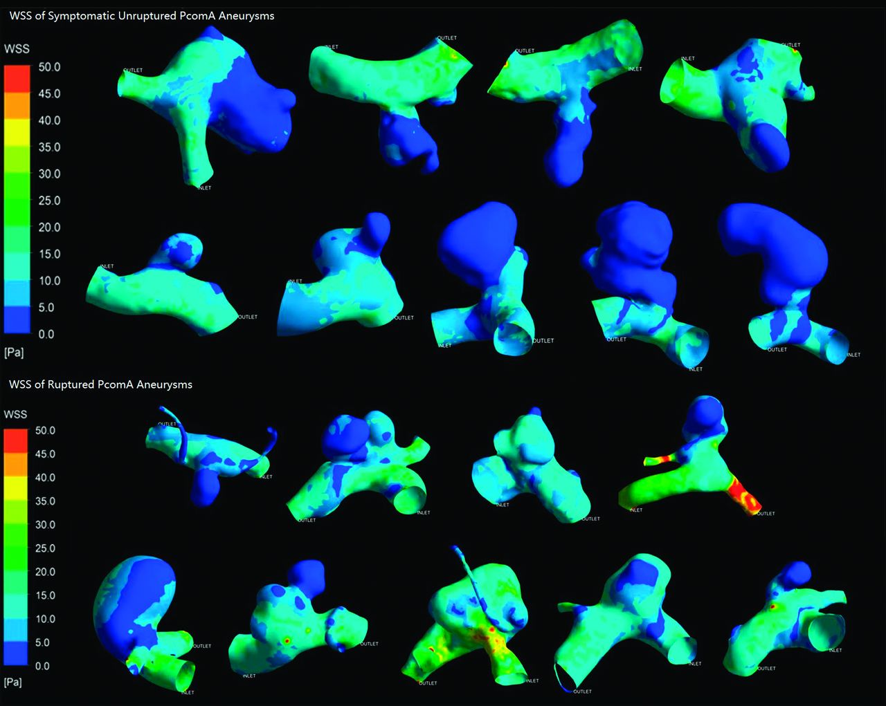
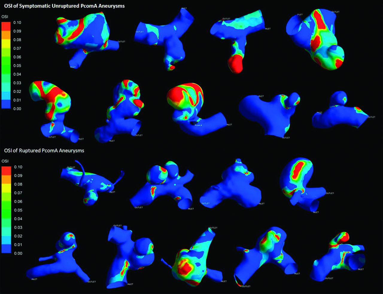
All results are detailed in the Table. Figs 2 and 3 depict the contour of time-averaged WSS and OSI. Because most parameters did not conform to both the normality and homogeneity of variance tests, we then performed a statistical analysis of all the parameters by use of the Mann-Whitney U test. All results are recorded as median (25% percentile, 75% percentile) or median ± interquartile range. The symptomatic unruptured PcomA aneurysms were very similar to ruptured ones in morphology and hemodynamics. There was no significant statistical difference between the 2 groups in any morphologic parameters, such as Va, Vv, size, AR, and SR. AR in the symptomatic unruptured group was slightly higher than that in the ruptured group (1.35 ± 1.12 versus 0.97 ± 0.49, P = .050). We demonstrated a lower WSS in the symptomatic unruptured group (5.27 ± 3.20 versus 8.11 ± 4.93, P = .024). For other hemodynamic parameters, including WSS of the parent artery wall, LSA, and OSI, there were no significant differences between the 2 groups.

View this table:
  • View inline
  • View popup

Statistical analysis of morphologic and hemodynamic parameters

Fig 2.
  • Download figure
  • Open in new tab
  • Download powerpoint
Fig 2.

Wall shear stress of symptomatic unruptured and ruptured posterior communicating artery aneurysms.

Fig 3.
  • Download figure
  • Open in new tab
  • Download powerpoint
Fig 3.

Oscillatory shear index of symptomatic unruptured and ruptured posterior communicating artery aneurysms.

Discussion

With the development of CFD techniques, more neurointerventionalists are realizing their clinical utility and are trying to use these powerful tools to guide neurointerventional practice in treating IAs. The morphologic and hemodynamic differences between ruptured and unruptured aneurysms were compared among different individuals to evaluate the rupture risk of IAs.9⇓⇓⇓–13 However, most of time, we only obtain the postrupture details of an aneurysm and its parent artery. Once an aneurysm ruptures, sudden expansion of a pseudosac or ruptured points may cause great change in intra-aneurysmal hemodynamic environment and mitigate the blood flow to stabilize the ruptured aneurysm. Accordingly, the best way to evaluate the rupture risk of IA is to study the aneurysm in the ready-to-rupture state. A few cases of gross blood-flow characteristics of IA just before rupture have been reported,14,15 but without comparison with ruptured aneurysms. The exact morphology and hemodynamics just before aneurysm rupture are not clear yet, but both studies of aneurysms before their rupture demonstrated low WSS on the aneurysm sacs. Some studies revealed lower WSS on ruptured aneurysms than on asymptomatic unruptured ones.5,7,16 A CFD study of hemodynamic differences between unruptured and ruptured IAs during observation has been reported recently. Thirteen aneurysms that ruptured during the course of follow-up observation were included in this study, and averaged WSS in the unruptured group was lower than in the ruptured group in the MCA. In our study, we also noticed a lower WSS in the unruptured PcomA aneurysms with oculomotor nerve palsy than in the ruptured ones (5.27 ± 3.20 versus 8.11 ± 4.93, P = .024), which is similar to the qualitative trend in the Takao et al study.17 Also, the AR was larger in the symptomatic unruptured group, and the P value of AR was very close to statistical significance (1.35 ± 1.12 versus 0.97 ± 0.49, P = .050). This means that compared with ruptured PcomA aneurysms, symptomatic unruptured ones have a more irregular shape. Destructive remodeling may take place, which facilitates IA rupture caused by pathobiologic responses induced by low WSS, such as matrix metalloproteinase production by macrophages,18,19 increased inflammatory cell infiltration,20 and increased reactive oxygen species.21 A recent study performed on an animal model also proved that high AR appears more deleterious to the aneurysm wall with pathologically low WSS.22 Above all, in this case series, it inferred that low WSS might play an important role in aneurysm rupture.

In another study we performed, we analyzed morphologic and hemodynamic parameters of mirror PcomA aneurysms.23 We found that lower WSS, higher LSA, and higher OSI contribute to aneurysm rupture. However, our results in this study only demonstrated that WSS of the aneurysm wall was significantly different between the 2 groups, instead of LSA or OSI hemodynamically. This discrepancy may be caused by small sample size, and it may reveal the particularity of these special symptomatic aneurysms in some ways. We should note that all unruptured aneurysms in the previous study were asymptomatic ones. This indicates that PcomA aneurysms with oculomotor nerve palsy, such as symptomatic IAs, might be different from asymptomatic unruptured or ruptured ones, which deserve separate study. Oculomotor nerve palsy is an indication for urgent treatment. This consensus is mainly based on vast clinical observations and physician clinical experience. Statistical analysis of morphologic and hemodynamic parameters revealed that unruptured PcomA aneurysms with oculomotor nerve palsy had almost the same morphologic and hemodynamic characteristics as that of ruptured ones. From this point of view, urgent treatment should be reasonable to prevent severe consequences after aneurysm rupture.

The small sample size is due to the fact that a PcomA aneurysm that has sudden unilateral oculomotor nerve palsy before it ruptures is relatively rare. That was the main limitation of this study. Larger case series are needed for further study and to generalize the result. As a retrospective study, we could not use the patient-specific boundary conditions to perform the CFD simulation. The same and specified initialization settings and inlet boundary conditions made hemodynamic results depend on the geometry of the luminal models. Also, traction-free outlet boundary conditions were another limitation to this study, but proper assumptions of outlet boundary conditions must be further investigated.7,24 In some cases, it was arbitrary to ignore the influence of the poorly developed posterior communicating artery, the diameter of which was <0.5 mm.

Conclusions

From morphologic and hemodynamic perspectives, we demonstrated that PcomA aneurysms with oculomotor nerve palsy had characteristics similar to those of ruptured ones, except for lower WSS on the aneurysm wall, which might indicate an important role in aneurysm rupture.

Acknowledgments

We gratefully acknowledge Shanghai Supercomputer Center for their helpful provision of the calculation software. We are also grateful for the generous support of Jianping Xiang from Toshiba Stroke Research Center, State University of New York at Buffalo, Buffalo New York; Shengzhang Wang, from the Department of Mechanics and Engineering Science, Fudan University, Shanghai, China, and Pengfei Luo, from the Department of Burns, Changhai Hospital, Second Military Medical University, Shanghai, China.

Footnotes

  • Y. Yu and J. Xu are co-first authors.

  • This study was supported by National Natural Science Foundation of China (Grant No.81171092, J. Liu; Grant No.81171093, Q. Huang; Grant No. 81000494, P. Yang); Rising-star Plan and Key Project of Shanghai Science and Technology Committee, China (Grant No. 11QA1408400, Q. Huang; Grant No. 11JC1415800, J. Liu); the Dawn Project of Shanghai, China (Grant No. 11CG043, P. Yang); Australian Research Council (Grant Nos. DP110102985 and LP0990263, Y. Qian).

Indicates open access to non-subscribers at www.ajnr.org

REFERENCES

  1. 1.↵
    1. Jeong W,
    2. Rhee K
    . Hemodynamics of cerebral aneurysms: computational analyses of aneurysm progress and treatment. Comput Mathematic Methods Med 2012;2012:782801
  2. 2.↵
    1. Sforza DM,
    2. Putman CM,
    3. Cebral JR
    . Hemodynamics of cerebral aneurysms. Annu Rev Fluid Mech 2009;41:91–107
    CrossRefPubMed
  3. 3.↵
    1. Scholtes F,
    2. Martin D
    . Strategical implications of aneurysmal cranial nerve compression. Neuro-Chirurgie 2012;58:146–55
    CrossRefPubMed
  4. 4.↵
    1. Langner S,
    2. Schroeder HW,
    3. Hosten N,
    4. et al
    . [Diagnosing neurovascular compression syndromes]. RoFo 2012;184:220–28
    CrossRefPubMed
  5. 5.↵
    1. Shojima M,
    2. Oshima M,
    3. Takagi K,
    4. et al
    . Magnitude and role of wall shear stress on cerebral aneurysm: computational fluid dynamic study of 20 middle cerebral artery aneurysms. Stroke 2004;35:2500–05
    Abstract/FREE Full Text
  6. 6.↵
    1. Tremmel M,
    2. Dhar S,
    3. Levy EI,
    4. et al
    . Influence of intracranial aneurysm-to-parent vessel size ratio on hemodynamics and implication for rupture: results from a virtual experimental study. Neurosurgery 2009;64:622–30
    CrossRefPubMed
  7. 7.↵
    1. Jou LD,
    2. Lee DH,
    3. Morsi H,
    4. et al
    . Wall shear stress on ruptured and unruptured intracranial aneurysms at the internal carotid artery. AJNR Am J Neuroradiol 2008;29:1761–67
    Abstract/FREE Full Text
  8. 8.↵
    1. He X,
    2. Ku DN
    . Pulsatile flow in the human left coronary artery bifurcation: average conditions. J Biomech Eng 1996;118:74–82
    CrossRefPubMed
  9. 9.↵
    1. Dhar S,
    2. Tremmel M,
    3. Mocco J,
    4. et al
    . Morphology parameters for intracranial aneurysm rupture risk assessment. Neurosurgery 2008;63:185–96
    CrossRefPubMed
  10. 10.↵
    1. Cebral JR,
    2. Mut F,
    3. Weir J,
    4. et al
    . Quantitative characterization of the hemodynamic environment in ruptured and unruptured brain aneurysms. AJNR Am J Neuroradiol 2011;32:145–51
    Abstract/FREE Full Text
  11. 11.↵
    1. Beck J,
    2. Rohde S,
    3. Berkefeld J,
    4. et al
    . Size and location of ruptured and unruptured intracranial aneurysms measured by 3-dimensional rotational angiography. Surg Neurol 2006;65:18–25
    CrossRefPubMed
  12. 12.↵
    1. Saitua F,
    2. Acuna R,
    3. Herrera P
    . Percutaneous endoscopic gastrostomy: the technique of choice? J Pediatr Surg 2003;38:1512–15
    CrossRefPubMed
  13. 13.↵
    1. Xiang J,
    2. Natarajan SK,
    3. Tremmel M,
    4. et al
    . Hemodynamic-morphologic discriminants for intracranial aneurysm rupture. Stroke 2011;42:144–52
    Abstract/FREE Full Text
  14. 14.↵
    1. Sforza DM,
    2. Putman CM,
    3. Scrivano E,
    4. et al
    . Blood-flow characteristics in a terminal basilar tip aneurysm prior to its fatal rupture. AJNR Am J Neuroradiol 2010;31:1127–31
    Abstract/FREE Full Text
  15. 15.↵
    1. Cebral JR,
    2. Hendrickson S,
    3. Putman CM
    . Hemodynamics in a lethal basilar artery aneurysm just before its rupture. AJNR Am J Neuroradiol 2009;30:95–98
    Abstract/FREE Full Text
  16. 16.↵
    1. Goubergrits L,
    2. Schaller J,
    3. Kertzscher U,
    4. et al
    . Statistical wall shear stress maps of ruptured and unruptured middle cerebral artery aneurysms. J R Soc Interface 2012;9:677–88
    Abstract/FREE Full Text
  17. 17.↵
    1. Takao H,
    2. Murayama Y,
    3. Otsuka S,
    4. et al
    . Hemodynamic differences between unruptured and ruptured intracranial aneurysms during observation. Stroke 2012;43:1436–39
    Abstract/FREE Full Text
  18. 18.↵
    1. Newby AC
    . Metalloproteinase expression in monocytes and macrophages and its relationship to atherosclerotic plaque instability. Arterioscler Thromb Vasc Biol 2008;28:2108–14
    Abstract/FREE Full Text
  19. 19.↵
    1. Galis ZS,
    2. Sukhova GK,
    3. Lark MW,
    4. et al
    . Increased expression of matrix metalloproteinases and matrix degrading activity in vulnerable regions of human atherosclerotic plaques. J Clin Invest 1994;94:2493–503
    CrossRefPubMed
  20. 20.↵
    1. Gui T,
    2. Shimokado A,
    3. Sun Y,
    4. et al
    . Diverse roles of macrophages in atherosclerosis: from inflammatory biology to biomarker discovery. Mediat Inflamm 2012;2012:693083
  21. 21.↵
    1. Chiu JJ,
    2. Chien S
    . Effects of disturbed flow on vascular endothelium: pathophysiological basis and clinical perspectives. Physiol Rev 2011;91:327–87
    Abstract/FREE Full Text
  22. 22.↵
    1. Zeng Z,
    2. Durka MJ,
    3. Kallmes DF,
    4. et al
    . Can aspect ratio be used to categorize intra-aneurysmal hemodynamics? A study of elastase induced aneurysms in rabbit. J Biomech 2011;44:2809–16
    CrossRefPubMed
  23. 23.↵
    1. Xu J,
    2. Yu Y,
    3. Wu X,
    4. et al
    . Morphological and hemodynamic analysis of mirror posterior communicating artery aneurysms. PloS One. 2013;8:e55413
    CrossRefPubMed
  24. 24.↵
    1. Castro M,
    2. Putman C,
    3. Radaelli A,
    4. et al
    . Hemodynamics and rupture of terminal cerebral aneurysms. Acad Radiol 2009;16:1201–07
    CrossRefPubMed
  • Received December 2, 2012.
  • Accepted after revision February 15, 2013.
  • © 2013 by American Journal of Neuroradiology
PreviousNext
Back to top

In this issue

American Journal of Neuroradiology: 34 (11)
American Journal of Neuroradiology
Vol. 34, Issue 11
1 Nov 2013
  • Table of Contents
  • Index by author
  • Complete Issue (PDF)
Advertisement
Print
Download PDF
Email Article

Thank you for your interest in spreading the word on American Journal of Neuroradiology.

NOTE: We only request your email address so that the person you are recommending the page to knows that you wanted them to see it, and that it is not junk mail. We do not capture any email address.

Enter multiple addresses on separate lines or separate them with commas.
Analysis of Morphologic and Hemodynamic Parameters for Unruptured Posterior Communicating Artery Aneurysms with Oculomotor Nerve Palsy
(Your Name) has sent you a message from American Journal of Neuroradiology
(Your Name) thought you would like to see the American Journal of Neuroradiology web site.
CAPTCHA
This question is for testing whether or not you are a human visitor and to prevent automated spam submissions.
Cite this article
Y. Yu, J. Xu, Y. Fang, X. Wu, P. Yang, C. Jiang, Y. Qian, J. Liu, Q. Huang
Analysis of Morphologic and Hemodynamic Parameters for Unruptured Posterior Communicating Artery Aneurysms with Oculomotor Nerve Palsy
American Journal of Neuroradiology Nov 2013, 34 (11) 2187-2191; DOI: 10.3174/ajnr.A3575

Citation Manager Formats

  • BibTeX
  • Bookends
  • EasyBib
  • EndNote (tagged)
  • EndNote 8 (xml)
  • Medlars
  • Mendeley
  • Papers
  • RefWorks Tagged
  • Ref Manager
  • RIS
  • Zotero
0 Responses
Respond to this article
Share
Bookmark this article
Analysis of Morphologic and Hemodynamic Parameters for Unruptured Posterior Communicating Artery Aneurysms with Oculomotor Nerve Palsy
Y. Yu, J. Xu, Y. Fang, X. Wu, P. Yang, C. Jiang, Y. Qian, J. Liu, Q. Huang
American Journal of Neuroradiology Nov 2013, 34 (11) 2187-2191; DOI: 10.3174/ajnr.A3575
del.icio.us logo Twitter logo Facebook logo Mendeley logo
  • Tweet Widget
  • Facebook Like
  • Google Plus One
Purchase

Jump to section

  • Article
    • Abstract
    • ABBREVIATIONS:
    • Materials and Methods
    • Results
    • Discussion
    • Conclusions
    • Acknowledgments
    • Footnotes
    • REFERENCES
  • Figures & Data
  • Info & Metrics
  • Responses
  • References
  • PDF

Related Articles

  • No related articles found.
  • PubMed
  • Google Scholar

Cited By...

  • Clinical, morphological, and hemodynamic independent characteristic factors for rupture of posterior communicating artery aneurysms
  • Morphological and hemodynamic analysis of posterior communicating artery aneurysms prone to rupture: a matched case-control study
  • Crossref (6)
  • Google Scholar

This article has been cited by the following articles in journals that are participating in Crossref Cited-by Linking.

  • Association of wall shear stress with intracranial aneurysm rupture: systematic review and meta-analysis
    Geng Zhou, Yueqi Zhu, Yanling Yin, Ming Su, Minghua Li
    Scientific Reports 2017 7 1
  • Clinical, morphological, and hemodynamic independent characteristic factors for rupture of posterior communicating artery aneurysms
    Ying Zhang, Linkai Jing, Jian Liu, Chuanhui Li, Jixing Fan, Shengzhang Wang, Haiyun Li, Xinjian Yang
    Journal of NeuroInterventional Surgery 2016 8 8
  • Morphological and hemodynamic analysis of posterior communicating artery aneurysms prone to rupture: a matched case–control study
    Guoli Duan, Nan Lv, Jianhua Yin, Jinyu Xu, Bo Hong, Yi Xu, Jianmin Liu, Qinghai Huang
    Journal of NeuroInterventional Surgery 2016 8 1
  • Morphological Characteristics of Ruptured Brain Aneurysms: A Systematic Literature Review and Meta‐Analysis
    Sebastian Sanchez, Meredith Hickerson, Rishi R. Patel, Dana Ghazaleh, Ranita Tarchand, Geeta S. Paranjape, Holly Pope, Santiago Ortega‐Gutierrez, John M. Pederson, David Hasan, Madhavan L. Raghavan, Edgar A. Samaniego
    Stroke: Vascular and Interventional Neurology 2023 3 2
  • Haemodynamics in a patient-specific intracranial aneurysm according to experimental and numerical approaches: A comparison of PIV, CFD and PC-MRI
    Yujie Li, Yuuya Yoneyama, Haruo Isoda, Masaki Terada, Takafumi Kosugi, Takashi Kosugi, Mingzi Zhang, Makoto Ohta
    Technology and Health Care 2021 29 2
  • The effect of downstream resistance on flow diverter treatment of a cerebral aneurysm at a bifurcation: A joint computational-experimental study
    Anderson C. O. Tsang, Billy Y. S. Yiu, Abraham Y. S. Tang, W. C. Chung, Gilberto K. K. Leung, Alexander K. K. Poon, Alfred C. H. Yu, Simon S. M. Lai, K. W. Chow
    Journal of Hydrodynamics 2018 30 5

More in this TOC Section

  • Rescue Reentry in Carotid Near-Occlusion
  • Contour Neurovascular System: Five Year Follow Up
  • Effect of SARS-CoV2 on Endovascular Thrombectomy
Show more NEUROINTERVENTION

Similar Articles

Advertisement

Indexed Content

  • Current Issue
  • Accepted Manuscripts
  • Article Preview
  • Past Issues
  • Editorials
  • Editor's Choice
  • Fellows' Journal Club
  • Letters to the Editor
  • Video Articles

Cases

  • Case Collection
  • Archive - Case of the Week
  • Archive - Case of the Month
  • Archive - Classic Case

More from AJNR

  • Trainee Corner
  • Imaging Protocols
  • MRI Safety Corner
  • Book Reviews

Multimedia

  • AJNR Podcasts
  • AJNR Scantastics

Resources

  • Turnaround Time
  • Submit a Manuscript
  • Submit a Video Article
  • Submit an eLetter to the Editor/Response
  • Manuscript Submission Guidelines
  • Statistical Tips
  • Fast Publishing of Accepted Manuscripts
  • Graphical Abstract Preparation
  • Imaging Protocol Submission
  • Evidence-Based Medicine Level Guide
  • Publishing Checklists
  • Author Policies
  • Become a Reviewer/Academy of Reviewers
  • News and Updates

About Us

  • About AJNR
  • Editorial Board
  • Editorial Board Alumni
  • Alerts
  • Permissions
  • Not an AJNR Subscriber? Join Now
  • Advertise with Us
  • Librarian Resources
  • Feedback
  • Terms and Conditions
  • AJNR Editorial Board Alumni

American Society of Neuroradiology

  • Not an ASNR Member? Join Now

© 2025 by the American Society of Neuroradiology All rights, including for text and data mining, AI training, and similar technologies, are reserved.
Print ISSN: 0195-6108 Online ISSN: 1936-959X

Powered by HighWire