Advertisement

Main menu

  • Home
  • Content
    • Current Issue
    • Accepted Manuscripts
    • Article Preview
    • Past Issue Archive
    • Video Articles
    • AJNR Case Collection
    • Case of the Week Archive
    • Case of the Month Archive
    • Classic Case Archive
  • Special Collections
    • AJNR Awards
    • Low-Field MRI
    • Alzheimer Disease
    • ASNR Foundation Special Collection
    • Photon-Counting CT
    • View All
  • Multimedia
    • AJNR Podcasts
    • AJNR SCANtastic
    • Trainee Corner
    • MRI Safety Corner
    • Imaging Protocols
  • For Authors
    • Submit a Manuscript
    • Submit a Video Article
    • Submit an eLetter to the Editor/Response
    • Manuscript Submission Guidelines
    • Statistical Tips
    • Fast Publishing of Accepted Manuscripts
    • Graphical Abstract Preparation
    • Imaging Protocol Submission
    • Author Policies
  • About Us
    • About AJNR
    • Editorial Board
    • Editorial Board Alumni
  • More
    • Become a Reviewer/Academy of Reviewers
    • Subscribers
    • Permissions
    • Alerts
    • Feedback
    • Advertisers
    • ASNR Home

User menu

  • Alerts
  • Log in

Search

  • Advanced search
American Journal of Neuroradiology

American Journal of Neuroradiology

ASHNR American Society of Functional Neuroradiology ASHNR American Society of Pediatric Neuroradiology ASSR
  • Alerts
  • Log in

Advanced Search

  • Home
  • Content
    • Current Issue
    • Accepted Manuscripts
    • Article Preview
    • Past Issue Archive
    • Video Articles
    • AJNR Case Collection
    • Case of the Week Archive
    • Case of the Month Archive
    • Classic Case Archive
  • Special Collections
    • AJNR Awards
    • Low-Field MRI
    • Alzheimer Disease
    • ASNR Foundation Special Collection
    • Photon-Counting CT
    • View All
  • Multimedia
    • AJNR Podcasts
    • AJNR SCANtastic
    • Trainee Corner
    • MRI Safety Corner
    • Imaging Protocols
  • For Authors
    • Submit a Manuscript
    • Submit a Video Article
    • Submit an eLetter to the Editor/Response
    • Manuscript Submission Guidelines
    • Statistical Tips
    • Fast Publishing of Accepted Manuscripts
    • Graphical Abstract Preparation
    • Imaging Protocol Submission
    • Author Policies
  • About Us
    • About AJNR
    • Editorial Board
    • Editorial Board Alumni
  • More
    • Become a Reviewer/Academy of Reviewers
    • Subscribers
    • Permissions
    • Alerts
    • Feedback
    • Advertisers
    • ASNR Home
  • Follow AJNR on Twitter
  • Visit AJNR on Facebook
  • Follow AJNR on Instagram
  • Join AJNR on LinkedIn
  • RSS Feeds

AJNR Awards, New Junior Editors, and more. Read the latest AJNR updates

Research ArticleBrain
Open Access

Classification of Cerebral Arteriovenous Malformations and Intranidal Flow Patterns by Color-Encoded 4D-Hybrid-MRA

T. Illies, N.D. Forkert, T. Ries, J. Regelsberger and J. Fiehler
American Journal of Neuroradiology January 2013, 34 (1) 46-53; DOI: https://doi.org/10.3174/ajnr.A3204
T. Illies
aFrom the Departments of Diagnostic and Interventional Neuroradiology (T.I., T.R., J.F.)
  • Find this author on Google Scholar
  • Find this author on PubMed
  • Search for this author on this site
N.D. Forkert
bComputational Neuroscience (N.D.F.)
  • Find this author on Google Scholar
  • Find this author on PubMed
  • Search for this author on this site
T. Ries
aFrom the Departments of Diagnostic and Interventional Neuroradiology (T.I., T.R., J.F.)
  • Find this author on Google Scholar
  • Find this author on PubMed
  • Search for this author on this site
J. Regelsberger
cNeurosurgery (J.R.), University Medical Center Hamburg-Eppendorf, Hamburg, Germany.
  • Find this author on Google Scholar
  • Find this author on PubMed
  • Search for this author on this site
J. Fiehler
aFrom the Departments of Diagnostic and Interventional Neuroradiology (T.I., T.R., J.F.)
  • Find this author on Google Scholar
  • Find this author on PubMed
  • Search for this author on this site
  • Article
  • Figures & Data
  • Supplemental
  • Info & Metrics
  • Responses
  • References
  • PDF
Loading

Article Figures & Data

Figures

  • Fig 1.
    • Download figure
    • Open in new tab
    • Download powerpoint
    Fig 1.

    Patient 5. Homogeneous nidus perfusion (type 1). Inflow time point range is set from 15 to 19 seconds in the first image to 18–19 seconds in the last to generate the effect of blood flow. Corresponding to the inflow from the PCA and outflow into the transversal sinus, the nidus is perfused in an anterolateral-to-posteromedial direction.

  • Fig 2.
    • Download figure
    • Open in new tab
    • Download powerpoint
    Fig 2.

    Patient 9. Inhomogeneous nidus perfusion (type 2). Inflow time point setting from 18 to 20 seconds in the first image to 21–27 seconds in the last image to generate the effect of blood flow. Diversion of the central inflow into the nidus can be observed to the lateral and medial sides.
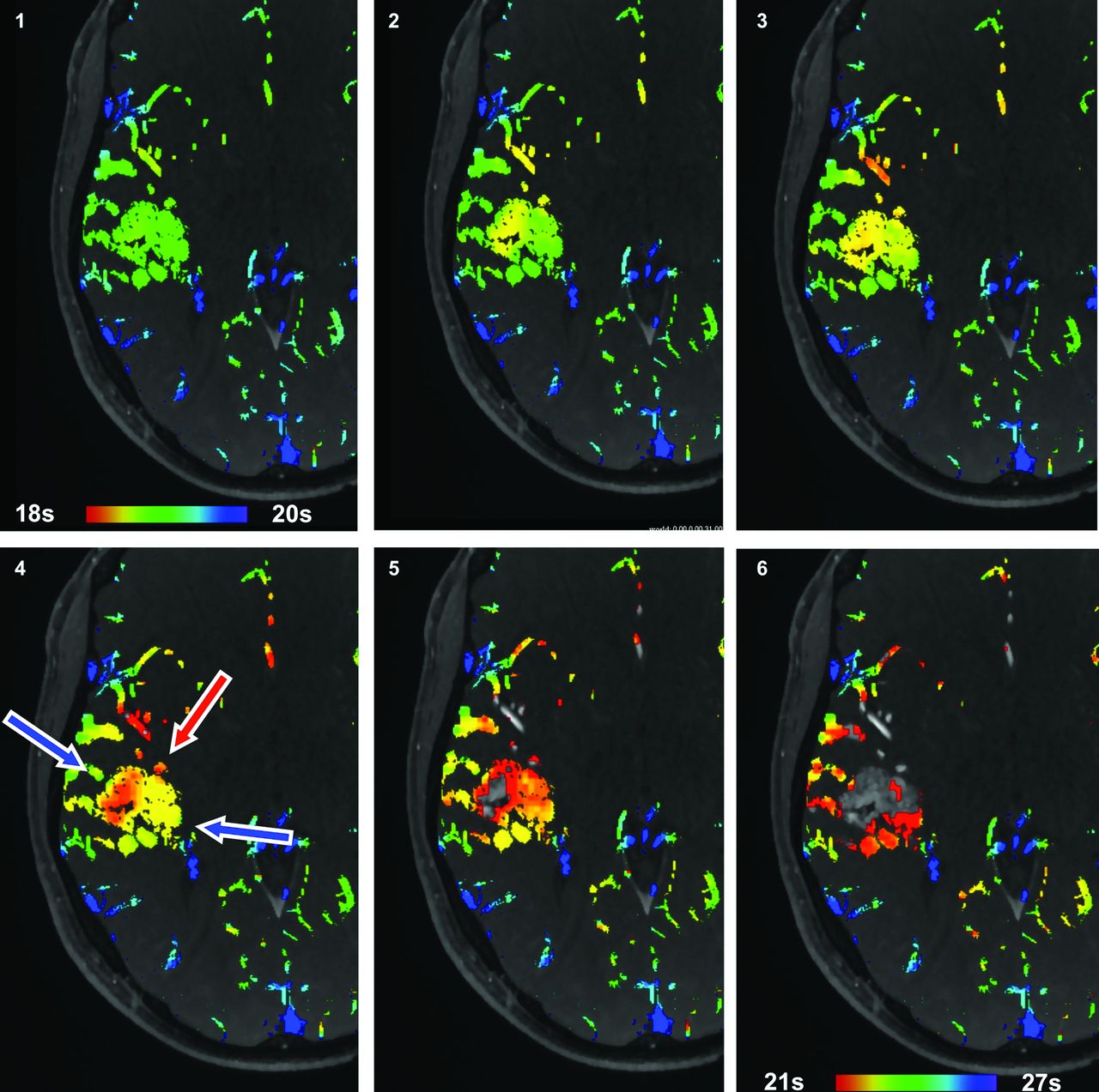
  • Fig 3.
    • Download figure
    • Open in new tab
    • Download powerpoint
    Fig 3.

    Patient 6. Inhomogeneous nidus perfusion (type 3). Inflow time point setting from 18 to 20 seconds in the first image to 21–23 seconds in the last image to generate the effect of blood flow. Corresponding to different feeding territories, inflow is seen in the paraventricular region and in the MCA territory anteriorly and laterally (red arrows) with multidirectional flow diversion toward drainage (blue arrows).
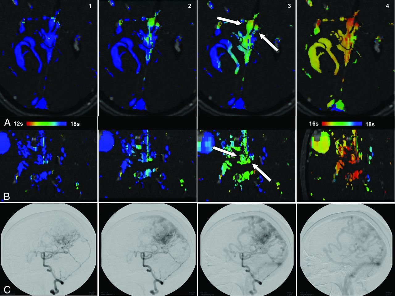
  • Fig 4.
    • Download figure
    • Open in new tab
    • Download powerpoint
    Fig 4.

    Patient 23. Axial (A) and coronal (B) color-encoded images show the right (ipsilateral to the AVM) internal cerebral vein dilated with symmetric inflow time points. Note the early inflow in the deep venous system in comparison with the confluens sinuum (column 3). On DSA, a deep venous drainage is not seen (C). Also note the huge aneurysmal dilation of cortical drainage with relatively late filling from its medial portion.

  • Fig 5.
    • Download figure
    • Open in new tab
    • Download powerpoint
    Fig 5.

    Patient 4. A, Two dilated superficial draining veins can be seen, which drain early into the transverse sinus and superior sagittal sinus (pictures 1 and 4). On picture 5, a dilated vein with delayed drainage into the internal cerebral veins is seen. B, DSA shows faint and retarded drainage into the deep venous system (picture 5).
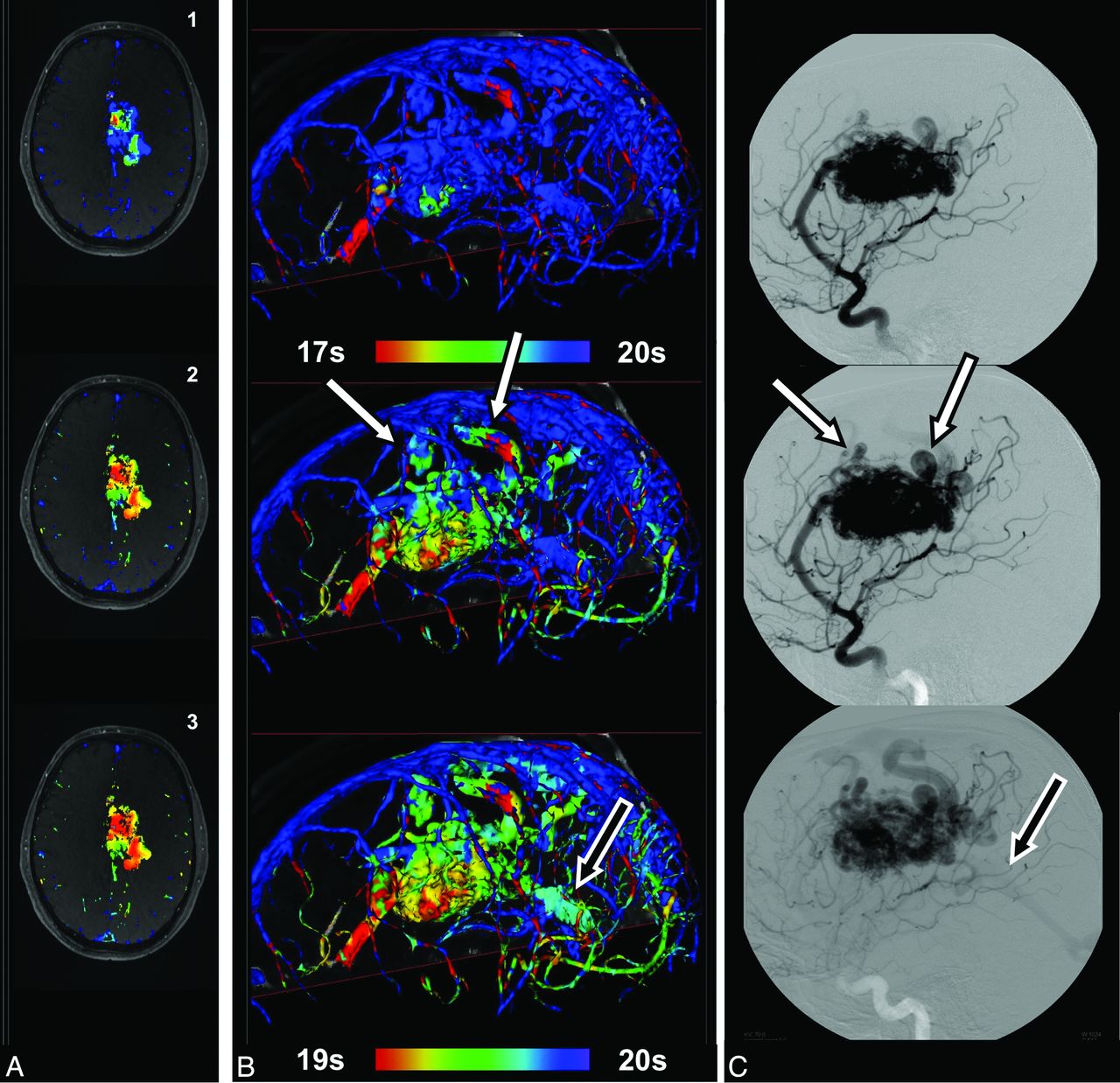
  • Fig 6.
    • Download figure
    • Open in new tab
    • Download powerpoint
    Fig 6.

    Patient 19. Brain arteriovenous malformation fed by the ACA and PCA. Initial drainage into veins draining into the superior sagittal sinus (white arrows), while the posteromedial part of the nidus begins to drain later into the vein of Galen (B and C, single black arrow). Corresponding inhomogeneous intranidal flow pattern with a late inflow time point in the posteromedial part of the AVM nidus (type 2) (A) is seen.

PreviousNext
Back to top

In this issue

American Journal of Neuroradiology: 34 (1)
American Journal of Neuroradiology
Vol. 34, Issue 1
1 Jan 2013
  • Table of Contents
  • Index by author
Advertisement
Print
Download PDF
Email Article

Thank you for your interest in spreading the word on American Journal of Neuroradiology.

NOTE: We only request your email address so that the person you are recommending the page to knows that you wanted them to see it, and that it is not junk mail. We do not capture any email address.

Enter multiple addresses on separate lines or separate them with commas.
Classification of Cerebral Arteriovenous Malformations and Intranidal Flow Patterns by Color-Encoded 4D-Hybrid-MRA
(Your Name) has sent you a message from American Journal of Neuroradiology
(Your Name) thought you would like to see the American Journal of Neuroradiology web site.
CAPTCHA
This question is for testing whether or not you are a human visitor and to prevent automated spam submissions.
Cite this article
T. Illies, N.D. Forkert, T. Ries, J. Regelsberger, J. Fiehler
Classification of Cerebral Arteriovenous Malformations and Intranidal Flow Patterns by Color-Encoded 4D-Hybrid-MRA
American Journal of Neuroradiology Jan 2013, 34 (1) 46-53; DOI: 10.3174/ajnr.A3204

Citation Manager Formats

  • BibTeX
  • Bookends
  • EasyBib
  • EndNote (tagged)
  • EndNote 8 (xml)
  • Medlars
  • Mendeley
  • Papers
  • RefWorks Tagged
  • Ref Manager
  • RIS
  • Zotero
0 Responses
Respond to this article
Share
Bookmark this article
Classification of Cerebral Arteriovenous Malformations and Intranidal Flow Patterns by Color-Encoded 4D-Hybrid-MRA
T. Illies, N.D. Forkert, T. Ries, J. Regelsberger, J. Fiehler
American Journal of Neuroradiology Jan 2013, 34 (1) 46-53; DOI: 10.3174/ajnr.A3204
del.icio.us logo Twitter logo Facebook logo Mendeley logo
  • Tweet Widget
  • Facebook Like
  • Google Plus One
Purchase

Jump to section

  • Article
    • Abstract
    • ABBREVIATIONS:
    • Materials and Methods
    • Results
    • Discussion
    • Conclusions
    • Footnotes
    • References
  • Figures & Data
  • Supplemental
  • Info & Metrics
  • Responses
  • References
  • PDF

Related Articles

  • PubMed
  • Google Scholar

Cited By...

  • Application of a Novel Brain Arteriovenous Malformation Endovascular Grading Scale for Transarterial Embolization
  • Value of 4D MR Angiography at 3T Compared with DSA for the Follow-Up of Treated Brain Arteriovenous Malformation
  • Fast 4D Flow MRI Re-Emerges as a Potential Clinical Tool for Neuroradiology
  • Intracranial 4D Flow MRI: Toward Individualized Assessment of Arteriovenous Malformation Hemodynamics and Treatment-Induced Changes
  • Crossref (19)
  • Google Scholar

This article has been cited by the following articles in journals that are participating in Crossref Cited-by Linking.

  • Intracranial 4D Flow MRI: Toward Individualized Assessment of Arteriovenous Malformation Hemodynamics and Treatment-Induced Changes
    S.A. Ansari, S. Schnell, T. Carroll, P. Vakil, M.C. Hurley, C. Wu, J. Carr, B.R. Bendok, H. Batjer, M. Markl
    American Journal of Neuroradiology 2013 34 10
  • Digital subtraction angiography in cerebrovascular disease: current practice and perspectives on diagnosis, acute treatment and prognosis
    Shirin Shaban, Bella Huasen, Abilash Haridas, Murray Killingsworth, John Worthington, Pascal Jabbour, Sonu Menachem Maimonides Bhaskar
    Acta Neurologica Belgica 2022 122 3
  • Value of 4D MR Angiography at 3T Compared with DSA for the Follow-Up of Treated Brain Arteriovenous Malformation
    S. Soize, F. Bouquigny, K. Kadziolka, C. Portefaix, L. Pierot
    American Journal of Neuroradiology 2014 35 10
  • Cerebral arteriovenous malformation: Complex 3D hemodynamics and 3D blood flow alterations during staged embolization
    Michael Markl, Can Wu, Michael C. Hurley, Sameer A. Ansari, Timothy J. Carroll, Rudy J. Rahme, Salah G. Aoun, James Carr, Hunt Batjer, Bernard R. Bendok
    Journal of Magnetic Resonance Imaging 2013 38 4
  • Time-resolved Spin-labeled MR Angiography for the Depiction of Cerebral Arteriovenous Malformations: A Comparison of Techniques
    Hélène Raoult, Elise Bannier, Benjamin Robert, Christian Barillot, Peter Schmitt, Jean-Yves Gauvrit
    Radiology 2014 271 2
  • Evaluation of Intracranial Arteriovenous Malformations With Four-Dimensional Arterial-Spin Labeling–Based 3-T Magnetic Resonance Angiography
    Yasuhiko Iryo, Toshinori Hirai, Masanobu Nakamura, Takayuki Kawano, Yasuyuki Kaku, Yuki Ohmori, Yutaka Kai, Minako Azuma, Shinichiro Nishimura, Yoshinori Shigematsu, Mika Kitajima, Yasuyuki Yamashita
    Journal of Computer Assisted Tomography 2016 40 2
  • Skeletonization method for vessel delineation of arteriovenous malformation
    D. Babin, A. Pižurica, L. Velicki, V. Matić, I. Galić, H. Leventić, V. Zlokolica, W. Philips
    Computers in Biology and Medicine 2018 93
  • From2Dto4DPhase‐ContrastMRIin the Neurovascular System: Will It Be a Quantum Jump or a Fancy Decoration?
    Sung Won Youn, Jongmin Lee
    Journal of Magnetic Resonance Imaging 2022 55 2
  • MR Selective Flow-Tracking Cartography: A Postprocessing Procedure Applied to Four-dimensional Flow MR Imaging for Complete Characterization of Cranial Dural Arteriovenous Fistulas
    Myriam Edjlali, Pauline Roca, Cécile Rabrait, Denis Trystram, Christine Rodriguez-Régent, Kevin M. Johnson, Oliver Wieben, Patrick Turski, Jean-François Meder, Olivier Naggara, Catherine Oppenheim
    Radiology 2014 270 1

More in this TOC Section

  • Statin Therapy Does Not Affect the Radiographic and Clinical Profile of Patients with TIA and Minor Stroke
  • Usefulness of Quantitative Susceptibility Mapping for the Diagnosis of Parkinson Disease
  • White Matter Alterations in the Brains of Patients with Active, Remitted, and Cured Cushing Syndrome: A DTI Study
Show more BRAIN

Similar Articles

Advertisement

Indexed Content

  • Current Issue
  • Accepted Manuscripts
  • Article Preview
  • Past Issues
  • Editorials
  • Editor's Choice
  • Fellows' Journal Club
  • Letters to the Editor
  • Video Articles

Cases

  • Case Collection
  • Archive - Case of the Week
  • Archive - Case of the Month
  • Archive - Classic Case

More from AJNR

  • Trainee Corner
  • Imaging Protocols
  • MRI Safety Corner
  • Book Reviews

Multimedia

  • AJNR Podcasts
  • AJNR Scantastics

Resources

  • Turnaround Time
  • Submit a Manuscript
  • Submit a Video Article
  • Submit an eLetter to the Editor/Response
  • Manuscript Submission Guidelines
  • Statistical Tips
  • Fast Publishing of Accepted Manuscripts
  • Graphical Abstract Preparation
  • Imaging Protocol Submission
  • Evidence-Based Medicine Level Guide
  • Publishing Checklists
  • Author Policies
  • Become a Reviewer/Academy of Reviewers
  • News and Updates

About Us

  • About AJNR
  • Editorial Board
  • Editorial Board Alumni
  • Alerts
  • Permissions
  • Not an AJNR Subscriber? Join Now
  • Advertise with Us
  • Librarian Resources
  • Feedback
  • Terms and Conditions
  • AJNR Editorial Board Alumni

American Society of Neuroradiology

  • Not an ASNR Member? Join Now

© 2025 by the American Society of Neuroradiology All rights, including for text and data mining, AI training, and similar technologies, are reserved.
Print ISSN: 0195-6108 Online ISSN: 1936-959X

Powered by HighWire