Advertisement

Main menu

  • Home
  • Content
    • Current Issue
    • Accepted Manuscripts
    • Article Preview
    • Past Issue Archive
    • Video Articles
    • AJNR Case Collection
    • Case of the Week Archive
    • Case of the Month Archive
    • Classic Case Archive
  • Special Collections
    • Advancing NeuroMRI with High-Relaxivity Contrast Agents
    • AJNR Awards
    • Low-Field MRI
    • Alzheimer Disease
    • ASNR Foundation Special Collection
    • Photon-Counting CT
    • View All
  • Multimedia
    • AJNR Podcasts
    • AJNR SCANtastic
    • Trainee Corner
    • MRI Safety Corner
    • Imaging Protocols
  • For Authors
    • Submit a Manuscript
    • Submit a Video Article
    • Submit an eLetter to the Editor/Response
    • Manuscript Submission Guidelines
    • Statistical Tips
    • Fast Publishing of Accepted Manuscripts
    • Graphical Abstract Preparation
    • Imaging Protocol Submission
    • Author Policies
  • About Us
    • About AJNR
    • Editorial Board
    • Editorial Board Alumni
  • More
    • Become a Reviewer/Academy of Reviewers
    • Subscribers
    • Permissions
    • Alerts
    • Feedback
    • Advertisers
    • ASNR Home

User menu

  • Alerts
  • Log in

Search

  • Advanced search
American Journal of Neuroradiology

American Journal of Neuroradiology

ASHNR American Society of Functional Neuroradiology ASHNR American Society of Pediatric Neuroradiology ASSR
  • Alerts
  • Log in

Advanced Search

  • Home
  • Content
    • Current Issue
    • Accepted Manuscripts
    • Article Preview
    • Past Issue Archive
    • Video Articles
    • AJNR Case Collection
    • Case of the Week Archive
    • Case of the Month Archive
    • Classic Case Archive
  • Special Collections
    • Advancing NeuroMRI with High-Relaxivity Contrast Agents
    • AJNR Awards
    • Low-Field MRI
    • Alzheimer Disease
    • ASNR Foundation Special Collection
    • Photon-Counting CT
    • View All
  • Multimedia
    • AJNR Podcasts
    • AJNR SCANtastic
    • Trainee Corner
    • MRI Safety Corner
    • Imaging Protocols
  • For Authors
    • Submit a Manuscript
    • Submit a Video Article
    • Submit an eLetter to the Editor/Response
    • Manuscript Submission Guidelines
    • Statistical Tips
    • Fast Publishing of Accepted Manuscripts
    • Graphical Abstract Preparation
    • Imaging Protocol Submission
    • Author Policies
  • About Us
    • About AJNR
    • Editorial Board
    • Editorial Board Alumni
  • More
    • Become a Reviewer/Academy of Reviewers
    • Subscribers
    • Permissions
    • Alerts
    • Feedback
    • Advertisers
    • ASNR Home
  • Follow AJNR on Twitter
  • Visit AJNR on Facebook
  • Follow AJNR on Instagram
  • Join AJNR on LinkedIn
  • RSS Feeds

AJNR Awards, New Junior Editors, and more. Read the latest AJNR updates


Improved Turnaround Times | Median time to first decision: 12 days

Research ArticleNeurointervention

C-Arm CT Measurement of Cerebral Blood Volume Using Intra-Arterial Injection of Contrast Medium: An Experimental Study in Canines

R. Yasuda, K. Royalty, K. Pulfer, D. Consigny and C.M. Strother
American Journal of Neuroradiology October 2012, 33 (9) 1696-1701; DOI: https://doi.org/10.3174/ajnr.A3077
R. Yasuda
aFrom the Department of Neurosurgery, Mie University Graduate School of Medicine (R.Y.), Tsu, Mie, Japan
  • Find this author on Google Scholar
  • Find this author on PubMed
  • Search for this author on this site
K. Royalty
bSiemens Medical Solutions (K.R.), Hoffman Estates, Illinois
  • Find this author on Google Scholar
  • Find this author on PubMed
  • Search for this author on this site
K. Pulfer
cDepartment of Radiology, University of Wisconsin School of Medicine and Public Health (K.P., D.C., C.M.S.), Madison, Wisconsin.
  • Find this author on Google Scholar
  • Find this author on PubMed
  • Search for this author on this site
D. Consigny
cDepartment of Radiology, University of Wisconsin School of Medicine and Public Health (K.P., D.C., C.M.S.), Madison, Wisconsin.
  • Find this author on Google Scholar
  • Find this author on PubMed
  • Search for this author on this site
C.M. Strother
cDepartment of Radiology, University of Wisconsin School of Medicine and Public Health (K.P., D.C., C.M.S.), Madison, Wisconsin.
  • Find this author on Google Scholar
  • Find this author on PubMed
  • Search for this author on this site
  • Article
  • Figures & Data
  • Info & Metrics
  • Responses
  • References
  • PDF
Loading

Article Figures & Data

Figures

  • Tables
  • Fig 1.
    • Download figure
    • Open in new tab
    • Download powerpoint
    Fig 1.

    Bland-Altman plots. Modalities compared are shown at the top of each plot.
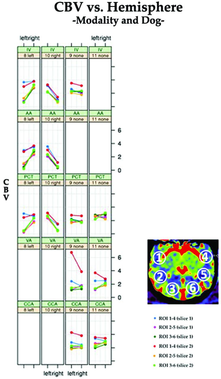
  • Fig 2.
    • Download figure
    • Open in new tab
    • Download powerpoint
    Fig 2.

    Scatterplots comparing injection sites and CBV values in normal and abnormal hemispheres in 2 normal animals and in 2 with ischemia. Analysis was not completed for the VA and CCA injections in the animals with a stroke. Each color depicts a different region of interest/section combination type (see key). The upper panel title contains the dog number and stroke laterality, and the lower panel title contains the type of CT injection. Line segments reflect pairing of ROIs across hemispheres. The left column is from dog #8, the second column from the left is from dog #10, the third column from the left is from dog #9, and the right column is from dog #11. Because dog #8 had a stroke, analysis for C-arm VA injection was not performed in this dog.

Tables

  • Figures
    • View popup
    Table 1:

    Injection protocols

    ModalityContrastSaline ChaseTotal Injection Volume (ml)
    Concentration (%)Volume (ml)Injection Rate (ml/s)Volume (ml)Injection Rate (ml/s)
    PCT100121.0101.522
    C-arm IV100251.0101.035
    C-arm AA15–2011.25–154.0–75
    C-arm CCA10–304.3–152.5–43–50
    C-arm VA10–303–9.91.5–2.5–30–33
    • View popup
    Table 2:

    Dogs and C-arm injections performed

    Dog #Stroke CreationC-Arm Injection Sites
    1NormalCCA(L), VA
    2LCCA(L), VA, IV
    3NormalCCA(L), VA
    4L, RAA, IV, VA, CCA(R)
    5RVA, IV
    6LAA, IV, VA, CCA(R)
    7L, RAA, IV, VA
    8LAA, IV, VA
    9NormalCCA(L), CCA(R), VA, IV
    10RAA, IV
    11NormalCCA(L), CCA(R), VA
    12L, RAA, IV
    • Note:—PCT was performed in all dogs.

    • L indicates left; R, right.

    • View popup
    Table 3:

    Observer performance in evaluating CBV maps from PCT and C-arm studies

    PCTC-Arm IVC-Arm AAC-Arm CCAC-Arm VA
    Observer 1
        True-positive8762
        False-positive
        False-negative123
        True-negative4174
    Observer 2
        True-positive8853
        False-positive
        False-negative122
        True-negative4174
    • View popup
    Table 4:

    Mean CBV values in each modality

    ModalityNormal HemisphereStroke HemisphereP valuea
    PCT2.38 ± 0.54 (n = 14)1.01 ± 0.85 (n = 10)<0.001
    C-arm IV2.33 ± 0.67 (n = 8)1.00 ± 0.76 (n = 10)<0.001
    C-arm AA2.47 ± 0.76 (n = 4)1.02 ± 0.93 (n = 8)<0.001
    C-arm CCA2.40 ± 0.73 (n = 7)––
    C-arm VA2.23 ± 1.21 (n = 10)––
    • ↵a General estimating equation model. The numbers in the parentheses represent hemispheres studied.

    • View popup
    Table 5:

    Bland-Altman analysis

    Bias (SD)95% Limits of Agreement
    LowerUpper
    PCT vs C-arm IV−0.031 (0.447)−1.2181.155
    PCT vs C-arm AA−0.092 (0.605)−1.6301.446
    PCT vs C-arm CCA0.048 (0.912)−2.0552.153
    PCT vs C-arm VA0.180 (1.463)−3.1053.465
PreviousNext
Back to top

In this issue

American Journal of Neuroradiology: 33 (9)
American Journal of Neuroradiology
Vol. 33, Issue 9
1 Oct 2012
  • Table of Contents
  • Index by author
Advertisement
Print
Download PDF
Email Article

Thank you for your interest in spreading the word on American Journal of Neuroradiology.

NOTE: We only request your email address so that the person you are recommending the page to knows that you wanted them to see it, and that it is not junk mail. We do not capture any email address.

Enter multiple addresses on separate lines or separate them with commas.
C-Arm CT Measurement of Cerebral Blood Volume Using Intra-Arterial Injection of Contrast Medium: An Experimental Study in Canines
(Your Name) has sent you a message from American Journal of Neuroradiology
(Your Name) thought you would like to see the American Journal of Neuroradiology web site.
CAPTCHA
This question is for testing whether or not you are a human visitor and to prevent automated spam submissions.
Cite this article
R. Yasuda, K. Royalty, K. Pulfer, D. Consigny, C.M. Strother
C-Arm CT Measurement of Cerebral Blood Volume Using Intra-Arterial Injection of Contrast Medium: An Experimental Study in Canines
American Journal of Neuroradiology Oct 2012, 33 (9) 1696-1701; DOI: 10.3174/ajnr.A3077

Citation Manager Formats

  • BibTeX
  • Bookends
  • EasyBib
  • EndNote (tagged)
  • EndNote 8 (xml)
  • Medlars
  • Mendeley
  • Papers
  • RefWorks Tagged
  • Ref Manager
  • RIS
  • Zotero
0 Responses
Respond to this article
Share
Bookmark this article
C-Arm CT Measurement of Cerebral Blood Volume Using Intra-Arterial Injection of Contrast Medium: An Experimental Study in Canines
R. Yasuda, K. Royalty, K. Pulfer, D. Consigny, C.M. Strother
American Journal of Neuroradiology Oct 2012, 33 (9) 1696-1701; DOI: 10.3174/ajnr.A3077
del.icio.us logo Twitter logo Facebook logo Mendeley logo
  • Tweet Widget
  • Facebook Like
  • Google Plus One
Purchase

Jump to section

  • Article
    • Abstract
    • ABBREVIATIONS:
    • Materials and Methods
    • Results
    • Discussion
    • Conclusions
    • Footnotes
    • References
  • Figures & Data
  • Info & Metrics
  • Responses
  • References
  • PDF

Related Articles

  • No related articles found.
  • PubMed
  • Google Scholar

Cited By...

  • Flat-detector computed tomography PBV map in the evaluation of presurgical embolization for hypervascular brain tumors
  • Exploring the Value of Using Color-Coded Quantitative DSA Evaluation on Bilateral Common Carotid Arteries in Predicting the Reliability of Intra-Ascending Aorta Flat Detector CT-CBV Maps
  • C-Arm CT Measurement of Cerebral Blood Volume and Cerebral Blood Flow Using a Novel High-Speed Acquisition and a Single Intravenous Contrast Injection
  • Crossref
  • Google Scholar

This article has not yet been cited by articles in journals that are participating in Crossref Cited-by Linking.

More in this TOC Section

  • Prophylactic Endovascular Treatments for CBS
  • Smoking & Aneurysm Outcomes: A Meta-Analysis
  • “Buddy-wire anchoring” technique for TVE
Show more NEUROINTERVENTION

Similar Articles

Advertisement

Indexed Content

  • Current Issue
  • Accepted Manuscripts
  • Article Preview
  • Past Issues
  • Editorials
  • Editor's Choice
  • Fellows' Journal Club
  • Letters to the Editor
  • Video Articles

Cases

  • Case Collection
  • Archive - Case of the Week
  • Archive - Case of the Month
  • Archive - Classic Case

More from AJNR

  • Trainee Corner
  • Imaging Protocols
  • MRI Safety Corner
  • Book Reviews

Multimedia

  • AJNR Podcasts
  • AJNR Scantastics

Resources

  • Turnaround Time
  • Submit a Manuscript
  • Submit a Video Article
  • Submit an eLetter to the Editor/Response
  • Manuscript Submission Guidelines
  • Statistical Tips
  • Fast Publishing of Accepted Manuscripts
  • Graphical Abstract Preparation
  • Imaging Protocol Submission
  • Evidence-Based Medicine Level Guide
  • Publishing Checklists
  • Author Policies
  • Become a Reviewer/Academy of Reviewers
  • News and Updates

About Us

  • About AJNR
  • Editorial Board
  • Editorial Board Alumni
  • Alerts
  • Permissions
  • Not an AJNR Subscriber? Join Now
  • Advertise with Us
  • Librarian Resources
  • Feedback
  • Terms and Conditions
  • AJNR Editorial Board Alumni

American Society of Neuroradiology

  • Not an ASNR Member? Join Now

© 2025 by the American Society of Neuroradiology All rights, including for text and data mining, AI training, and similar technologies, are reserved.
Print ISSN: 0195-6108 Online ISSN: 1936-959X

Powered by HighWire