
Article Figures & Data
Figures
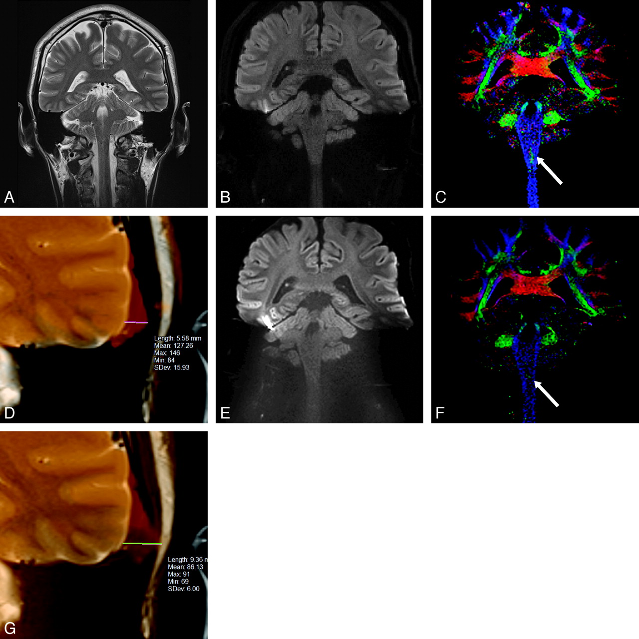
- Fig 1.
40-year-old healthy male volunteer. Coronal MR images of brain obtained at the same level: A, T2-weighted image, (B) RS-EPI diffusion trace image, (C) RS-EPI color-coded fiber orientation map, (D) the fusion image of RS-EPI and T2-weighted image of left temporal lobe, (E) SS-EPI diffusion trace image, (F) SS-EPI color-coded fiber orientation map, (G) the fusion image of SS-EPI and T2-weighted image of left temporal lobe. The natural lateral contour of the left temporal lobe in the T2-weighted image (A) becomes prominently distorted in the SS-EPI image (E), but the convex shape manages to be preserved in the RS-EPI image (B). The decussation of the internal arcuate fibers is seen at the medulla level on the RS-EPI image (C, arrow), more clearly compared with the SS-EPI image (F, arrow). The distance between the contours of the left temporal base is 5.58 mm for RS-EPI (D), and 9.36 mm for SS-EPI (G).
- Fig 2.
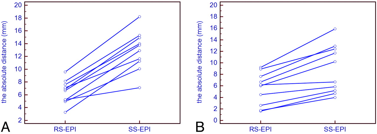
A, Distortion measurements at the level of the temporal base. B, Distortion measurements at the level of the cerebellum. The absolute distances between the contours of the temporal base (A) and the cerebellum (B), visible on the diffusion trace images for RS-EPI and SS-EPI and the corresponding T2-weighted images, are shown. The amount of distortion is significantly reduced with RS-EPI compared with SS-EPI (P <.01).


Tables
Scale (0–4) 4 Perfect contour continuation, preserving the contrast of both white and gray matter; convexity (of the cerebrum and cerebellum) is almost the same as that of T2-weighted image 3 Good contour continuation, preserving the contrast of both white and gray matter; convexity (of the cerebrum and cerebellum) is preserved but is less pronounced compared with that of T2-weighted image 2 Poor contour continuation; discontinuity can be seen in some parts; convexity is lost 1 Contour continuation is lost; concave contour is observed 0 Very concave (opposite of the natural convexity of cerebrum and cerebellum), with significant distortion Scale (0–4) 4 Excellent; very good color contrast and discriminates with surrounding structure 3 Relatively good; very good color contrast and discriminates with surrounding structure 2 Enough good contrast with surrounding structure 1 Subtle, faint contrast with surrounding structure 0 Colors at border area are mixed; impossible to make diagnosis RS-EPI SS-EPI P Value Contours of temporal base Lateral 2.2 ± 0.5 0.4 ± 0.5 <0.01 Inferior 1.7 ± 0.6 0.0 ± 0.0 <0.01 Parietal lobe 3.9 ± 0.2 3.7 ± 0.3 0.14 Cerebellum Lateral 2.6 ± 0.8 0.4 ± 0.3 <0.01 Inferior 2.7 ± 0.7 0.6 ± 0.4 <0.01 Brain stem Center 3.4 ± 0.5 2.2 ± 0.9 <0.01 Margin 3.7 ± 0.3 3.0 ± 0.7 0.06 Trigeminal nerve 3.0 ± 0.7 1.8 ± 0.8 <0.01 Acoustic nerve 2.3 ± 0.6 1.1 ± 0.6 <0.01 RS-EPI SS-EPI P Value Cerebellar vermis 3.4 ± 0.5 2.2 ± 0.7 <0.05 Temporal gyrus 3.3 ± 0.4 1.2 ± 0.8 <0.01 Decussation of the internal arcuate fibers 3.3 ± 0.9 1.6 ± 0.8 <0.01 Cerebellar peduncle Superior 4.0 ± 0.0 3.9 ± 0.3 0.34 Middle 3.6 ± 0.3 2.8 ± 0.7 0.06 Cerebral fornix 4.0 ± 0.1 3.7 ± 0.6 0.24 Laterality of basal ganglia 3.6 ± 0.3 2.8 ± 0.6 <0.05 Brain stem 3.4 ± 0.6 2.5 ± 0.7 0.07 Pyramidal tract 3.7 ± 0.3 3.2 ± 0.5 0.06
In this issue


Jump to section
Related Articles
Cited By...
- No citing articles found.