
Article Figures & Data
Figures
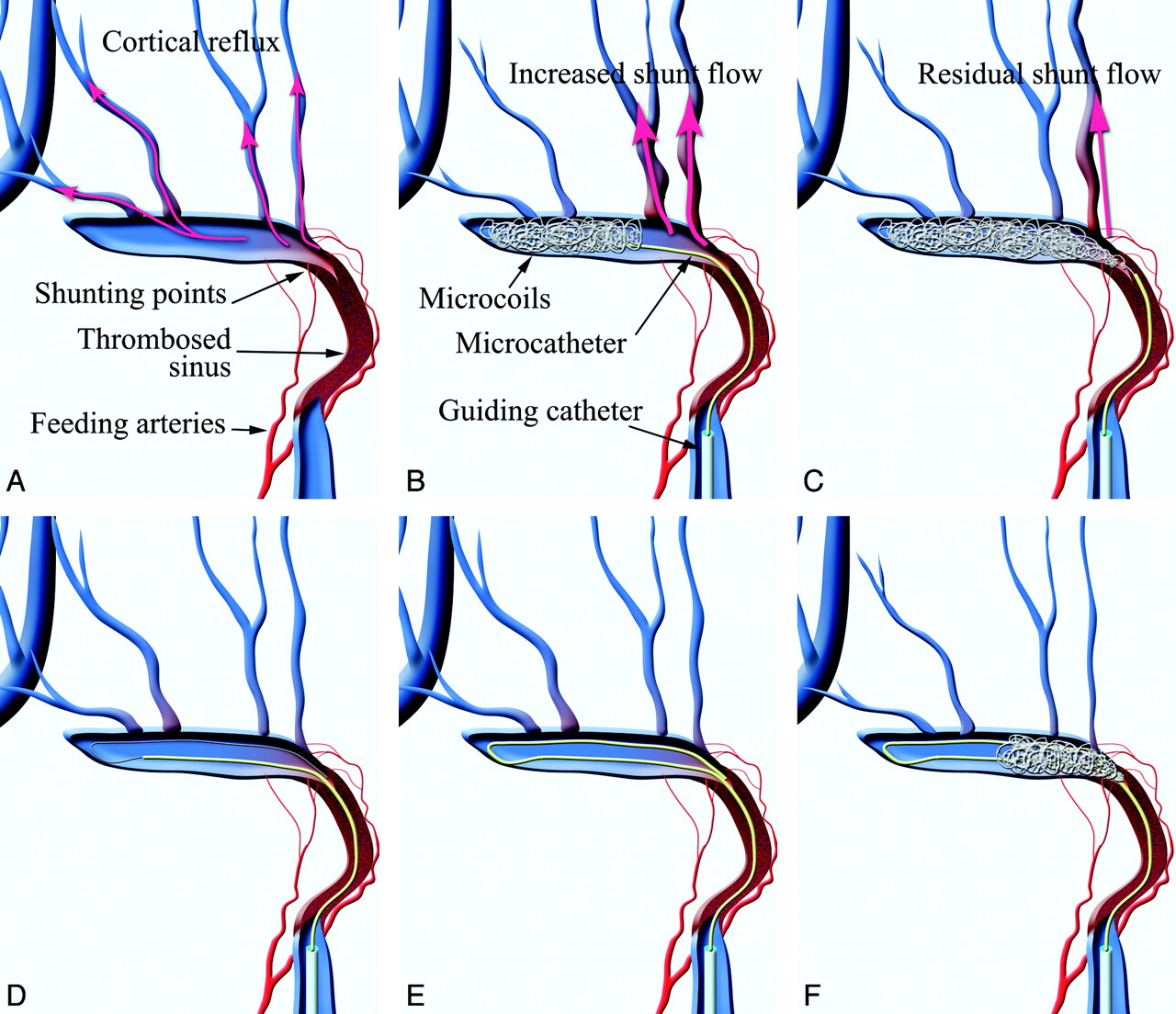
- Fig 1.
Schematic drawings of the risk of increasing and/or residual cortical reflux while placing coils from the distal-to-proximal segment and the turn-back embolization technique for DAVFs. A, In a TSS-DAVF with sinus occlusion like the Cognard type IIa+b or III, shunt surgery points and dangerous cortical reflux, such as the vein of Labbe and temporal veins, tend to be located at the proximal part of the transverse sinus, which is close to the occluded sinus. B and C, For such cases, standard transvenous embolization through the thrombosed sinus from the distal normal segment to the proximal abnormal segment has a risk of increased cortical venous reflux while packing from the distal to proximal part (B) or a risk of residual reflux due to insufficient packing of the shunt surgery point (C). D, In such cases, a transfemoral transvenous approach through the occluded ipsilateral sigmoid sinus can be performed. After antegrade navigation of a microcatheter into the sinus, a microguidewire is deflected at the distal end of the occluded sinus. E, Then the microcatheter is advanced to the shunt surgery point over the microguidewire. F, Microcoils can be placed into the shunt surgery point at the initial stage of sinus packing. This technique can minimize the risk of insufficient occlusion of the shunt flow when placing coils from an abnormal-to-normal segment rather than from a normal-to-abnormal segment.
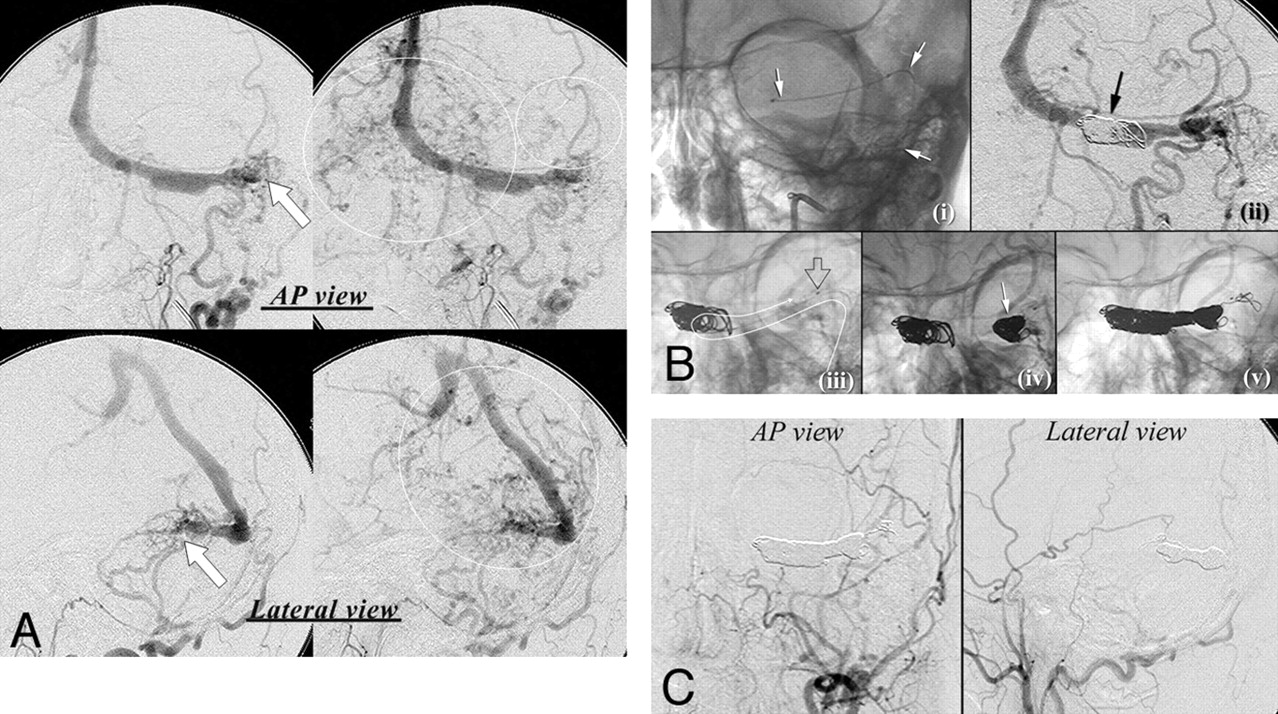
- Fig 2.
A 52-year-old man with hemianopsia and ataxia. A, The DAVF is fed mainly by the left occipital artery and drains into the straight sinus (arrows) on diagnostic angiography (upper images, frontal view; lower images, lateral view). The left transverse sinus does not communicate with the right transverse sinus. Marked cortical venous reflux into the vein of Labbe and the deep venous system is also seen (circle). B, Turn-back embolization method. i, AP view of the fluorogram. A microcatheter is navigated through the occluded ipsilateral TSS (arrow). ii, AP view of angiography. Because there is no toehold to turn the catheter, some coils not occluding the main drainage route are placed without stemming drainage flow (arrow). iii, AP view of the fluorogram. The microcatheter is turned back to the shunt surgery point at the proximal part of the TSS. The white curved arrow indicates the course of the microcatheter. The open arrow shows the tip of the microcatheter. iv, AP view of the fluorogram. The shunt surgery point is densely packed at the initial step of sinus packing (arrow). v, Fluorogram immediately after packing of the involved TSS. The TSS was packed with a drawing microcatheter after coiling of the shunt surgery point. C, Digital subtraction angiography immediately after embolization shows complete obliteration of the DAVF.
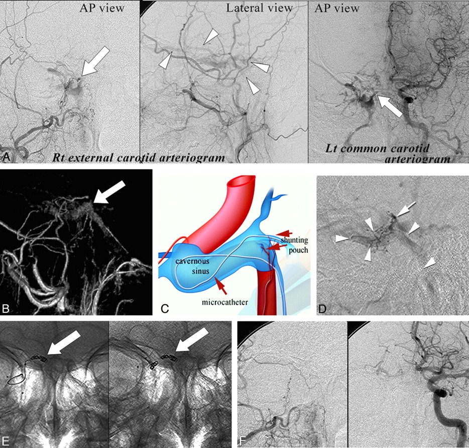
- Fig 3.
A 55-year-old man with double vision. A, Right external carotid angiography and left common carotid angiography show the CS-DAVF (arrow) fed by the ascending pharyngeal artery and the artery of the foramen rotundum. Arrowheads indicate drainage veins. B, The shunt surgery venous pouch can be identified on surface rendering of rotational digital subtraction angiography at the posteromedial wall of the right cavernous sinus. C, Schematic drawing of navigation of the microcatheter into the shunt surgery venous pouch. Because the shunt surgery venous pouch is acutely angulated from the accessible ipsilateral inferior petrosal sinus, a microcatheter is navigated into the pouch by the turn-back technique as shown in the schematic drawing. D, Lateral view of the selective shunt surgery pouch venography. Arrowheads indicate the course of the microcatheter; the arrow indicates the tip of the microcatheter. E, AP view of the fluorogram. After navigation of the microcatheter, detachable microcoils are selectively placed in the shunt surgery venous pouch without sinus packing (arrows). F, Anterior views of the right external and left common carotid arteriogram immediately after the procedure show complete obliteration of the DVAF.


In this issue


Jump to section
Related Articles
Cited By...
- No citing articles found.