Advertisement

Main menu

  • Home
  • Content
    • Current Issue
    • Accepted Manuscripts
    • Article Preview
    • Past Issue Archive
    • Video Articles
    • AJNR Case Collection
    • Case of the Week Archive
    • Case of the Month Archive
    • Classic Case Archive
  • Special Collections
    • AJNR Awards
    • Low-Field MRI
    • Alzheimer Disease
    • ASNR Foundation Special Collection
    • Photon-Counting CT
    • View All
  • Multimedia
    • AJNR Podcasts
    • AJNR SCANtastic
    • Trainee Corner
    • MRI Safety Corner
    • Imaging Protocols
  • For Authors
    • Submit a Manuscript
    • Submit a Video Article
    • Submit an eLetter to the Editor/Response
    • Manuscript Submission Guidelines
    • Statistical Tips
    • Fast Publishing of Accepted Manuscripts
    • Graphical Abstract Preparation
    • Imaging Protocol Submission
    • Author Policies
  • About Us
    • About AJNR
    • Editorial Board
    • Editorial Board Alumni
  • More
    • Become a Reviewer/Academy of Reviewers
    • Subscribers
    • Permissions
    • Alerts
    • Feedback
    • Advertisers
    • ASNR Home

User menu

  • Alerts
  • Log in

Search

  • Advanced search
American Journal of Neuroradiology

American Journal of Neuroradiology

ASHNR American Society of Functional Neuroradiology ASHNR American Society of Pediatric Neuroradiology ASSR
  • Alerts
  • Log in

Advanced Search

  • Home
  • Content
    • Current Issue
    • Accepted Manuscripts
    • Article Preview
    • Past Issue Archive
    • Video Articles
    • AJNR Case Collection
    • Case of the Week Archive
    • Case of the Month Archive
    • Classic Case Archive
  • Special Collections
    • AJNR Awards
    • Low-Field MRI
    • Alzheimer Disease
    • ASNR Foundation Special Collection
    • Photon-Counting CT
    • View All
  • Multimedia
    • AJNR Podcasts
    • AJNR SCANtastic
    • Trainee Corner
    • MRI Safety Corner
    • Imaging Protocols
  • For Authors
    • Submit a Manuscript
    • Submit a Video Article
    • Submit an eLetter to the Editor/Response
    • Manuscript Submission Guidelines
    • Statistical Tips
    • Fast Publishing of Accepted Manuscripts
    • Graphical Abstract Preparation
    • Imaging Protocol Submission
    • Author Policies
  • About Us
    • About AJNR
    • Editorial Board
    • Editorial Board Alumni
  • More
    • Become a Reviewer/Academy of Reviewers
    • Subscribers
    • Permissions
    • Alerts
    • Feedback
    • Advertisers
    • ASNR Home
  • Follow AJNR on Twitter
  • Visit AJNR on Facebook
  • Follow AJNR on Instagram
  • Join AJNR on LinkedIn
  • RSS Feeds

AJNR Awards, New Junior Editors, and more. Read the latest AJNR updates

Research ArticleBrain

Dual-Energy CT in the Evaluation of Intracerebral Hemorrhage of Unknown Origin: Differentiation between Tumor Bleeding and Pure Hemorrhage

S.J. Kim, H.K. Lim, H.Y. Lee, C.G. Choi, D.H. Lee, D.C. Suh, S.M. Kim, J.K. Kim and B. Krauss
American Journal of Neuroradiology May 2012, 33 (5) 865-872; DOI: https://doi.org/10.3174/ajnr.A2890
S.J. Kim
  • Find this author on Google Scholar
  • Find this author on PubMed
  • Search for this author on this site
H.K. Lim
  • Find this author on Google Scholar
  • Find this author on PubMed
  • Search for this author on this site
H.Y. Lee
  • Find this author on Google Scholar
  • Find this author on PubMed
  • Search for this author on this site
C.G. Choi
  • Find this author on Google Scholar
  • Find this author on PubMed
  • Search for this author on this site
D.H. Lee
  • Find this author on Google Scholar
  • Find this author on PubMed
  • Search for this author on this site
D.C. Suh
  • Find this author on Google Scholar
  • Find this author on PubMed
  • Search for this author on this site
S.M. Kim
  • Find this author on Google Scholar
  • Find this author on PubMed
  • Search for this author on this site
J.K. Kim
  • Find this author on Google Scholar
  • Find this author on PubMed
  • Search for this author on this site
B. Krauss
  • Find this author on Google Scholar
  • Find this author on PubMed
  • Search for this author on this site
  • Article
  • Figures & Data
  • Info & Metrics
  • Responses
  • References
  • PDF
Loading

Article Figures & Data

Figures

  • Tables
  • Fig 1.
    • Download figure
    • Open in new tab
    • Download powerpoint
    Fig 1.

    Diagram of image postprocessing of DECT for analysis of hematoma. Enhanced average images, which are equivalent to conventional enhanced images, are produced by mixing and averaging the CT numbers of corresponding voxels at a 7:3 ratio of A and B tube images. Using dual-energy material decomposition software, we separated the iodine component from the images obtained from the A and B tubes; this iodine component is shown in iodine-overlay images and displayed in red. Virtual noncontrast images are generated by removing iodine from the source images. Fusion images are obtained by mixing iodine overlay images and virtual noncontrast images, typically at a ratio of 50:50.
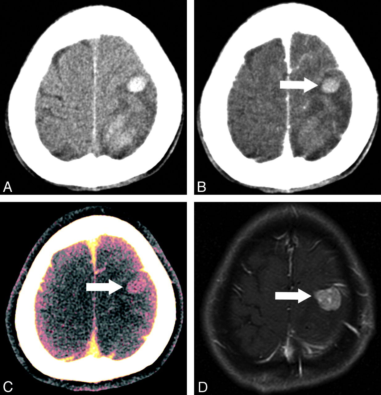
  • Fig 2.
    • Download figure
    • Open in new tab
    • Download powerpoint
    Fig 2.

    A 35-year-old man with known renal cell carcinoma and pulmonary metastasis. A, True noncontrast CT shows hemorrhage in the right frontal lobe. B, Enhanced average image obtained by mixing 80- and 140-kV images. A small area of enhancement is suspected in the anterior portion of the hematoma (arrow). Iodine overlay image (C) and virtual noncontrast image (D) obtained by postprocessing of 80- and 140-kV images by using dual-energy material decomposition software. The iodine component is displayed in red in the iodine-overlay image, and the enhancing tumor portion is clearly identified (arrow). High attenuation of hemorrhage is not visualized in this image set, but high attenuation of the bony structure is preserved (C). Virtual noncontrast image (D) shows the hematoma clearly, but the image is smoothed compared with the true noncontrast image in A. E, Fusion image obtained by mixing of iodine-overlay and virtual noncontrast images clearly shows an enhancing mass in the anterior portion of the hematoma (arrow). The brain lesion was histologically confirmed as a hemorrhagic metastasis from renal cell carcinoma.
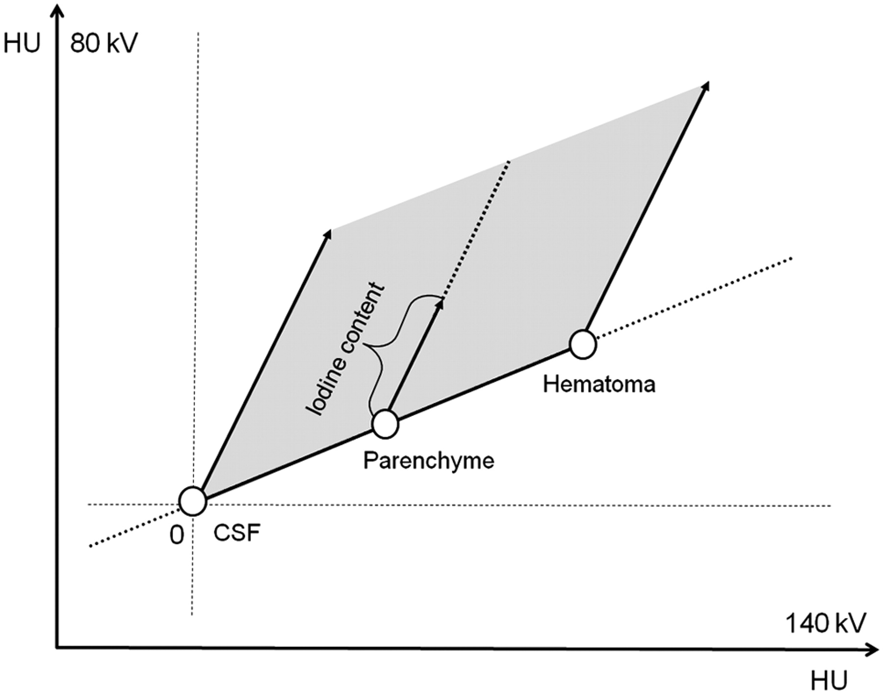
  • Fig 3.
    • Download figure
    • Open in new tab
    • Download powerpoint
    Fig 3.

    Mechanism of differentiating hematoma and iodine. Measured voxel values are plotted on the 2-energy scatterplot. CSF, brain parenchyma, and hematoma have similar CT numbers on both 80- and 140-kV images. In contrast, iodine has nearly a 2 times higher CT number on 80 kV images compared with 140 kV. Therefore, voxels in the area of gray shading represent various amounts of iodine content.

  • Fig 4.
    • Download figure
    • Open in new tab
    • Download powerpoint
    Fig 4.

    A 37-year-old woman with known hepatocellular carcinoma. A, True noncontrast CT image shows acute ICH in the left frontal lobe. Part of a separate lesion is noted in the left parietal lobe. B, Enhanced average image at the corresponding level fails to show enhancement in the frontal lobe lesion (arrow). The parietal lobe lesion showed irregular rim enhancement in the main body of the hematoma (not shown here). C, DECT fusion image clearly shows the frontal lobe lesion in red, representing the iodine component (arrow). D, The parietal lobe lesion was surgically removed and confirmed as a metastasis, but the frontal lobe lesion was not treated initially. Gadolinium-enhanced MR imaging performed 1 month later clearly shows the lesion in the left frontal lobe; its size has increased (arrow).

  • Fig 5.
    • Download figure
    • Open in new tab
    • Download powerpoint
    Fig 5.

    A false-negative result of DECT in a 48-year-old man with known hepatocellular carcinoma. A, True noncontrast CT shows a hematoma in the right occipital lobe. Small dotlike enhancement is noted in the medial aspect of hematoma on an enhanced average image (B) and a fusion image (C). However, no definite mass lesion is identified. D, On the enhanced T1-weighted image, some tubular enhancement is noted (arrow), but a tumorous lesion is not definite. E and F, One-month follow-up axial and sagittal postcontrast MR images show multiple enhancing nodules (arrows) around the hematoma (arrowheads). These strongly suggest the possibility of previous ICH caused by underlying metastatic tumor.

  • Fig 6.
    • Download figure
    • Open in new tab
    • Download powerpoint
    Fig 6.

    A false-positive result of DECT in a 50-year-old man with right-sided weakness commencing 3 days earlier. A, True noncontrast CT shows a hematoma in the left frontal lobe superior frontal gyrus. A thin irregular rim-enhancing lesion is shown on the enhanced average image (B) and fusion image (C) (arrows). D, T2-weighted sagittal image shows a multilocular dark-signal-intensity lesion mixed with high signal intensity with a dark-signal-intensity rim, suggesting multiple-stage hemorrhage. On the basis of images B and C, a hemorrhagic tumor was suspected, but the diagnosis was changed after reviewing MR imaging, which suggested the possibility of a cavernous malformation. Diagnosis of a cavernous malformation was confirmed by surgery.

  • Fig 7.
    • Download figure
    • Open in new tab
    • Download powerpoint
    Fig 7.

    ROC curves for fusion, EA, and EA + TNC images. Fusion images show the highest performance with an AUC of 0.964.

Tables

  • Figures
    • View popup
    Table 1:

    Underlying pathology of ICH

    Underlying PathologyPatient No.
    Tumor17
        Metastases, including melanomas13 (4 melanomas)
        Primary (astrocytomaa/PNET/meningioma)4 (1/1/2)
    Nontumor39
        Unrecognized hypertensive9
        Infarct, HT/coagulopathy/trauma2/8/4
        AVM/DAVF/CM3/1/3
        Amyloid angiopathy/Moyamoya disease/vasculitis1/1/2
        Dural sinus thrombosis/occlusion5
    Total56
    • Note:—PNET indicates primitive neuroectodermal tumor; infarct, HT, infarct, hemorrhagic transformation; DAVF, dural arteriovenous fistula; CM, cavernous malformation.

    • ↵a Gemistocytic astrocytoma grade II.

    • View popup
    Table 2:

    Sensitivity, specificity, PPV, and NPV of each image series

    Sensitivity (%) (CI)Specificity (%) (CI)PPV (%) (CI)NPV (%)(CI)
    Fusion images94.497.494.497.4
    (72.6–99.1)(86.5–99.6)(72.6–99.1)(86.5–99.6)
    EA images61.192.378.683.7
    (35.8–82.6)(79.1–98.3)(49.2–95.1)(69.3–3.2)
    EA + TNC images66.789.775.085.4
    (41.0–6.6)(75.8–97.1)(47.6–92.6)(70.8–94.4)
    • Note:—CI indicates confidence interval; PPV, positive predictive value; NPV, negative predictive value.

    • View popup
    Table 3:

    Comparison of CT numbers of ICH and white matter, SD of CSF, and CNR on TNC and VNC images (mean ± SD)a

    TNC ImagesVNC ImagesP Value
    ICH71.5 ± 7.362.8 ± 9.6.000
    White matter31.0 ± 2.031.2 ± 3.0.675
    SD of CSF6.1 ± 1.53.7 ± 0.6.000
    CNR7.0 ± 1.98.8 ± 3.0.000
    • ↵a Values are HU in ICH, white matter, SD of CSF, and ratio in CNR.

PreviousNext
Back to top

In this issue

American Journal of Neuroradiology: 33 (5)
American Journal of Neuroradiology
Vol. 33, Issue 5
1 May 2012
  • Table of Contents
  • Index by author
Advertisement
Print
Download PDF
Email Article

Thank you for your interest in spreading the word on American Journal of Neuroradiology.

NOTE: We only request your email address so that the person you are recommending the page to knows that you wanted them to see it, and that it is not junk mail. We do not capture any email address.

Enter multiple addresses on separate lines or separate them with commas.
Dual-Energy CT in the Evaluation of Intracerebral Hemorrhage of Unknown Origin: Differentiation between Tumor Bleeding and Pure Hemorrhage
(Your Name) has sent you a message from American Journal of Neuroradiology
(Your Name) thought you would like to see the American Journal of Neuroradiology web site.
CAPTCHA
This question is for testing whether or not you are a human visitor and to prevent automated spam submissions.
Cite this article
S.J. Kim, H.K. Lim, H.Y. Lee, C.G. Choi, D.H. Lee, D.C. Suh, S.M. Kim, J.K. Kim, B. Krauss
Dual-Energy CT in the Evaluation of Intracerebral Hemorrhage of Unknown Origin: Differentiation between Tumor Bleeding and Pure Hemorrhage
American Journal of Neuroradiology May 2012, 33 (5) 865-872; DOI: 10.3174/ajnr.A2890

Citation Manager Formats

  • BibTeX
  • Bookends
  • EasyBib
  • EndNote (tagged)
  • EndNote 8 (xml)
  • Medlars
  • Mendeley
  • Papers
  • RefWorks Tagged
  • Ref Manager
  • RIS
  • Zotero
0 Responses
Respond to this article
Share
Bookmark this article
Dual-Energy CT in the Evaluation of Intracerebral Hemorrhage of Unknown Origin: Differentiation between Tumor Bleeding and Pure Hemorrhage
S.J. Kim, H.K. Lim, H.Y. Lee, C.G. Choi, D.H. Lee, D.C. Suh, S.M. Kim, J.K. Kim, B. Krauss
American Journal of Neuroradiology May 2012, 33 (5) 865-872; DOI: 10.3174/ajnr.A2890
del.icio.us logo Twitter logo Facebook logo Mendeley logo
  • Tweet Widget
  • Facebook Like
  • Google Plus One
Purchase

Jump to section

  • Article
    • Abstract
    • ABBREVIATIONS:
    • Materials and Methods
    • Results
    • Discussion
    • Conclusions
    • Footnotes
    • References
  • Figures & Data
  • Info & Metrics
  • Responses
  • References
  • PDF

Related Articles

  • No related articles found.
  • PubMed
  • Google Scholar

Cited By...

  • Change in Emergency Department Length of Stay following Routine Adoption of Dual-Energy CT to Differentiate Intracranial Hemorrhage from Calcification
  • Discrimination of Hemorrhage and Contrast Media in a Head Phantom on Photon-Counting Detector CT Data
  • Residual Thromboembolic Material in Cerebral Arteries after Endovascular Stroke Therapy Can Be Identified by Dual-Energy CT
  • Discrimination of Tumorous Intracerebral Hemorrhage from Benign Causes Using CT Densitometry
  • Dual-Energy Computed Tomography
  • Second European Stroke Science Workshop
  • Crossref (74)
  • Google Scholar

This article has been cited by the following articles in journals that are participating in Crossref Cited-by Linking.

  • Dual-Energy CT: New Horizon in Medical Imaging
    Hyun Woo Goo, Jin Mo Goo
    Korean Journal of Radiology 2017 18 4
  • Material Separation Using Dual-Energy CT: Current and Emerging Applications
    Manuel Patino, Andrea Prochowski, Mukta D. Agrawal, Frank J. Simeone, Rajiv Gupta, Peter F. Hahn, Dushyant V. Sahani
    RadioGraphics 2016 36 4
  • Principles and applications of multienergy CT: Report of AAPM Task Group 291
    Cynthia H. McCollough, Kirsten Boedeker, Dianna Cody, Xinhui Duan, Thomas Flohr, Sandra S. Halliburton, Jiang Hsieh, Rick R. Layman, Norbert J. Pelc
    Medical Physics 2020 47 7
  • Dual-Energy CT: Oncologic Applications
    Carlo Nicola De Cecco, Anna Darnell, Marco Rengo, Giuseppe Muscogiuri, Davide Bellini, Carmen Ayuso, Andrea Laghi
    American Journal of Roentgenology 2012 199 5_supplement
  • Dual-energy CT: theoretical principles and clinical applications
    Andrea Agostini, Alessandra Borgheresi, Alberto Mari, Chiara Floridi, Federico Bruno, Marina Carotti, Nicolò Schicchi, Antonio Barile, Stefania Maggi, Andrea Giovagnoni
    La radiologia medica 2019 124 12
  • Dual-Energy CT: What the Neuroradiologist Should Know
    Alida A. Postma, Marco Das, Annika A. R. Stadler, Joachim E. Wildberger
    Current Radiology Reports 2015 3 5
  • Recent developments of dual-energy CT in oncology
    David Simons, Marc Kachelrieß, Heinz-Peter Schlemmer
    European Radiology 2014 24 4
  • Dual-Energy CT of the Brain and Intracranial Vessels
    Alida A. Postma, Paul A. M. Hofman, Annika A. R. Stadler, Robert J. van Oostenbrugge, Maud P. M. Tijssen, Joachim E. Wildberger
    American Journal of Roentgenology 2012 199 5_supplement
  • Dual-Energy CT in Emergency Neuroimaging: Added Value and Novel Applications
    Christopher A. Potter, Aaron D. Sodickson
    RadioGraphics 2016 36 7
  • Dual-Energy Head CT Enables Accurate Distinction of Intraparenchymal Hemorrhage from Calcification in Emergency Department Patients
    Ranliang Hu, Laleh Daftari Besheli, Joseph Young, Markus Wu, Stuart Pomerantz, Michael H. Lev, Rajiv Gupta
    Radiology 2016 280 1

More in this TOC Section

  • Multimodal CT Provides Improved Performance for Lacunar Infarct Detection
  • Optimal MRI Sequence for Identifying Occlusion Location in Acute Stroke: Which Value of Time-Resolved Contrast-Enhanced MRA?
  • Evaluating the Effects of White Matter Multiple Sclerosis Lesions on the Volume Estimation of 6 Brain Tissue Segmentation Methods
Show more BRAIN

Similar Articles

Advertisement

Indexed Content

  • Current Issue
  • Accepted Manuscripts
  • Article Preview
  • Past Issues
  • Editorials
  • Editor's Choice
  • Fellows' Journal Club
  • Letters to the Editor
  • Video Articles

Cases

  • Case Collection
  • Archive - Case of the Week
  • Archive - Case of the Month
  • Archive - Classic Case

More from AJNR

  • Trainee Corner
  • Imaging Protocols
  • MRI Safety Corner
  • Book Reviews

Multimedia

  • AJNR Podcasts
  • AJNR Scantastics

Resources

  • Turnaround Time
  • Submit a Manuscript
  • Submit a Video Article
  • Submit an eLetter to the Editor/Response
  • Manuscript Submission Guidelines
  • Statistical Tips
  • Fast Publishing of Accepted Manuscripts
  • Graphical Abstract Preparation
  • Imaging Protocol Submission
  • Evidence-Based Medicine Level Guide
  • Publishing Checklists
  • Author Policies
  • Become a Reviewer/Academy of Reviewers
  • News and Updates

About Us

  • About AJNR
  • Editorial Board
  • Editorial Board Alumni
  • Alerts
  • Permissions
  • Not an AJNR Subscriber? Join Now
  • Advertise with Us
  • Librarian Resources
  • Feedback
  • Terms and Conditions
  • AJNR Editorial Board Alumni

American Society of Neuroradiology

  • Not an ASNR Member? Join Now

© 2025 by the American Society of Neuroradiology All rights, including for text and data mining, AI training, and similar technologies, are reserved.
Print ISSN: 0195-6108 Online ISSN: 1936-959X

Powered by HighWire