
Article Figures & Data
Figures
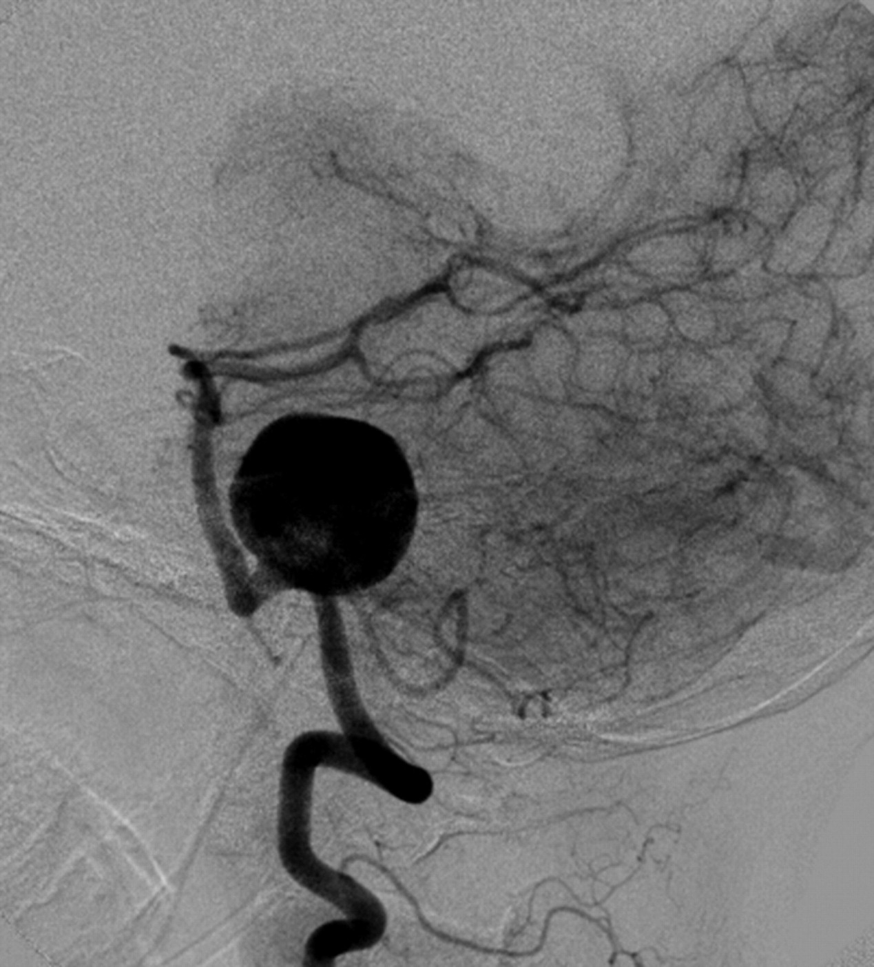
- Fig 1.
Lateral projection from angiography performed from a catheter positioned within the left vertebral artery demonstrates a very large (23-mm) wide-neck aneurysm arising from the mid-V4 segment of the vertebral artery. The aneurysm circumferentially incorporates a 10-mm-length segment of the parent artery. The PICA arises from the proximal aspect of the aneurysm neck. The neck of the aneurysm does not incorporate the vertebrobasilar junction.
- Fig 2.
Lateral projection from an angiogram obtained immediately after successful deployment of a single 4 × 20 mm PED construct across the aneurysm neck. Contrast material is stagnant within the aneurysm fundus, persisting into the venous phase of angiography. Contrast material was noted to washout from the aneurysm fundus into the PICA, which also remained faintly opacified throughout the venous phase.
- Fig 3.
A, Maximum intensity projection from an MRA performed on POD 9 demonstrates near-complete thrombosis of the aneurysm fundus. The left PICA remains patent, filling from a tiny niche of residual flow in the region of the aneurysm neck (arrow). B, Axial postcontrast T1-weighted sequence demonstrates linear enhancement of the aneurysm wall marginating the intra-aneurysmal thrombus. A focal niche of enhancement corresponds to the small neck remnant, which allows the patency of the PICA (arrow).
- Fig 4.
Noncontrast head CT scan obtained on POD 20 demonstrates aneurysmal rupture with dissection through the brain stem and into the fourth ventricle. A small amount of subarachnoid hemorrhage is noted within the prepontine cistern.
- Fig 5.
A, Base of the brain stem showing a large saccular aneurysm arising from the midportion of the left vertebral artery and embedded in the cerebellopontine angle, compressing the ipsilateral cranial nerves VII and VIII. Note that postdissection remnants of acute subarachnoid hemorrhage surround the aneurysm. Note distention of the segment of the artery at the base of the aneurysm by the stent. Cranial nerves are designated by Roman numerals. Lines with numbers indicate the plane of section of whole mounts of the aneurysm and vertebral artery as illustrated in B (Masson trichrome). B, Cross-sections of the aneurysm show incorporation of the vertebral artery into its wall (levels 1 and 2) and exit (level 1). Location of the stent (asterisk) is outlined by the luminal thrombus that partially fills the aneurysm. Fresh blood tracks along a lateral wall to the rupture through the dome (arrow). Boxes with letters represent microscopic fields shown in Fig 6.
- Fig 6.
Microscopic features in the wall of the aneurysm corresponding to the boxed regions in Fig 5B. A, Junction of the arterial wall with the aneurysm, showing mural thrombus adjacent to fresh blood in the ostium of the aneurysm (asterisk). B, High power of the area in the box demonstrates organization of the thrombus by sprouts of capillaries (A and B, hematoxylin-orcein-phloxine-saffron). C, Site of rupture shows frayed tearing of the wall, suggesting dislodgment of any focal mural necrosis by the hemorrhage (Masson trichrome). D, Segment of the wall displays focal attenuation (asterisk) and necrosis with attenuated infiltrates of acute inflammatory cells along the outer meningeal portion of the wall. E, High power of the wall shows focal loss of fibroblasts (asterisk); arrowheads indicate remaining fibroblasts (D and E, HE). F, In other regions of the wall, dissecting microhemorrhage (arrow) is associated with the mural necrosis. G, Micro-hemorrhage associated with mural necrosis seen in high power. (F, Masson trichrome; G, HE).


In this issue


Jump to section
Related Articles
Cited By...
- Hemodynamic Analysis of Postoperative Rupture of Unruptured Intracranial Aneurysms after Placement of Flow-Diverting Stents: A Matched Case-Control Study
- Flow diversion for the treatment of posterior inferior cerebellar artery aneurysms: a novel classification and strategies
- Ultrasound for the evaluation of stenosis after flow diversion
- Republished: The first North American use of the Pipeline Flex flow diverter
- Gene expression comparison of flow diversion and coiling in an experimental aneurysm model
- Rupture of giant vertebrobasilar aneurysm following flow diversion: mechanical stretch as a potential mechanism for early aneurysm rupture
- Prospective Study of Early MRI Appearances following Flow-Diverting Stent Placement for Intracranial Aneurysms
- The first North American use of the Pipeline Flex flow diverter
- Rupture of giant vertebrobasilar aneurysm following flow diversion: mechanical stretch as a potential mechanism for early aneurysm rupture
- Incidence of Microemboli and Correlation with Platelet Inhibition in Aneurysmal Flow Diversion
- Canadian Experience with the Pipeline Embolization Device for Repair of Unruptured Intracranial Aneurysms
- Pipeline Embolization Device in Aneurysmal Subarachnoid Hemorrhage
- Safety of the Pipeline Embolization Device in Treatment of Posterior Circulation Aneurysms