Advertisement

Main menu

  • Home
  • Content
    • Current Issue
    • Accepted Manuscripts
    • Article Preview
    • Past Issue Archive
    • Video Articles
    • AJNR Case Collection
    • Case of the Week Archive
    • Case of the Month Archive
    • Classic Case Archive
  • Special Collections
    • Advancing NeuroMRI with High-Relaxivity Contrast Agents
    • AJNR Awards
    • Low-Field MRI
    • Alzheimer Disease
    • ASNR Foundation Special Collection
    • Photon-Counting CT
    • View All
  • Multimedia
    • AJNR Podcasts
    • AJNR SCANtastic
    • Trainee Corner
    • MRI Safety Corner
    • Imaging Protocols
  • For Authors
    • Submit a Manuscript
    • Submit a Video Article
    • Submit an eLetter to the Editor/Response
    • Manuscript Submission Guidelines
    • Statistical Tips
    • Fast Publishing of Accepted Manuscripts
    • Graphical Abstract Preparation
    • Imaging Protocol Submission
    • Author Policies
  • About Us
    • About AJNR
    • Editorial Board
    • Editorial Board Alumni
  • More
    • Become a Reviewer/Academy of Reviewers
    • Subscribers
    • Permissions
    • Alerts
    • Feedback
    • Advertisers
    • ASNR Home

User menu

  • Alerts
  • Log in

Search

  • Advanced search
American Journal of Neuroradiology

American Journal of Neuroradiology

ASHNR American Society of Functional Neuroradiology ASHNR American Society of Pediatric Neuroradiology ASSR
  • Alerts
  • Log in

Advanced Search

  • Home
  • Content
    • Current Issue
    • Accepted Manuscripts
    • Article Preview
    • Past Issue Archive
    • Video Articles
    • AJNR Case Collection
    • Case of the Week Archive
    • Case of the Month Archive
    • Classic Case Archive
  • Special Collections
    • Advancing NeuroMRI with High-Relaxivity Contrast Agents
    • AJNR Awards
    • Low-Field MRI
    • Alzheimer Disease
    • ASNR Foundation Special Collection
    • Photon-Counting CT
    • View All
  • Multimedia
    • AJNR Podcasts
    • AJNR SCANtastic
    • Trainee Corner
    • MRI Safety Corner
    • Imaging Protocols
  • For Authors
    • Submit a Manuscript
    • Submit a Video Article
    • Submit an eLetter to the Editor/Response
    • Manuscript Submission Guidelines
    • Statistical Tips
    • Fast Publishing of Accepted Manuscripts
    • Graphical Abstract Preparation
    • Imaging Protocol Submission
    • Author Policies
  • About Us
    • About AJNR
    • Editorial Board
    • Editorial Board Alumni
  • More
    • Become a Reviewer/Academy of Reviewers
    • Subscribers
    • Permissions
    • Alerts
    • Feedback
    • Advertisers
    • ASNR Home
  • Follow AJNR on Twitter
  • Visit AJNR on Facebook
  • Follow AJNR on Instagram
  • Join AJNR on LinkedIn
  • RSS Feeds

AJNR Awards, New Junior Editors, and more. Read the latest AJNR updates


Improved Turnaround Times | Median time to first decision: 12 days

Research ArticleBrain

Evaluation of Aqueductal Stenosis by 3D Sampling Perfection with Application-Optimized Contrasts Using Different Flip Angle Evolutions Sequence: Preliminary Results with 3T MR Imaging

O. Algin and B. Turkbey
American Journal of Neuroradiology April 2012, 33 (4) 740-746; DOI: https://doi.org/10.3174/ajnr.A2833
O. Algin
  • Find this author on Google Scholar
  • Find this author on PubMed
  • Search for this author on this site
B. Turkbey
  • Find this author on Google Scholar
  • Find this author on PubMed
  • Search for this author on this site
  • Article
  • Figures & Data
  • Info & Metrics
  • Responses
  • References
  • PDF
Loading

Article Figures & Data

Figures

  • Tables
  • Fig 1.
    • Download figure
    • Open in new tab
    • Download powerpoint
    Fig 1.

    A 23-year-old woman (control). A, Sagittal 3D-SPACE with VFAM image shows a normal aqueduct (grade 0, arrow). B, Thin-section axial reformatted 3D-SPACE with VFAM image demonstrates hypointense flow-void signal intensity consistent with an open aqueduct (arrow). C and D, Sagittal (C) and axial (D) PC-MRI are well-matched with 3D-SPACE sequence results (arrows).

  • Fig 2.
    • Download figure
    • Open in new tab
    • Download powerpoint
    Fig 2.

    A 35-year-old man with partial aqueductal stenosis (patient 12). A, Sagittal T2-weighted image shows a narrowed aqueduct (arrow). B, Sagittal 3D-SPACE with VFAM image clearly demonstrates a prominent hypointense signal intensity in the cerebral aqueduct (arrow). The hypointense signal intensity (also called flow-void sign) on the 3D-SPACE MR image indicates the absence of a complete stenosis. C, Coronal oblique curved reconstructed 3D-SPACE image demonstrates a narrow but open aqueduct (arrow). D and E, Axial (D) and sagittal (E) PC-MRI indicate a narrowed but open aqueduct, consistent with partial aqueductal stenosis (arrows).
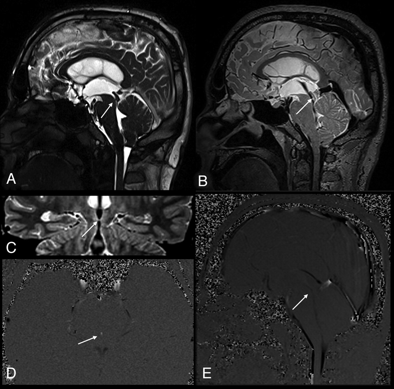
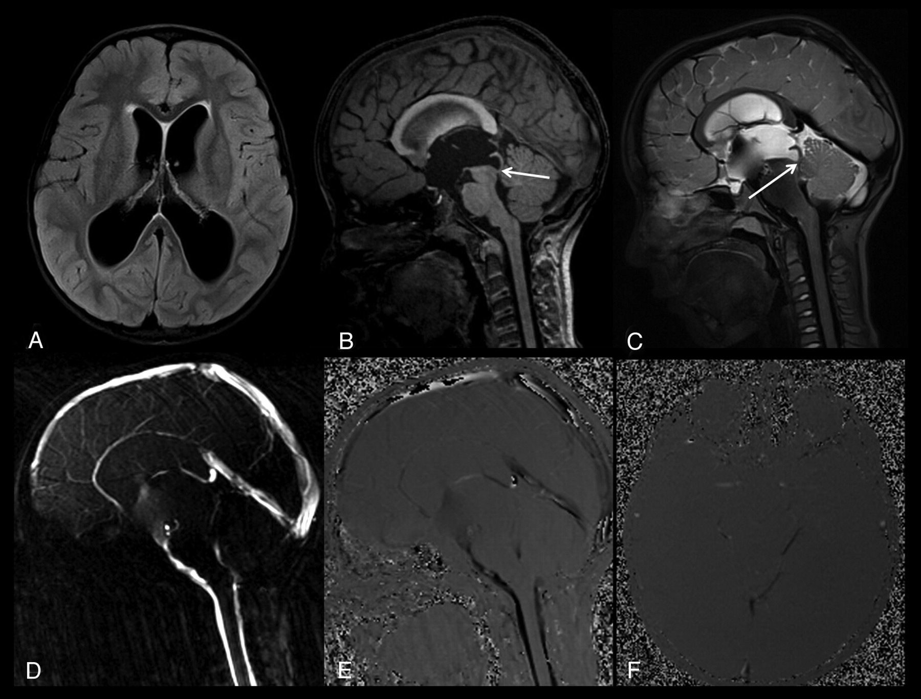
  • Fig 3.
    • Download figure
    • Open in new tab
    • Download powerpoint
    Fig 3.

    A 7-year-old boy with a complete aqueductal stenosis and hydrocephalus (patient 1). A, Axial FLAIR image shows a compensated hydrocephalus. B, Sagittal thin-section T1-weighted image demonstrates a narrowed distal aqueduct and a prestenotic aqueductal dilation (arrow). C, Sagittal 3D-SPACE with VFAM MR image shows a restricted hyperintense CSF flow proximal to the stenotic segment, whereas the unrestricted flow of CSF distal to the stenosis appears hypointense (arrow). Sagittal (D and E) and axial (F) PC-MRI shows a complete aqueductal stenosis, consistent with 3D-SPACE images.
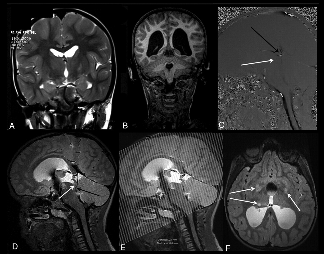
  • Fig 4.
    • Download figure
    • Open in new tab
    • Download powerpoint
    Fig 4.

    A 9-year-old boy with complete aqueductal stenosis and neurofibromatosis type 1 (patient 21). A and B, Coronal T2-weighted MR image obtained 2 years ago shows no evidence of hydrocephalus (A), whereas a recent T1-weighted MR image shows a progressive hydrocephalus (B). C, Sagittal PC-MRI obtained at the level of the aqueduct shows absence of aqueductal flow (white arrow). Sagittal PC-MRI shows a flow representing the deep venous system in the posteroinferior part of the aqueduct (black arrow, C). The pulsation secondary to the venous flow in the quadrigeminal cistern can mimic the aqueductal flow. D, Sagittal 3D-SPACE with VFAM MR image clearly demonstrates the venous structures located in the third ventricular outlet–the quadrigeminal cistern (black arrow). This image demonstrates a complete aqueductal stenosis (white arrow, D). E and F, Axial thin-section (1 mm) reformatted images obtained from sagittal 3D-SPACE images (E) show multiple hamartomas (arrows, F).

  • Fig 5.
    • Download figure
    • Open in new tab
    • Download powerpoint
    Fig 5.

    Timing diagram of the 3D-SPACE with different flip angles (3D-SPACE with VFAM). The diagram shows 1 TR of the pulse sequence. The first line shows the variation in the heights of the radio-frequency pulses during the echo-train. Gs is the section-selection gradient, which varies to select different sections in k-space. Gk and Gr are the phase-encoding and readout gradients, respectively. The timing diagram was drawn by John P. Mugler III and modified with his permission.

Tables

  • Figures
    • View popup
    Table 1:

    The demographic and clinical characteristics and MRI scores of the patient group

    No.SexAge (yr)PC-MRISPACEConsensus ResultsEHAdditional Illness
    1M7222Web-synechiaNF-1
    2F61000IdiopathicDWM
    3M59000IdiopathicDWM
    4M18000FVOO–
    5F69000TVAC–
    6F57000CoHDWM
    7F63000Idiopathic–
    8M69000Idiopathic–
    9M27222Web–
    10F50222Web–
    11M40222TG–
    12M35111TG–
    13M25222Idiopathic–
    14F54000FVOOChiari 1
    15M62000Idiopathic–
    16F34122TG–
    17M24000IdiopathicNF-1
    18M46000Idiopathic–
    19F43211Web–
    20M21000Idiopathic–
    21M9222Web, gliosisNF-1
    • Note:—DWM indicates Dandy-Walker malformation; FVOO, fourth ventricular outlet obstruction; TVAC, third ventricular arachnoid cyst; CoH, complex hydrocephalus; NF-1, neurofibromatosis type 1; EH, etiology of hydrocephalus; TG, tectal glioma.

    • View popup
    Table 2:

    The agreement level in consensus-based scores versus PC-MRI and SPACE scores

    Consensus VersusκConfidence Interval (95%)Agreement Level (No.) (%)
    PC-MRI0.828(0.618–1.000)19 (90.48)a
    SPACE1.000(1.000–1.000)21 (100)a
    • ↵a The number of times that tests agreed among themselves.

PreviousNext
Back to top

In this issue

American Journal of Neuroradiology: 33 (4)
American Journal of Neuroradiology
Vol. 33, Issue 4
1 Apr 2012
  • Table of Contents
  • Index by author
Advertisement
Print
Download PDF
Email Article

Thank you for your interest in spreading the word on American Journal of Neuroradiology.

NOTE: We only request your email address so that the person you are recommending the page to knows that you wanted them to see it, and that it is not junk mail. We do not capture any email address.

Enter multiple addresses on separate lines or separate them with commas.
Evaluation of Aqueductal Stenosis by 3D Sampling Perfection with Application-Optimized Contrasts Using Different Flip Angle Evolutions Sequence: Preliminary Results with 3T MR Imaging
(Your Name) has sent you a message from American Journal of Neuroradiology
(Your Name) thought you would like to see the American Journal of Neuroradiology web site.
CAPTCHA
This question is for testing whether or not you are a human visitor and to prevent automated spam submissions.
Cite this article
O. Algin, B. Turkbey
Evaluation of Aqueductal Stenosis by 3D Sampling Perfection with Application-Optimized Contrasts Using Different Flip Angle Evolutions Sequence: Preliminary Results with 3T MR Imaging
American Journal of Neuroradiology Apr 2012, 33 (4) 740-746; DOI: 10.3174/ajnr.A2833

Citation Manager Formats

  • BibTeX
  • Bookends
  • EasyBib
  • EndNote (tagged)
  • EndNote 8 (xml)
  • Medlars
  • Mendeley
  • Papers
  • RefWorks Tagged
  • Ref Manager
  • RIS
  • Zotero
0 Responses
Respond to this article
Share
Bookmark this article
Evaluation of Aqueductal Stenosis by 3D Sampling Perfection with Application-Optimized Contrasts Using Different Flip Angle Evolutions Sequence: Preliminary Results with 3T MR Imaging
O. Algin, B. Turkbey
American Journal of Neuroradiology Apr 2012, 33 (4) 740-746; DOI: 10.3174/ajnr.A2833
del.icio.us logo Twitter logo Facebook logo Mendeley logo
  • Tweet Widget
  • Facebook Like
  • Google Plus One
Purchase

Jump to section

  • Article
    • Abstract
    • ABBREVIATIONS:
    • Materials and Methods
    • Results
    • Discussion
    • Conclusions
    • Acknowledgments
    • Footnotes
    • References
  • Figures & Data
  • Info & Metrics
  • Responses
  • References
  • PDF

Related Articles

  • No related articles found.
  • PubMed
  • Google Scholar

Cited By...

  • Utility of 3D-T2 space MRI sequence in diagnosing a rare cause of lower backache: horseshoe cord and meningocoele manque in a case of composite split cord malformation
  • Optimal Diagnostic Indices for Idiopathic Normal Pressure Hydrocephalus Based on the 3D Quantitative Volumetric Analysis for the Cerebral Ventricle and Subarachnoid Space
  • Crossref
  • Google Scholar

This article has not yet been cited by articles in journals that are participating in Crossref Cited-by Linking.

More in this TOC Section

  • Usefulness of Quantitative Susceptibility Mapping for the Diagnosis of Parkinson Disease
  • White Matter Alterations in the Brains of Patients with Active, Remitted, and Cured Cushing Syndrome: A DTI Study
  • Qualitative and Quantitative Analysis of MR Imaging Findings in Patients with Middle Cerebral Artery Stroke Implanted with Mesenchymal Stem Cells
Show more Brain

Similar Articles

Advertisement

Indexed Content

  • Current Issue
  • Accepted Manuscripts
  • Article Preview
  • Past Issues
  • Editorials
  • Editor's Choice
  • Fellows' Journal Club
  • Letters to the Editor
  • Video Articles

Cases

  • Case Collection
  • Archive - Case of the Week
  • Archive - Case of the Month
  • Archive - Classic Case

More from AJNR

  • Trainee Corner
  • Imaging Protocols
  • MRI Safety Corner
  • Book Reviews

Multimedia

  • AJNR Podcasts
  • AJNR Scantastics

Resources

  • Turnaround Time
  • Submit a Manuscript
  • Submit a Video Article
  • Submit an eLetter to the Editor/Response
  • Manuscript Submission Guidelines
  • Statistical Tips
  • Fast Publishing of Accepted Manuscripts
  • Graphical Abstract Preparation
  • Imaging Protocol Submission
  • Evidence-Based Medicine Level Guide
  • Publishing Checklists
  • Author Policies
  • Become a Reviewer/Academy of Reviewers
  • News and Updates

About Us

  • About AJNR
  • Editorial Board
  • Editorial Board Alumni
  • Alerts
  • Permissions
  • Not an AJNR Subscriber? Join Now
  • Advertise with Us
  • Librarian Resources
  • Feedback
  • Terms and Conditions
  • AJNR Editorial Board Alumni

American Society of Neuroradiology

  • Not an ASNR Member? Join Now

© 2025 by the American Society of Neuroradiology All rights, including for text and data mining, AI training, and similar technologies, are reserved.
Print ISSN: 0195-6108 Online ISSN: 1936-959X

Powered by HighWire