Advertisement

Main menu

  • Home
  • Content
    • Current Issue
    • Accepted Manuscripts
    • Article Preview
    • Past Issue Archive
    • Video Articles
    • AJNR Case Collection
    • Case of the Week Archive
    • Case of the Month Archive
    • Classic Case Archive
  • Special Collections
    • AJNR Awards
    • Low-Field MRI
    • Alzheimer Disease
    • ASNR Foundation Special Collection
    • Photon-Counting CT
    • View All
  • Multimedia
    • AJNR Podcasts
    • AJNR SCANtastic
    • Trainee Corner
    • MRI Safety Corner
    • Imaging Protocols
  • For Authors
    • Submit a Manuscript
    • Submit a Video Article
    • Submit an eLetter to the Editor/Response
    • Manuscript Submission Guidelines
    • Statistical Tips
    • Fast Publishing of Accepted Manuscripts
    • Graphical Abstract Preparation
    • Imaging Protocol Submission
    • Author Policies
  • About Us
    • About AJNR
    • Editorial Board
    • Editorial Board Alumni
  • More
    • Become a Reviewer/Academy of Reviewers
    • Subscribers
    • Permissions
    • Alerts
    • Feedback
    • Advertisers
    • ASNR Home

User menu

  • Alerts
  • Log in

Search

  • Advanced search
American Journal of Neuroradiology

American Journal of Neuroradiology

ASHNR American Society of Functional Neuroradiology ASHNR American Society of Pediatric Neuroradiology ASSR
  • Alerts
  • Log in

Advanced Search

  • Home
  • Content
    • Current Issue
    • Accepted Manuscripts
    • Article Preview
    • Past Issue Archive
    • Video Articles
    • AJNR Case Collection
    • Case of the Week Archive
    • Case of the Month Archive
    • Classic Case Archive
  • Special Collections
    • AJNR Awards
    • Low-Field MRI
    • Alzheimer Disease
    • ASNR Foundation Special Collection
    • Photon-Counting CT
    • View All
  • Multimedia
    • AJNR Podcasts
    • AJNR SCANtastic
    • Trainee Corner
    • MRI Safety Corner
    • Imaging Protocols
  • For Authors
    • Submit a Manuscript
    • Submit a Video Article
    • Submit an eLetter to the Editor/Response
    • Manuscript Submission Guidelines
    • Statistical Tips
    • Fast Publishing of Accepted Manuscripts
    • Graphical Abstract Preparation
    • Imaging Protocol Submission
    • Author Policies
  • About Us
    • About AJNR
    • Editorial Board
    • Editorial Board Alumni
  • More
    • Become a Reviewer/Academy of Reviewers
    • Subscribers
    • Permissions
    • Alerts
    • Feedback
    • Advertisers
    • ASNR Home
  • Follow AJNR on Twitter
  • Visit AJNR on Facebook
  • Follow AJNR on Instagram
  • Join AJNR on LinkedIn
  • RSS Feeds

AJNR Awards, New Junior Editors, and more. Read the latest AJNR updates

Case ReportCase Report

MR Imaging Findings in the Reticular Formation in Siblings with MPV17-Related Mitochondrial Depletion Syndrome

A.N. Merkle, D.R. Nascene and A.M. McKinney
American Journal of Neuroradiology March 2012, 33 (3) E34-E35; DOI: https://doi.org/10.3174/ajnr.A2455
A.N. Merkle
  • Find this author on Google Scholar
  • Find this author on PubMed
  • Search for this author on this site
D.R. Nascene
  • Find this author on Google Scholar
  • Find this author on PubMed
  • Search for this author on this site
A.M. McKinney
  • Find this author on Google Scholar
  • Find this author on PubMed
  • Search for this author on this site
  • Article
  • Figures & Data
  • Info & Metrics
  • Responses
  • References
  • PDF
Loading

Abstract

SUMMARY: Hepatocerebral MPV17-MDS is quite rare (<30 confirmed cases), with limited findings described on MR imaging. We report 2 siblings having abnormalities within the reticular formation of the lower brain stem and within the reticulospinal tracts at the cervicocranial junction on T2WI. The presence of these MR imaging findings (relative to previous reports) raises the possibility that they represent subtle but characteristic findings corresponding to clinically observed abnormalities of tone encountered with this recently described disorder.

ABBREVIATIONS:

1H-MR spectroscopy
proton MR spectroscopy
MDS
mitochondrial depletion syndrome
MPV17-MDS
MPV17-related mitochondrial depletion syndrome
T1WI
T1-weighted imaging
T2WI
T2-weighted imaging

MPV17-related hepatocerebral MDS is a rare congenital autosomal recessive disorder typically characterized by hepatic failure, failure to thrive, and neurologic findings (dependent on the age of onset), such as hypotonia and dystonic movements.1,2 It is 1 of several described MDSs recently confirmed by dedicated laboratory and genetic testing.2 Presentation usually occurs in the first months of life, with the life span typically limited to months.1,2 The MR imaging literature describing MPV17-MDS is sparse and nonspecific, but the variable findings that have been reported range from normal-to-diffuse white matter abnormalities, which may resemble a leukodystrophy or hypomyelination.1 Particular regions noted in case reports include the cerebellar white matter, middle cerebellar peduncles, substantia nigra, and a single patient with subtle involvement of the dorsal brain stem.2⇓–4 Herein, we describe 2 infant siblings with MPV17-MDS exhibiting nearly identical involvement on T2WI at the cervicomedullary junction and within the lower dorsal brain stem.

Case Reports

Case 1

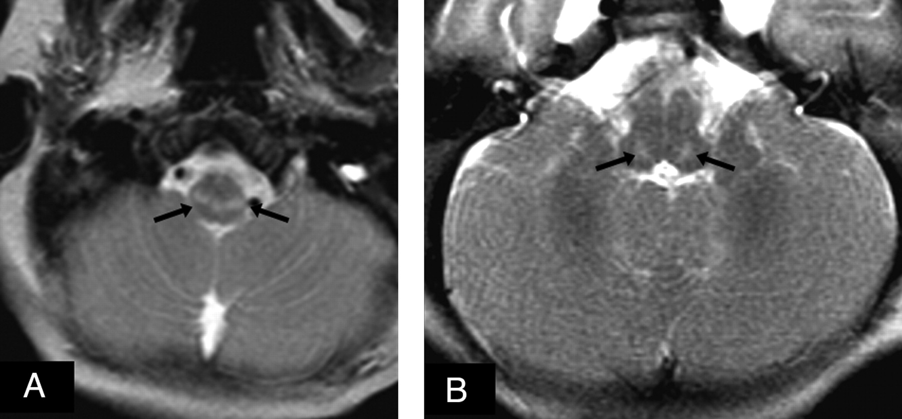
A 3-month-old male infant with no significant perinatal/neonatal history presented with progressive deficiencies in feeding, jaundice, failure to thrive, and mild hypotonia; elevated liver enzymes were subsequently detected. Thereafter, a 1.5T MR imaging at 5 months of age demonstrated mild moderately delayed myelination, as represented on T1WI by limited hyperintensity within the anterior limb of the internal capsule, the corpus callosum splenium and lack of significant occipital white matter hyperintensity. There were hyperintense abnormalities on T2WI bilaterally within the reticular formation of the lower dorsal brain stem and the reticulospinal tracts of the cervicomedullary junction (Fig 1), without reduced diffusion. Findings of point-resolved 1H-MR spectroscopy of the basal ganglia and periventricular white matter appeared normal for his age, without a lactate peak and with equivalent N-acetylaspartate and choline peaks. A head CT at 6.5 months of age did not demonstrate any significant abnormalities or overt atrophy. Hepatomegaly was clinically apparent and confirmed by CT. Muscle biopsies were performed but were negative for the more common mitochondrial disorders. The patient died at 7 months of age due to hepatic failure. Liver necropsy was nondiagnostic due to inadequate samples. The parents subsequently had 2 developmentally normal girls.

Fig 1.
  • Download figure
  • Open in new tab
  • Download powerpoint
Fig 1.

At 5 months of age, the male sibling had mild moderately delayed myelination on T1WI (not shown). A and B, There was abnormal T2 hyperintensity (arrows) extending from the reticulospinal tracts at the cervicomedullary junction up to the reticular formation of the medulla and pons, without reduced diffusion on diffusion-weighted imaging (not shown). 1H-MR spectroscopy (not shown) did not demonstrate a lactate peak.

Case 2

Approximately 2 years following the second developmentally normal girl, a daughter was born with no significant perinatal/neonatal concerns but presented at 4–5 weeks of age with liver dysfunction, jaundice, mild hypotonia, and failure to thrive. At this time, an MR imaging demonstrated myelination appropriate for her age on T1WI, and the MR imaging findings were initially interpreted as negative. Nevertheless, retrospective review (after obtaining and comparing with the first infant's images) demonstrated mild T2 hyperintensity of the dorsal lower brain stem within the reticular formation and within the reticulospinal tracts of the cervicomedullary junction, without reduced diffusion (Fig 2). 1H-MR spectroscopy was not performed. A muscle biopsy was negative for the more common mitochondrial disorders, and an extensive blood work-up was negative for leukodystrophies. A liver biopsy was considered nondiagnostic due to inadequate samples. The patient died at 4 months of age. A liver postmortem examination with samples evaluated by quantitative real-time polymerase chain reaction confirmed mitochondrial DNA copy number at 16% of the expected value for her age, and MPV17 gene sequencing analysis was positive for 2 heterozygous mutations.

Fig 2.
  • Download figure
  • Open in new tab
  • Download powerpoint
Fig 2.

At 5 weeks of age, the female sibling did not have overt hypomyelination on T1WI (not shown). A–C, However, there was abnormal hyperintensity on T2WI (arrows), extending from the reticulospinal tracts at the cervicomedullary junction up to the reticular formation of the medulla and pons in a distribution similar to that of her sibling.

Discussion

Spinazzola et al (2006)5 first described the mutation in the nuclear MPV17 gene, which leads to a defective inner mitochondrial membrane protein of unknown function and subsequent mitochondrial DNA depletion. At this time, there have been fewer than 30 patients described with confirmed MPV17-MDS.1,2 Because the clinical findings can be variable and nonspecific for MDS, there must be a suspicion for this disease to initiate the dedicated genetic testing that is necessary for diagnosis; therefore, characteristic MR imaging findings that could alert a clinician to this disorder would have diagnostic utility.

Although the reticular formation is a poorly defined area on MR imaging, it is associated with major subnetworks, including the reticulospinal tracts and descending reticular activating system, which are responsible for tone, posture, and balance and have connections with the cerebellum, which is implicated in this disorder.1,6 Lesions involving such locations may correspond with the typical late neurologic presentation of MPV17-MDS, which often involves hypotonia or dystonic movements.1,2 In the limited available reports of MPV17-MDS, the MR imaging findings have been described as normal (most commonly), “nonspecific white matter abnormalities,” “cytotoxic edema” involving the deep and subcortical white matter of the cerebrum, or “atrophy,” with only a few reports of specific regional abnormalities.1,3⇓–5,7 We reviewed the available images in these clinical reports and found only 1 with dorsal brain stem involvement, which we opine is localized within the reticular formation.3 Thus, the incidence of involvement of the reticular formation or reticulospinal tracts in MPV17-MDS is unknown.

Our description of 2 siblings with nearly identical involvement on MR imaging of the reticulospinal tracts and reticular formation, in conjunction with abnormalities in muscle tone, is most likely consistent with insult to these locations. The findings suggest that subtle but characteristic MR imaging findings could exist in MPV17-MDS. From a technical standpoint, these findings are at the caudal portion of the MR image and thus may not always be covered on axial images. In addition, the involved areas may only have subtle hyperintensity in the earlier stages that can be easily overlooked, as in patient 2. Thus, we concur with previous recommendations to use serial imaging to detect progression of subtle abnormalities on MR imaging or potential hypomyelination.3

References

  1. 1.↵
    1. El-Hattab A,
    2. Li F,
    3. Schmitt E,
    4. et al
    . MPV17-associated hepatocerebral mitochondrial DNA depletion syndrome: new patients and novel mutations. Mol Genet Metab 2010;99:300–08
    CrossRefPubMed
  2. 2.↵
    1. Spinazzola A,
    2. Invernizzi F,
    3. Carrara F,
    4. et al
    . Clinical and molecular features of mitochondrial DNA depletion syndromes. J Inherit Metab Dis 2009;32:143–58
    CrossRefPubMed
  3. 3.↵
    1. deBruyn J,
    2. Chan A,
    3. Bhargava R,
    4. et al
    . Liver failure in mitochondrial DNA depletion syndrome: the importance of serial neuroimaging in liver transplantation evaluation. J Pediatr Gastroenterol Nutr 2007;45:252–56
    CrossRefPubMed
  4. 4.↵
    1. Navarro-Sastre A,
    2. Martn-Hernndez E,
    3. Campos Y,
    4. et al
    . Lethal hepatopathy and leukodystrophy caused by a novel mutation in MPV17 gene: description of an alternative MPV17 spliced form. Mol Genet Metab 2008;94:234–39
    CrossRefPubMed
  5. 5.↵
    1. Spinazzola A,
    2. Viscomi C,
    3. Fernandez-Vizarra E,
    4. et al
    . MPV17 encodes an inner mitochondrial membrane protein and is mutated in infantile hepatic mitochondrial DNA depletion. Nat Genet 2006;38:570–75
    CrossRefPubMed
  6. 6.↵
    1. Purves D,
    2. Augustine GJ,
    3. Fitzpatrick D,
    4. et al
    . Neuroscience. 4th ed. Sunderland, Massachusetts: Sinaur Associates; 2008:426–30
  7. 7.↵
    1. Karadimas C,
    2. Vu T,
    3. Holve S,
    4. et al
    . Navajo neurohepatopathy is caused by a mutation in the MPV17 gene. Am J Hum Genet 2006;79:544–48
    CrossRefPubMed
  • Received October 26, 2010.
  • Accepted after revision November 2, 2010.
  • © 2012 by American Journal of Neuroradiology
View Abstract
PreviousNext
Back to top

In this issue

American Journal of Neuroradiology: 33 (3)
American Journal of Neuroradiology
Vol. 33, Issue 3
1 Mar 2012
  • Table of Contents
  • Index by author
Advertisement
Print
Download PDF
Email Article

Thank you for your interest in spreading the word on American Journal of Neuroradiology.

NOTE: We only request your email address so that the person you are recommending the page to knows that you wanted them to see it, and that it is not junk mail. We do not capture any email address.

Enter multiple addresses on separate lines or separate them with commas.
MR Imaging Findings in the Reticular Formation in Siblings with MPV17-Related Mitochondrial Depletion Syndrome
(Your Name) has sent you a message from American Journal of Neuroradiology
(Your Name) thought you would like to see the American Journal of Neuroradiology web site.
CAPTCHA
This question is for testing whether or not you are a human visitor and to prevent automated spam submissions.
Cite this article
A.N. Merkle, D.R. Nascene, A.M. McKinney
MR Imaging Findings in the Reticular Formation in Siblings with MPV17-Related Mitochondrial Depletion Syndrome
American Journal of Neuroradiology Mar 2012, 33 (3) E34-E35; DOI: 10.3174/ajnr.A2455

Citation Manager Formats

  • BibTeX
  • Bookends
  • EasyBib
  • EndNote (tagged)
  • EndNote 8 (xml)
  • Medlars
  • Mendeley
  • Papers
  • RefWorks Tagged
  • Ref Manager
  • RIS
  • Zotero
0 Responses
Respond to this article
Share
Bookmark this article
MR Imaging Findings in the Reticular Formation in Siblings with MPV17-Related Mitochondrial Depletion Syndrome
A.N. Merkle, D.R. Nascene, A.M. McKinney
American Journal of Neuroradiology Mar 2012, 33 (3) E34-E35; DOI: 10.3174/ajnr.A2455
del.icio.us logo Twitter logo Facebook logo Mendeley logo
  • Tweet Widget
  • Facebook Like
  • Google Plus One
Purchase

Jump to section

  • Article
    • Abstract
    • ABBREVIATIONS:
    • Case Reports
    • Discussion
    • References
  • Figures & Data
  • Info & Metrics
  • Responses
  • References
  • PDF

Related Articles

  • No related articles found.
  • PubMed
  • Google Scholar

Cited By...

  • No citing articles found.
  • Crossref (13)
  • Google Scholar

This article has been cited by the following articles in journals that are participating in Crossref Cited-by Linking.

  • Mitochondrial DNA Depletion Syndromes: Review and Updates of Genetic Basis, Manifestations, and Therapeutic Options
    Ayman W. El-Hattab, Fernando Scaglia
    Neurotherapeutics 2013 10 2
  • Clinical and Molecular Characteristics of Mitochondrial DNA Depletion Syndrome Associated with Neonatal Cholestasis and Liver Failure
    Abdulrahman Al-Hussaini, Eissa Faqeih, Ayman W. El-Hattab, Majid Alfadhel, Ali Asery, Badr Alsaleem, Eman Bakhsh, Ashraf Ali, Ali Alasmari, Khurram Lone, Ahmed Nahari, Wafaa Eyaid, Mohammed Al Balwi, Kate Craig, Anna Butterworth, Langping He, Robert W. Taylor
    The Journal of Pediatrics 2014 164 3
  • MPV17-related mitochondrial DNA maintenance defect: New cases and review of clinical, biochemical, and molecular aspects
    Ayman W. El-Hattab, Julia Wang, Hongzheng Dai, Mohammed Almannai, Christian Staufner, Majid Alfadhel, Michael J. Gambello, Pankaj Prasun, Saleem Raza, Hernando J. Lyons, Manal Afqi, Mohammed A. M. Saleh, Eissa A. Faqeih, Hamad I. Alzaidan, Abduljabbar Alshenqiti, Leigh Anne Flore, Jozef Hertecant, Stephanie Sacharow, Deborah S. Barbouth, Kei Murayama, Amit A. Shah, Henry C. Lin, Lee-Jun C. Wong
    Human Mutation 2018 39 4
  • Clinical, biochemical, cellular and molecular characterization of mitochondrial DNA depletion syndrome due to novel mutations in the MPV17 gene
    Johanna Uusimaa, Julie Evans, Conrad Smith, Anna Butterworth, Kate Craig, Neil Ashley, Chunyan Liao, Janet Carver, Alan Diot, Lorna Macleod, Iain Hargreaves, Abdulrahman Al-Hussaini, Eissa Faqeih, Ali Asery, Mohammed Al Balwi, Wafaa Eyaid, Areej Al-Sunaid, Deirdre Kelly, Indra van Mourik, Sarah Ball, Joanna Jarvis, Arundhati Mulay, Nedim Hadzic, Marianne Samyn, Alastair Baker, Shamima Rahman, Helen Stewart, Andrew AM Morris, Anneke Seller, Carl Fratter, Robert W Taylor, Joanna Poulton
    European Journal of Human Genetics 2014 22 2
  • Neuroimaging in mitochondrial disorders
    Caterina Garone, Michal Minczuk, Mario Mascalchi, Martino Montomoli, Renzo Guerrini
    Essays in Biochemistry 2018 62 3
  • Congenital Genetic Inborn Errors of Metabolism Presenting as an Adult or Persisting Into Adulthood: Neuroimaging in the More Common or Recognizable Disorders
    Shri H. Krishna, Alexander M. McKinney, Leandro T. Lucato
    Seminars in Ultrasound, CT and MRI 2014 35 2
  • Mitochondrial Depletion Syndromes in Children and Adults
    Josef Finsterer, Uwe Ahting
    Canadian Journal of Neurological Sciences / Journal Canadien des Sciences Neurologiques 2013 40 5
  • MPV17 Mutations Are Associated With a Quiescent Energetic Metabolic Profile
    Sandra Jacinto, Patrícia Guerreiro, Rita Machado de Oliveira, Teresa Cunha-Oliveira, Maria João Santos, Manuela Grazina, Ana Cristina Rego, Tiago F. Outeiro
    Frontiers in Cellular Neuroscience 2021 15
  • MPV17 Gene Variant Mutation Presenting as Leucoencephalopathy with Peripheral Neuropathy
    Ravindranadh Chowdary Mundlamuri, Pradeep Divate, Parthasarthy Satishchandra
    Neurology India 2021 69 6
  • Mitochondrial Disorders Caused by Nuclear Genes
    Ayman W. El-Hattab
    2013

More in this TOC Section

  • Atypical Diffusion-Restricted Lesion in 5-Fluorouracil Encephalopathy
  • Dural Infantile Hemangioma Masquerading as a Skull Vault Lesion
  • Multimodal Imaging of Spike Propagation: A Technical Case Report
Show more Case Reports

Similar Articles

Advertisement

Indexed Content

  • Current Issue
  • Accepted Manuscripts
  • Article Preview
  • Past Issues
  • Editorials
  • Editor's Choice
  • Fellows' Journal Club
  • Letters to the Editor
  • Video Articles

Cases

  • Case Collection
  • Archive - Case of the Week
  • Archive - Case of the Month
  • Archive - Classic Case

More from AJNR

  • Trainee Corner
  • Imaging Protocols
  • MRI Safety Corner
  • Book Reviews

Multimedia

  • AJNR Podcasts
  • AJNR Scantastics

Resources

  • Turnaround Time
  • Submit a Manuscript
  • Submit a Video Article
  • Submit an eLetter to the Editor/Response
  • Manuscript Submission Guidelines
  • Statistical Tips
  • Fast Publishing of Accepted Manuscripts
  • Graphical Abstract Preparation
  • Imaging Protocol Submission
  • Evidence-Based Medicine Level Guide
  • Publishing Checklists
  • Author Policies
  • Become a Reviewer/Academy of Reviewers
  • News and Updates

About Us

  • About AJNR
  • Editorial Board
  • Editorial Board Alumni
  • Alerts
  • Permissions
  • Not an AJNR Subscriber? Join Now
  • Advertise with Us
  • Librarian Resources
  • Feedback
  • Terms and Conditions
  • AJNR Editorial Board Alumni

American Society of Neuroradiology

  • Not an ASNR Member? Join Now

© 2025 by the American Society of Neuroradiology All rights, including for text and data mining, AI training, and similar technologies, are reserved.
Print ISSN: 0195-6108 Online ISSN: 1936-959X

Powered by HighWire