Advertisement

Main menu

  • Home
  • Content
    • Current Issue
    • Accepted Manuscripts
    • Article Preview
    • Past Issue Archive
    • Video Articles
    • AJNR Case Collection
    • Case of the Week Archive
    • Case of the Month Archive
    • Classic Case Archive
  • Special Collections
    • AJNR Awards
    • Low-Field MRI
    • Alzheimer Disease
    • ASNR Foundation Special Collection
    • Photon-Counting CT
    • View All
  • Multimedia
    • AJNR Podcasts
    • AJNR SCANtastic
    • Trainee Corner
    • MRI Safety Corner
    • Imaging Protocols
  • For Authors
    • Submit a Manuscript
    • Submit a Video Article
    • Submit an eLetter to the Editor/Response
    • Manuscript Submission Guidelines
    • Statistical Tips
    • Fast Publishing of Accepted Manuscripts
    • Graphical Abstract Preparation
    • Imaging Protocol Submission
    • Author Policies
  • About Us
    • About AJNR
    • Editorial Board
    • Editorial Board Alumni
  • More
    • Become a Reviewer/Academy of Reviewers
    • Subscribers
    • Permissions
    • Alerts
    • Feedback
    • Advertisers
    • ASNR Home

User menu

  • Alerts
  • Log in

Search

  • Advanced search
American Journal of Neuroradiology

American Journal of Neuroradiology

ASHNR American Society of Functional Neuroradiology ASHNR American Society of Pediatric Neuroradiology ASSR
  • Alerts
  • Log in

Advanced Search

  • Home
  • Content
    • Current Issue
    • Accepted Manuscripts
    • Article Preview
    • Past Issue Archive
    • Video Articles
    • AJNR Case Collection
    • Case of the Week Archive
    • Case of the Month Archive
    • Classic Case Archive
  • Special Collections
    • AJNR Awards
    • Low-Field MRI
    • Alzheimer Disease
    • ASNR Foundation Special Collection
    • Photon-Counting CT
    • View All
  • Multimedia
    • AJNR Podcasts
    • AJNR SCANtastic
    • Trainee Corner
    • MRI Safety Corner
    • Imaging Protocols
  • For Authors
    • Submit a Manuscript
    • Submit a Video Article
    • Submit an eLetter to the Editor/Response
    • Manuscript Submission Guidelines
    • Statistical Tips
    • Fast Publishing of Accepted Manuscripts
    • Graphical Abstract Preparation
    • Imaging Protocol Submission
    • Author Policies
  • About Us
    • About AJNR
    • Editorial Board
    • Editorial Board Alumni
  • More
    • Become a Reviewer/Academy of Reviewers
    • Subscribers
    • Permissions
    • Alerts
    • Feedback
    • Advertisers
    • ASNR Home
  • Follow AJNR on Twitter
  • Visit AJNR on Facebook
  • Follow AJNR on Instagram
  • Join AJNR on LinkedIn
  • RSS Feeds

AJNR Awards, New Junior Editors, and more. Read the latest AJNR updates

Research ArticleNeurointervention

Liquid Embolization Material Reduces the Delivered Radiation Dose: Clinical Myth or Reality?

F. Bing, R. Doucet, F. Lacroix, J.P. Bahary, T. Darsaut, D. Roy, F. Guilbert, J. Raymond and A. Weill
American Journal of Neuroradiology February 2012, 33 (2) 320-322; DOI: https://doi.org/10.3174/ajnr.A2943
F. Bing
  • Find this author on Google Scholar
  • Find this author on PubMed
  • Search for this author on this site
R. Doucet
  • Find this author on Google Scholar
  • Find this author on PubMed
  • Search for this author on this site
F. Lacroix
  • Find this author on Google Scholar
  • Find this author on PubMed
  • Search for this author on this site
J.P. Bahary
  • Find this author on Google Scholar
  • Find this author on PubMed
  • Search for this author on this site
T. Darsaut
  • Find this author on Google Scholar
  • Find this author on PubMed
  • Search for this author on this site
D. Roy
  • Find this author on Google Scholar
  • Find this author on PubMed
  • Search for this author on this site
F. Guilbert
  • Find this author on Google Scholar
  • Find this author on PubMed
  • Search for this author on this site
J. Raymond
  • Find this author on Google Scholar
  • Find this author on PubMed
  • Search for this author on this site
A. Weill
  • Find this author on Google Scholar
  • Find this author on PubMed
  • Search for this author on this site
  • Article
  • Figures & Data
  • Info & Metrics
  • Responses
  • References
  • PDF
Loading

Abstract

BACKGROUND AND PURPOSE: To be radiopaque, BAVM embolization products must contain high-atomic-number materials, which may also attenuate photon beams delivered with radiosurgery. This “shielding effect” has been invoked to explain why radiation therapy may be less effective for previously embolized BAVMs. To evaluate the impact of embolization material on radiation dose, we measured and compared the dose delivered to the center of an AVM model, before and following embolization with various materials in a LINAC.

MATERIALS AND METHODS: Two in vitro AVM models were constructed by drilling interconnected tubular perforations in plastic water phantoms to simulate nidal vessels. Phantoms were designed to allow the positioning of a radiation detector at their center. One model was embolized with Onyx 18 and a second model, with a combination of Indermil, Lipiodol, tungsten powder, and Onyx 18. The radiation delivered was compared between embolized and nonembolized controls following irradiation with a standard 250-cGy dose.

RESULTS: The mean dose of radiation delivered to the model embolized with Onyx alone was 244 ± 5 cGy before and 246 ± 5 cGy following embolization. The mean dose of radiation delivered to the model embolized with various agents was 242 ± 5 cGy before, and 254 ± 5 cGy after embolization.

CONCLUSIONS: Embolic material did not reduce the radiation dose delivered by a LINAC to the center of our experimental BAVM models. The shielding effect may be compensated by scattered and reflected radiation.

ABBREVIATIONS:

BAVM
brain arteriovenous malformation
LINAC
linear accelerator
n-BCA
n-butyl cyanoacrylate
Z
atomic number

The main goal of BAVM treatment is to prevent intracranial hemorrhage. The benefit of prophylactic treatment of unruptured BAVMs is uncertain.1 Therapeutic management may consist of 1 or a combination of different approaches, including endovascular embolization, surgery, and radiosurgery. The choice of treatment technique depends on the size and location of the BAVM. Embolization alone can occasionally cure BAVMs, but this technique is more frequently used to decrease the risks of surgery or reduce the size before radiation therapy.

BAVM embolization before radiation therapy remains, however, controversial. Some authors claim that embolization before radiation therapy reduces the final obliteration rate.2–6 Materials of high atomic number (Z) are added to embolic agents for radiopacity, which may attenuate the dose of radiation delivered in radiosurgical treatments through a “shielding effect,” thereby compromising a known determinant of successful BAVM obliteration.7

To evaluate the impact of embolization material on the radiation dose delivered, we constructed a BAVM model that permitted the measurement of radiation doses delivered before and following the deposition of embolization material in the simulated BAVM.

Materials and Methods

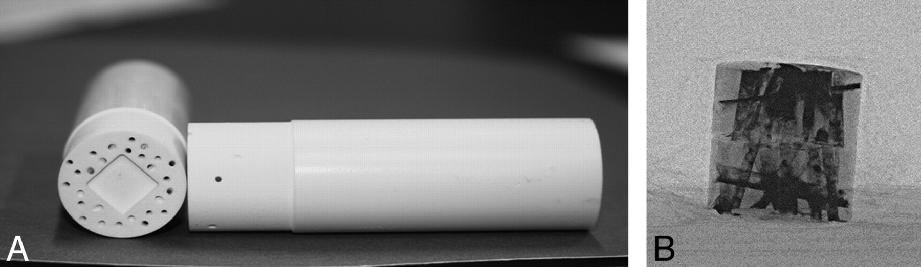
Two in vitro BAVM models were constructed by drilling interconnected tubular perforations to simulate nidal vessels in plastic water-calibration phantoms (plastic water, CIRS, Norfolk, Virginia) (Fig 1A). Models were configured to permit positioning of a water-equivalent detector (Gafchromic EBT film; ISP, Wayne, New Jersey) in the center of the phantom.

Fig 1.
  • Download figure
  • Open in new tab
  • Download powerpoint
Fig 1.

A, Cylindric AVM model: a plastic water phantom with a tubular perforation (model A). B, X-ray radiography of the model B showing the radiopacity of the embolic agent. The film is positioned in the center of the phantom.

The first model (A), measuring 2 × 2 × 4 cm, with 1-mm-diameter channels was embolized with Onyx 18 (ev3, Irvine, California), a commonly used liquid embolic agent containing ethylene-vinyl alcohol copolymer, dimethyl-sulfoxide, and tantalum (Z = 73) added for radiopacity. Onyx is presented in a micronized powder form (35% weight/volume). The second model (B), measuring 4 × 4 × 8 cm, with 1- to 5-mm-diameter channels, was embolized by using various and multiple agents in greater quantity: n-BCA (Indermil; Henkel Loctite, Dusseldorf, Germany) with ethiodized oil (Lipiodol, Andre Guerbet, Aulnay-sous-Bois, France) and Onyx 18. We also added tungsten (Z = 74) to the embolic material to obtain a more important shielding effect. In clinical practice, Lipiodol (Z = 53 for iodine), tantalum, or tungsten is added to n-BCA and accounts for the shielding effect. Plain radiographs of the BAVM models were taken to ensure that radiopaque embolization material filled the channels (Fig 1B).

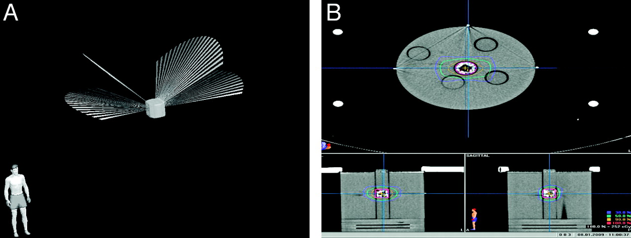
After appropriate positioning of the water-equivalent detector at the center of the phantom, we irradiated BAVM models with 250 cGy, by using a standard 4-arc stereotactic treatment plan on a LINAC (beam energy of 6 mV) (Fig 2A, -B) and compared measured radiation doses for the embolized models and the equivalent nonembolized controls. Each experiment was repeated twice.

Fig 2.
  • Download figure
  • Open in new tab
  • Download powerpoint
Fig 2.

A, Planification of standard treatment with 4 arcs of the AVM model A. B, Isodoses curves for AVM model A positioned in a brain phantom.

Results

The nonembolized model A controls had a mean dose of radiation delivered of 244 ± 5 cGy (247 and 240 cGy for individual experiments). Following embolization with Onyx 18, this model received a mean radiation dose of 246 ± 5 cGy (245 and 247 cGy). The limitations of measurement certainty of the film (±5 cGy) showed no definitive differences in the pre- and postembolization doses delivered to the model embolized with Onyx 18.

The model B control received a mean radiation dose of 242 ± 5 cGy (245 and 238 cGy) and 254.0 ± 5 cGy (256 and 252 cGy) after treatment of the model with multiple varied embolization agents in common clinical use. An unexpected slight increase in radiation dose following embolization was found for model B.

Discussion

Some nonrandomized cohort publications2–6 concluded that BAVM embolization before radiosurgery may have decreased the expected final rate of cure. One proposed reason was that high Z materials present in embolization products created a shielding effect responsible for the decrease in delivered dose. There have been supportive in vitro experiments to buttress this hypothesis: Andrade-Souza et al8 showed that a glue mixture (embucrilate + Lipiodol) was responsible for the downstream radiation reduction proportional to the Lipiodol concentration, but they only used a single beam. Against this hypothesis, Nath and Yue9 tested the shielding effect of metallic encapsulation and radiographic contrast agents for catheter-based intravascular brachytherapy, by using both photon- and β-emitting radionuclides, and showed that if the shielding effect was present when low-energy photon emitters were used, it became minimal for high-energy photons (range of energy used in radiation therapy). Wilczek et al10 tested the dose perturbation induced by different stents in brachytherapy and noticed that the shielding effect exists and depends on the strut thickness and mesh attenuation rather than on the atomic number of the metal. Shtraus et al11 found that the measured attenuation of Onyx placed in a bottle was 3% higher than the attenuation of water.

Light ion beams can also be used in radiosurgery, with clinically and radiobiologically interesting results, especially for large BAVMs.12 Measuring the dose pertubation by various high Z metallic devices used to shield critical structures, Das et al13 showed that electron beams gave a dual effect of dose reduction and dose enhancement, depending on the thickness and distance from the interface. Thin metal devices may enhance the dose on the transmission side and hence lose their shielding effect.

In our experiment, we did not find any attenuation of the photon beam in the center of our AVM model, either with Onyx alone or other high Z substances. The fact that photons are delivered through multiple arc beams and rebound of radiation on high Z material (backscatter effect) may explain the observed compensation for the theoretic shielding effect.

Without a randomized study, which would be very difficult to realize, it is impossible to prove that embolization has a negative effect on the outcome of BAVMs treated with radiosurgery. Nevertheless, if this effect exists, it can be explained by reasons other than a direct attenuation of the photon beam by high Z materials:

  • 1) Recanalization of the embolized nidus can decrease the cure rate.14 Natarajan et al15 reported a rate of 14.3% of recanalization with Onyx at the time of the 6-month follow-up examination.

  • 2) AVMs present a high angiogenic activity,16 which can be attenuated after radiation therapy.17 Embolization may induce hypoxia, which makes the tissue less radiosensitive and increases the angiogenic activity of the BAVM,18,19 rendering it an active instead of a static lesion.20,21 Moreover, it has been shown that hypoxia-inductible factor and vascular endothelial growth factor are expressed more frequently in embolized than in nonembolized BAVMs18 in surgical specimens.

  • 3) A good definition of the key target volume in radiosurgery is essential.22 CT and some MR imaging sequences may be useless when artifacts generated by embolic agents are substantial. As a consequence, definition of the margins of the nidus is at best an approximation, and the dose delivered to the BAVM margin is imprecise.11

Conclusions

Embolization material did not reduce the radiation dose delivered by a LINAC to the center of our experimental AVM models. The theoretic shielding effect from high Z materials used in embolization products is most likely compensated by backscatter effects and by multiple entry points of x-ray beams in current clinical use. If preradiosurgical embolization reduces the obliteration rate of the BAVMs, it may occur through other mechanisms.

Footnotes

  • Disclosures: Daniel Roy—Research Support (including provision of equipment or materials): Stryker/Micrus, Details: Approximately $20,000/year from each company for research support; Other Financial Relationships: Micrus, Details: $40,000 for academic support. Alain Weill—Research Support (including provision of equipment or materials): Micrus, ev3, Details: Micrus gives financial support for our academic fund (dedicated to research and salary of fellows); for this article, ev3 provided the Onyx at no charge (they had no influence on the article), Consultant: Micrus, ev3, Details: I have a proctoring contract with these 2 companies.

References

  1. 1.↵
    1. Mohr JP
    . A randomized trial of unruptured brain arteriovenous malformations (ARUBA). Acta Neurochir Suppl 2008; 103: 3– 4
    CrossRef
  2. 2.↵
    1. Schlienger M,
    2. Atlan D,
    3. Lefkopoulos D,
    4. et al
    . Linac radiosurgery for cerebral arteriovenous malformations: results in 169 patients. Int J Radiat Oncol Biol Phys 2000; 46: 1135– 42
    CrossRefPubMed
  3. 3.↵
    1. Pollock BE,
    2. Flickinger JC,
    3. Lunsford LD,
    4. et al
    . Factors associated with successful arteriovenous malformation radiosurgery. Neurosurgery 1998; 42: 1239– 44, discussion 1244–47
    CrossRefPubMed
  4. 4.↵
    1. Miyawaki L,
    2. Dowd C,
    3. Wara W,
    4. et al
    . Five year results of LINAC radiosurgery for arteriovenous malformations: outcome for large AVMS. Int J Radiat Oncol Biol Phys 1999; 44: 1089– 106
    CrossRefPubMed
  5. 5.↵
    1. Back AG,
    2. Vollmer D,
    3. Zeck O,
    4. et al
    . Retrospective analysis of unstaged and staged Gamma Knife surgery with and without preceding embolization for the treatment of arteriovenous malformations. J Neurosurg 2008; 109(suppl): 57– 64
    CrossRefPubMed
  6. 6.↵
    1. Andrade-Souza YM,
    2. Ramani M,
    3. Scora D,
    4. et al
    . Embolization before radiosurgery reduces the obliteration rate of arteriovenous malformations. Neurosurgery 2007; 60: 443– 51, discussion 451–52
    PubMed
  7. 7.↵
    1. Schwartz M,
    2. Sixel K,
    3. Young C,
    4. et al
    . Prediction of obliteration of arteriovenous malformations after radiosurgery: the obliteration prediction index. Can J Neurol Sci 1997; 24: 106– 09
    PubMed
  8. 8.↵
    1. Andrade-Souza YM,
    2. Ramani M,
    3. Beachey DJ,
    4. et al
    . Liquid embolisation material reduces the delivered radiation dose: a physical experiment. Acta Neurochir (Wien) 2008; 150: 161– 64, discussion 164. Epub 2008 Jan 23
    CrossRefPubMed
  9. 9.↵
    1. Nath R,
    2. Yue N
    . Shielding effects of metallic encapsulations and radiographic contrast agents for catheter-based intravascular brachytherapy. Cardiovasc Radiat Med 2001; 2: 93– 103
    CrossRefPubMed
  10. 10.↵
    1. Wilczek K,
    2. Petelenz B,
    3. Strzala A,
    4. et al
    . Dose perturbation caused by stents: experiments with a model 90Sr/90Y source. Cardiovasc Intervent Radiol 2007; 30: 981– 91
    CrossRefPubMed
  11. 11.↵
    1. Shtraus N,
    2. Schifter D,
    3. Corn BW,
    4. et al
    . Radiosurgical treatment planning of AVM following embolization with Onyx: possible dosage error in treatment planning can be averted. J Neurooncol 2010; 98: 271– 76
    CrossRefPubMed
  12. 12.↵
    1. Andisheh B,
    2. Brahme A,
    3. Bitaraf MA,
    4. et al
    . Clinical and radiobiological advantages of single-dose stereotactic light-ion radiation therapy for large intracranial arteriovenous malformations: technical note. J Neurosurg 2009; 111: 919– 26
    CrossRefPubMed
  13. 13.↵
    1. Das IJ,
    2. Cheng CW,
    3. Mitra RK,
    4. et al
    . Transmission and dose perturbations with high-Z materials in clinical electron beams. Med Phys 2004; 31: 3213– 21
    CrossRefPubMed
  14. 14.↵
    1. Kwon Y,
    2. Jeon SR,
    3. Kim JH,
    4. et al
    . Analysis of the causes of treatment failure in gamma knife radiosurgery for intracranial arteriovenous malformations. J Neurosurg 2000; 93(suppl 3): 104– 06
    PubMed
  15. 15.↵
    1. Natarajan SK,
    2. Ghodke B,
    3. Britz GW,
    4. et al
    . Multimodality treatment of brain arteriovenous malformations with microsurgery after embolization with Onyx: single-center experience and technical nuances. Neurosurgery 2008; 62: 1213– 25, discussion 1225–26
    CrossRefPubMed
  16. 16.↵
    1. Konya D,
    2. Yildirim O,
    3. Kurtkaya O,
    4. et al
    . Testing the angiogenic potential of cerebrovascular malformations by use of a rat cornea model: usefulness and novel assessment of changes over time. Neurosurgery 2005; 56: 1339– 45, discussion 1345–36
    CrossRefPubMed
  17. 17.↵
    1. Kilic K,
    2. Konya D,
    3. Kurtkaya O,
    4. et al
    . Inhibition of angiogenesis induced by cerebral arteriovenous malformations using gamma knife irradiation. J Neurosurg 2007; 106: 463– 69
    CrossRefPubMed
  18. 18.↵
    1. Sure U,
    2. Battenberg E,
    3. Dempfle A,
    4. et al
    . Hypoxia-inducible factor and vascular endothelial growth factor are expressed more frequently in embolized than in nonembolized cerebral arteriovenous malformations. Neurosurgery 2004; 55: 663– 69, discussion 669–70
    CrossRefPubMed
  19. 19.↵
    1. Akakin A,
    2. Ozkan A,
    3. Akgun E,
    4. et al
    . Endovascular treatment increases but gamma knife radiosurgery decreases angiogenic activity of arteriovenous malformations: an in vivo experimental study using a rat cornea model. Neurosurgery 2010; 66: 121– 29, discussion 129–30
    CrossRefPubMed
  20. 20.↵
    1. Achrol AS,
    2. Guzman R,
    3. Varga M,
    4. et al
    . Pathogenesis and radiobiology of brain arteriovenous malformations: implications for risk stratification in natural history and posttreatment course. Neurosurg Focus 2009; 26: E9
    PubMed
  21. 21.↵
    1. Mullan S,
    2. Mojtahedi S,
    3. Johnson DL,
    4. et al
    . Embryological basis of some aspects of cerebral vascular fistulas and malformations. J Neurosurg 1996; 85: 1– 8
    PubMed
  22. 22.↵
    1. Valle RD,
    2. Zenteno M,
    3. Jaramillo J,
    4. et al
    . Definition of the key target volume in radiosurgical management of arteriovenous malformations: a new dynamic concept based on angiographic circulation time. J Neurosurg 2008; 109(suppl): 41– 50
    PubMed
  • Received March 3, 2011.
  • Accepted after revision April 25, 2011.
  • © 2012 by American Journal of Neuroradiology
PreviousNext
Back to top

In this issue

American Journal of Neuroradiology: 33 (2)
American Journal of Neuroradiology
Vol. 33, Issue 2
1 Feb 2012
  • Table of Contents
  • Index by author
Advertisement
Print
Download PDF
Email Article

Thank you for your interest in spreading the word on American Journal of Neuroradiology.

NOTE: We only request your email address so that the person you are recommending the page to knows that you wanted them to see it, and that it is not junk mail. We do not capture any email address.

Enter multiple addresses on separate lines or separate them with commas.
Liquid Embolization Material Reduces the Delivered Radiation Dose: Clinical Myth or Reality?
(Your Name) has sent you a message from American Journal of Neuroradiology
(Your Name) thought you would like to see the American Journal of Neuroradiology web site.
CAPTCHA
This question is for testing whether or not you are a human visitor and to prevent automated spam submissions.
Cite this article
F. Bing, R. Doucet, F. Lacroix, J.P. Bahary, T. Darsaut, D. Roy, F. Guilbert, J. Raymond, A. Weill
Liquid Embolization Material Reduces the Delivered Radiation Dose: Clinical Myth or Reality?
American Journal of Neuroradiology Feb 2012, 33 (2) 320-322; DOI: 10.3174/ajnr.A2943

Citation Manager Formats

  • BibTeX
  • Bookends
  • EasyBib
  • EndNote (tagged)
  • EndNote 8 (xml)
  • Medlars
  • Mendeley
  • Papers
  • RefWorks Tagged
  • Ref Manager
  • RIS
  • Zotero
0 Responses
Respond to this article
Share
Bookmark this article
Liquid Embolization Material Reduces the Delivered Radiation Dose: Clinical Myth or Reality?
F. Bing, R. Doucet, F. Lacroix, J.P. Bahary, T. Darsaut, D. Roy, F. Guilbert, J. Raymond, A. Weill
American Journal of Neuroradiology Feb 2012, 33 (2) 320-322; DOI: 10.3174/ajnr.A2943
del.icio.us logo Twitter logo Facebook logo Mendeley logo
  • Tweet Widget
  • Facebook Like
  • Google Plus One
Purchase

Jump to section

  • Article
    • Abstract
    • ABBREVIATIONS:
    • Materials and Methods
    • Results
    • Discussion
    • Conclusions
    • Footnotes
    • References
  • Figures & Data
  • Info & Metrics
  • Responses
  • References
  • PDF

Related Articles

  • No related articles found.
  • PubMed
  • Google Scholar

Cited By...

  • Beam hardening artifacts of liquid embolic agents: comparison between Squid and Onyx
  • Onyx embolization prior to stereotactic radiosurgery for brain arteriovenous malformations: a single-center treatment algorithm
  • Crossref (32)
  • Google Scholar

This article has been cited by the following articles in journals that are participating in Crossref Cited-by Linking.

  • Stereotactic radiosurgery for cerebral arteriovenous malformations: evaluation of long-term outcomes in a multicenter cohort
    Robert M. Starke, Hideyuki Kano, Dale Ding, John Y. K. Lee, David Mathieu, Jamie Whitesell, John T. Pierce, Paul P. Huang, Douglas Kondziolka, Chun-Po Yen, Caleb Feliciano, Rafael Rodgriguez-Mercado, Luis Almodovar, Daniel R. Pieper, Inga S. Grills, Danilo Silva, Mahmoud Abbassy, Symeon Missios, Gene H. Barnett, L. Dade Lunsford, Jason P. Sheehan
    Journal of Neurosurgery 2017 126 1
  • Embolization-induced angiogenesis in cerebral arteriovenous malformations
    Thomas J. Buell, Dale Ding, Robert M. Starke, R. Webster Crowley, Kenneth C. Liu
    Journal of Clinical Neuroscience 2014 21 11
  • Radiosurgery for ruptured intracranial arteriovenous malformations
    Dale Ding, Chun-Po Yen, Robert M. Starke, Zhiyuan Xu, Jason P. Sheehan
    Journal of Neurosurgery 2014 121 2
  • Radiosurgery for low-grade intracranial arteriovenous malformations
    Dale Ding, Chun-Po Yen, Zhiyuan Xu, Robert M. Starke, Jason P. Sheehan
    Journal of Neurosurgery 2014 121 2
  • International multicenter cohort study of pediatric brain arteriovenous malformations. Part 2: Outcomes after stereotactic radiosurgery
    Robert M. Starke, Dale Ding, Hideyuki Kano, David Mathieu, Paul P. Huang, Caleb Feliciano, Rafael Rodriguez-Mercado, Luis Almodovar, Inga S. Grills, Danilo Silva, Mahmoud Abbassy, Symeon Missios, Douglas Kondziolka, Gene H. Barnett, L. Dade Lunsford, Jason P. Sheehan
    Journal of Neurosurgery: Pediatrics 2017 19 2
  • Effect of Prior Hemorrhage on Intracranial Arteriovenous Malformation Radiosurgery Outcomes
    Dale Ding, Chun-Po Yen, Robert M. Starke, Zhiyuan Xu, Jason P. Sheehan
    Cerebrovascular Diseases 2015 39 1
  • Treatment of Brain AVMs (TOBAS): study protocol for a pragmatic randomized controlled trial
    Tim E. Darsaut, Elsa Magro, Jean-Christophe Gentric, André Lima Batista, Chiraz Chaalala, David Roberge, Michel W. Bojanowski, Alain Weill, Daniel Roy, Jean Raymond
    Trials 2015 16 1
  • Stereotactic radiosurgery alone or combined with embolization for brain arteriovenous malformations: a systematic review and meta-analysis
    Dylan Russell, Travis Peck, Dale Ding, Ching-Jen Chen, Davis G. Taylor, Robert M. Starke, Cheng-Chia Lee, Jason P. Sheehan
    Journal of Neurosurgery 2018 128 5
  • Stereotactic radiosurgery for Spetzler-Martin Grade III arteriovenous malformations: an international multicenter study
    Dale Ding, Robert M. Starke, Hideyuki Kano, John Y. K. Lee, David Mathieu, John Pierce, Paul P. Huang, Caleb Feliciano, Rafael Rodriguez-Mercado, Luis Almodovar, Inga S. Grills, Danilo Silva, Mahmoud Abbassy, Symeon Missios, Douglas Kondziolka, Gene H. Barnett, L. Dade Lunsford, Jason P. Sheehan
    Journal of Neurosurgery 2017 126 3
  • Stereotactic radiosurgery for arteriovenous malformations after Onyx embolization: a case-control study
    Cheng-Chia Lee, Ching-Jen Chen, Benjamin Ball, David Schlesinger, Zhiyuan Xu, Chun-Po Yen, Jason Sheehan
    Journal of Neurosurgery 2015 123 1

More in this TOC Section

  • Contour Neurovascular System: Five Year Follow Up
  • Effect of SARS-CoV2 on Endovascular Thrombectomy
  • Optimizing Voxel Size in 3D Rotational Angiography
Show more NEUROINTERVENTION

Similar Articles

Advertisement

Indexed Content

  • Current Issue
  • Accepted Manuscripts
  • Article Preview
  • Past Issues
  • Editorials
  • Editor's Choice
  • Fellows' Journal Club
  • Letters to the Editor
  • Video Articles

Cases

  • Case Collection
  • Archive - Case of the Week
  • Archive - Case of the Month
  • Archive - Classic Case

More from AJNR

  • Trainee Corner
  • Imaging Protocols
  • MRI Safety Corner
  • Book Reviews

Multimedia

  • AJNR Podcasts
  • AJNR Scantastics

Resources

  • Turnaround Time
  • Submit a Manuscript
  • Submit a Video Article
  • Submit an eLetter to the Editor/Response
  • Manuscript Submission Guidelines
  • Statistical Tips
  • Fast Publishing of Accepted Manuscripts
  • Graphical Abstract Preparation
  • Imaging Protocol Submission
  • Evidence-Based Medicine Level Guide
  • Publishing Checklists
  • Author Policies
  • Become a Reviewer/Academy of Reviewers
  • News and Updates

About Us

  • About AJNR
  • Editorial Board
  • Editorial Board Alumni
  • Alerts
  • Permissions
  • Not an AJNR Subscriber? Join Now
  • Advertise with Us
  • Librarian Resources
  • Feedback
  • Terms and Conditions
  • AJNR Editorial Board Alumni

American Society of Neuroradiology

  • Not an ASNR Member? Join Now

© 2025 by the American Society of Neuroradiology All rights, including for text and data mining, AI training, and similar technologies, are reserved.
Print ISSN: 0195-6108 Online ISSN: 1936-959X

Powered by HighWire