
Article Figures & Data
Figures
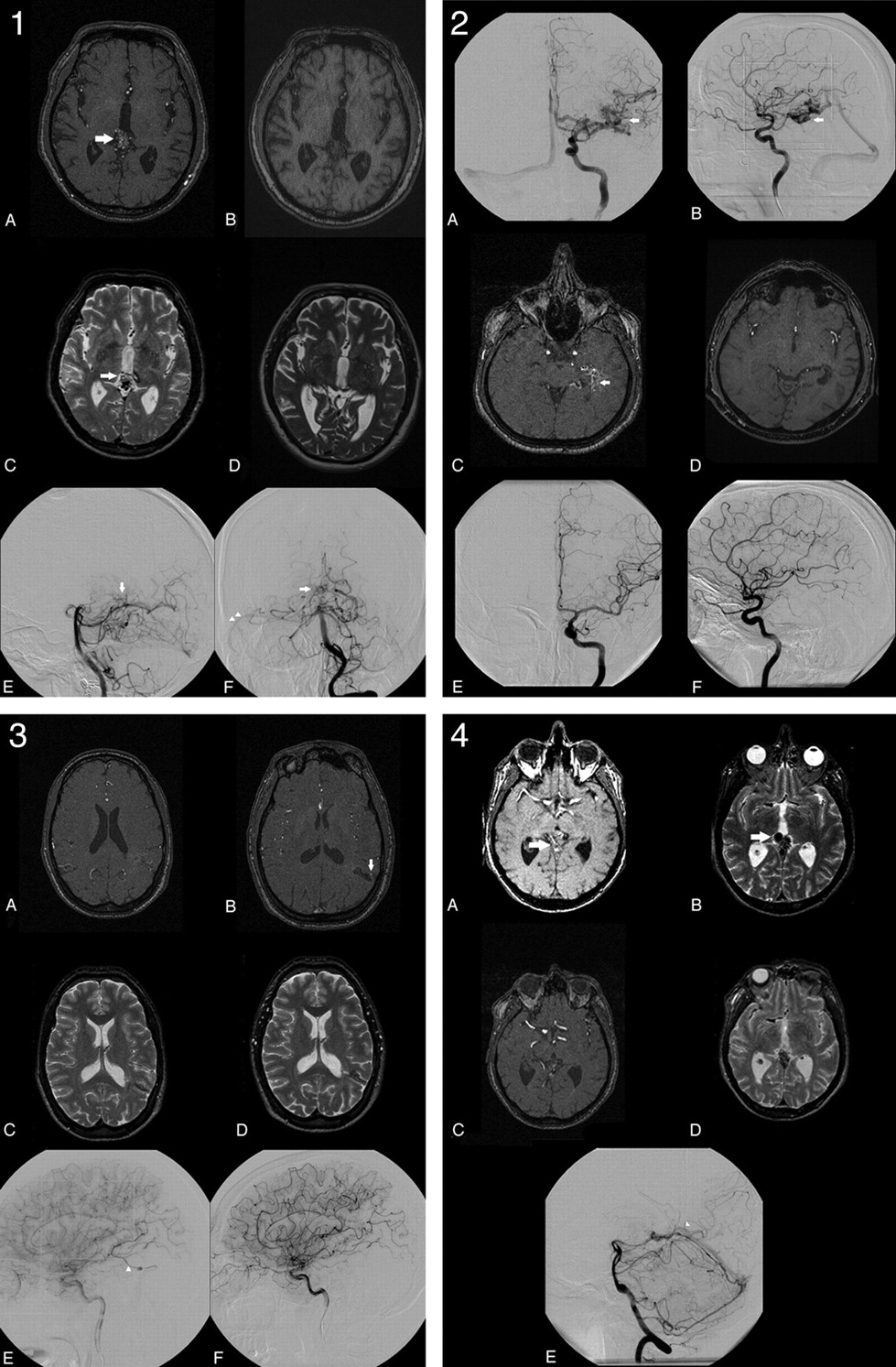
- Fig 1.
1, Overestimation of nidus obliteration: a 0.90-mL tectal bAVM, which is clearly visible on MR imaging1 (A and C white arrows). A−F, Both observers determined this bAVM to be obliterated on MR imaging2 (B and D) almost 3 years later. However DSA2, performed 6 weeks after MR imaging2, still demonstrates a small nidus (E and F, white arrows) with early venous drainage toward the lateral sinus (F, white arrowheads). 2, Successful determination of nidus obliteration in the case of a 3.68-mL-deep temporal bAVM (straight white arrow, A−C). On the basis of MR imaging2, both observers agreed that the nidus was obliterated 25 months after radiosurgery (D). Obliteration was angiographically confirmed 6 weeks later (E and F). 3, Underestimation of nidus obliteration: a small 0.17-mL central bAVM that was previously partially treated by endovascular embolization (A, C, and E). Because of hyperintensity on MR imaging2 (B, white vertical arrow), both observers judged this bAVM to be patent 1 year after radiosurgery (B and D). However DSA2 (F), performed 2 months after MR imaging2, demonstrates absence of the previously present early venous drainage (E, white arrowhead). 4, Successful determination of a remaining nidus in case of a patent 2.32-mL tectal bAVM (white horizontal arrow, A and B). On the basis of MR imaging2, both observers agreed that the nidus was patent 4 years after radiosurgery (C and D). The presence of a remnant nidus was angiographically confirmed 6 weeks later, when early venous drainage toward the straight sinus was observed (white arrowhead, E).
- Fig 2.
ROC demonstrates an area under the ROC curve for predicting obliteration of 0.81–0.83 for each individual observer.


Tables
Location No. (%) Presentation No. (%) Frontal 25 (20.8) Seizures 40 (33.3) Temporal 30 (25.0) Parenchymal hemorrhage 40 (33.3) Parietal 22 (18.3) Subarachnoid hemorrhage 5 (4.2) Occipital 14 (11.7) Intraventricular hemorrhage 8 (6.7) Basal ganglia 1 (0.8) Focal neurologic deficit 4 (3.3) Thalamic 13 (10.8) Headache 13 (10.8) Cerebellum 9 (7.5) Screening 10 (8.3) Brain stem 1 (0.8) Periventricular 1 (0.8) Corpus callosum 4 (3.3) DSA2c MRI2 Observer 1 (n = 117) MRI2 Observer 2 (n = 117) Patent PO DO Patent PO DO Patent 32 1 6 36 2 2 Obliterated 15 15 48 21 14 42 47 16 54 57 16 44 Observer 1 Observer 2 Sensitivity 0.52 0.52 Specificity 0.89 0.95 PPV 0.85 0.95 NPV 0.62 0.55 Prevalence 0.54 0.62 False-positive rate 0.10 0.04 False-negative rate 0.48 0.48 Overestimation of Nidus Obliteration on MRI2 (n = 6 patients, 8 observations) Underestimation of Nidus Obliteration on MRI2 (n = 49 patients, 65 observations) Mean age (yr) 29.3 (14.0–44.7)a 37.4 (33.1–41.7)a bAVM volume (mL) 4.3 (0.2–8.5)a 3.3 (2.3–4.2)a bAVM score 1.10 (0.57–1.63)a 1.11 (0.98–1.25)a SM gradation (%) I 1 (17%) 9 (18.4%) II 0 21 (42.9%) III 4 (67%) 16 (32.7%) IV 1 (17%) 2 (4.1%) V 0 0 Unclassifiable N/A 1 (2.0%) Drainage (%) Superficial 2 (33%) 32 (65.3%) Deep 4 (67%) 16 (32.7%) Unclassifiable N/A 1 (2.0%) Previous embolization (%) 2 (33%) 30 (61.2%) Treatment dose (cGy) 1900 (1643–2157)a 1935 (1874–1996)a Location (%) Corpus callosum N/A 1 (2%) Cerebellum N/A 6 (12.2%) Frontal N/A 11 (22.4%) Occipital 2 (33%) 7 (14.3%) Parietal 2 (33%) 11 (22.4%) Temporal 1 (17%) 10 (20.4%) Thalamic 1 (17%) 3 (6.1%) Interval MRI2-DSA2 (mo) 7.1 (4.1–10.1)a 2.5 (0.5–4.6)a -
Note:—N/A, not applicable, SM, Spetzler-Martin grade.
-
↵a The ranges in parentheses refer to the 95% CI.
-
In this issue


Jump to section
Related Articles
Cited By...
- Diagnostic Performance of TOF, 4D MRA, Arterial Spin-Labeling, and Susceptibility-Weighted Angiography Sequences in the Post-Radiosurgery Monitoring of Brain AVMs
- Are Dynamic Arterial Spin-Labeling MRA and Time-Resolved Contrast-Enhanced MRA Suited for Confirmation of Obliteration following Gamma Knife Radiosurgery of Brain Arteriovenous Malformations?
- Follow-Up MRI for Small Brain AVMs Treated by Radiosurgery: Is Gadolinium Really Necessary?
- Early Hemodynamic Response Assessment of Stereotactic Radiosurgery for a Cerebral Arteriovenous Malformation Using 4D Flow MRI
- Management of Brain Arteriovenous Malformations: A Scientific Statement for Healthcare Professionals From the American Heart Association/American Stroke Association
- Fast Contrast-Enhanced 4D MRA and 4D Flow MRI Using Constrained Reconstruction (HYPRFlow): Potential Applications for Brain Arteriovenous Malformations
- Further Examination of Diagnostic Performance in the Context of a Fellows' Journal Club Article
- Reply: