
Article Figures & Data
Figures
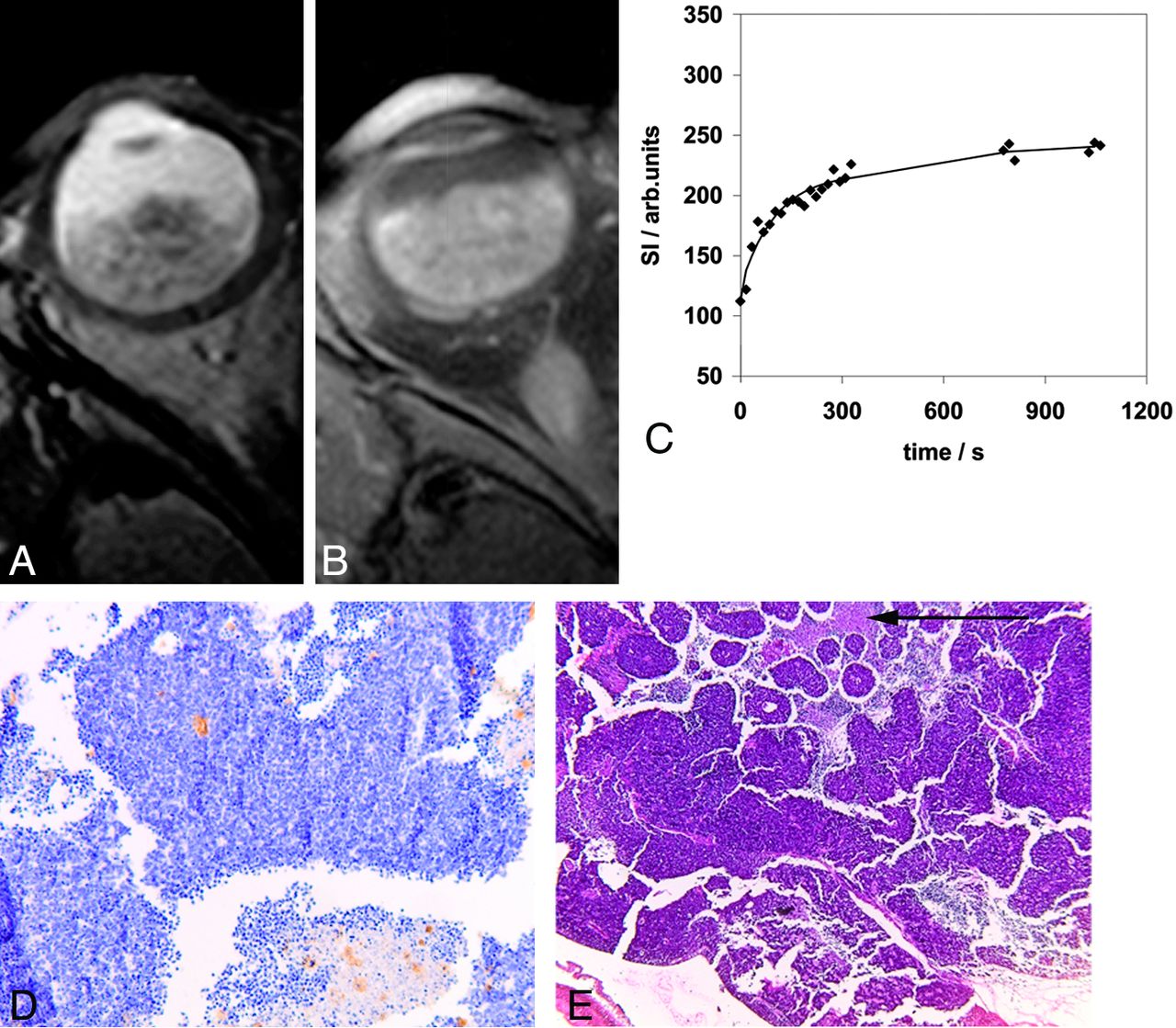
- Fig 1.
Retinoblastoma tumor in the right eye (patient 9) with transverse T2-weighted spin-echo (A) and transverse contrast-enhanced T1-weighted spin-echo (B) MR images. Curve-pattern analyses of the signal intensity curve of 1 observer (C) shows slow initial uptake of contrast (κ5min = 0.76) and slow further rising of the curve (κ17min = 1.06). Immunohistochemical staining with CD-31 (original magnification ×10) (D) shows brown-stained microvessels on a background of blue tumor cells with a MVD of 11. Hematoxilin-eosin staining (E) illustrates 30% necrotic areas (arrow).
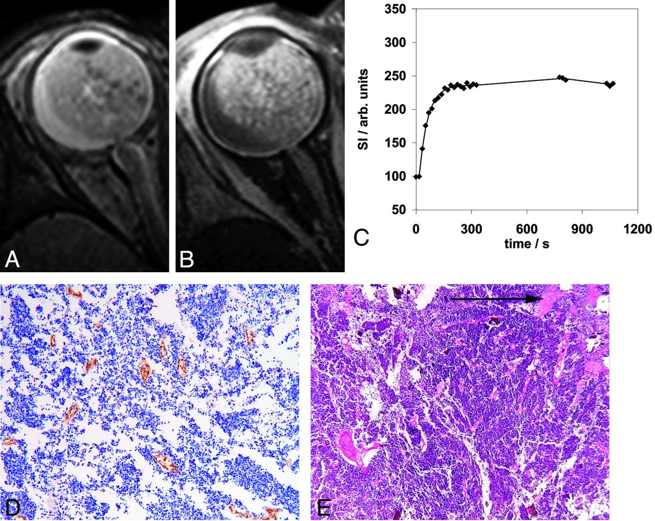
- Fig 2.
Retinoblastoma tumor in the right eye (patient 2) with transverse T2-weighted spin-echo (A) and transverse contrast-enhanced T1-weighted spin-echo (B) MR images. Curve-pattern analysis of the signal intensity curve of 1 observer (C) shows fast uptake of contrast agent (κ5min = 1.67) and early arrival at equilibrium (κ17min = 2.79). Immunohistochemical staining with CD-31 (original magnification ×10) (D) shows a high MVD of 21, and hematoxilin-eosin staining shows only 10% necrosis (arrow) (E).
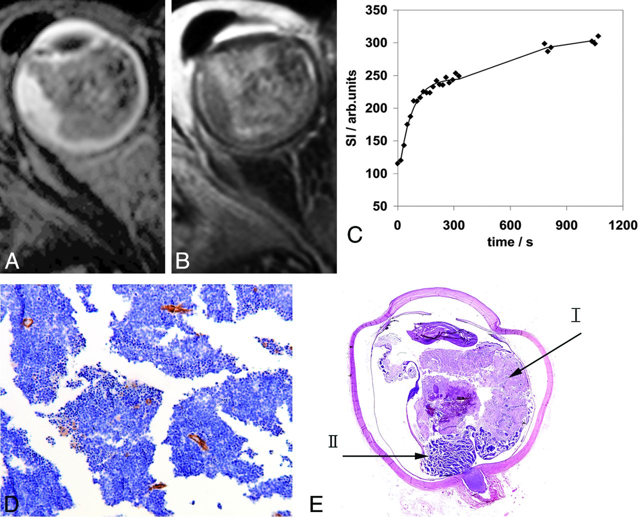
- Fig 3.
Retinoblastoma tumor in the right eye (patient 5) with transverse T2-weighted spin-echo (A) and transverse contrast-enhanced T1-weighted fat-suppressed spin-echo (B) MR images. Curve-pattern analysis of the signal intensity curve of 1 observer (C) shows a moderate uptake of contrast agent (κ5min = 1.22) and a continuing increase leading to κ17min = 0.84. Immunohistochemical staining with CD-31 (original magnification ×10) (D) shows an MVD of 14. Hematoxilin eosin staining (E) shows a large area of necrosis (70%) (arrow I) in vital tumor tissue (arrow II).
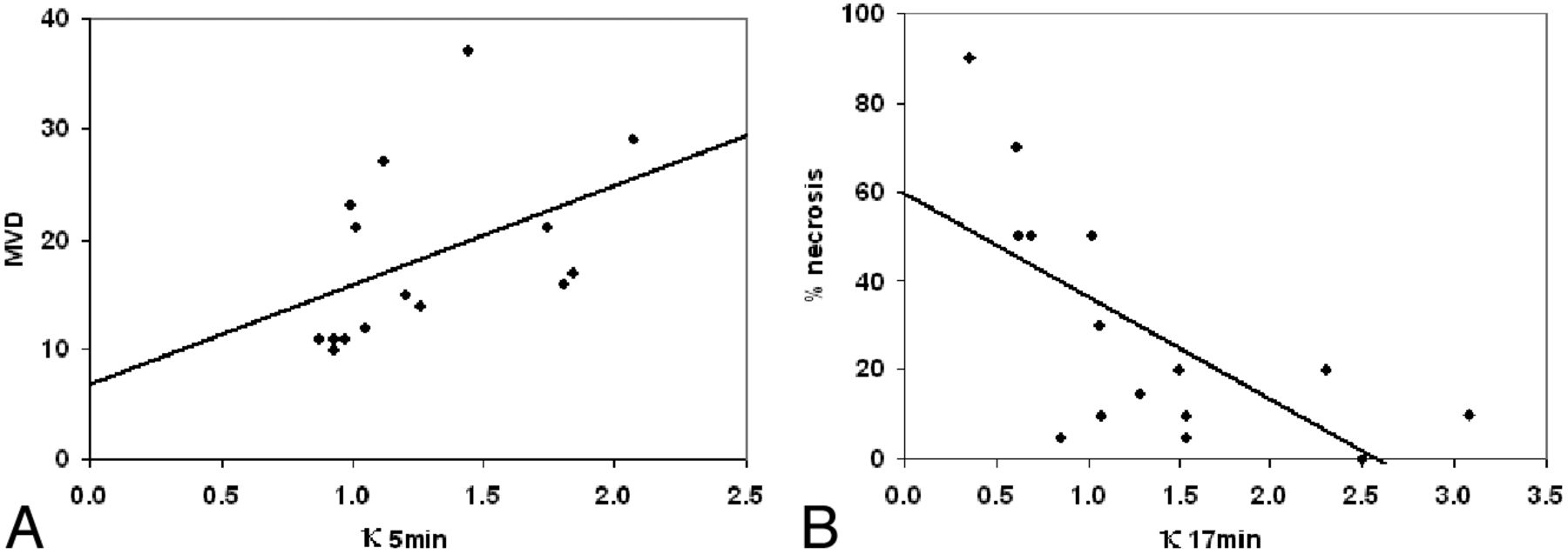
- Fig 4.
Graphs show the positive correlation (A) between κ5min and mean MVD (P = .008) and the negative correlation (B) between κ17min (a measure for late enhancement) and tumor necrosis (P = .002).


Tables
Patient Tumor Volume (mm3) κ5min κ17min MVD VEGF-Rb Flt-1 Optic Nerve Invasion Choroid Invasion Necrosis (%) 1 1690 2.07 1.53 29 Weak Weak No No 5 2 4782 1.74 3.08 21 Negative Positive Postlaminar No 10 3 656 1.01 0.35 21 Negative Negative (Pre)laminar No 90 4 2927 0.93 0.69 10 Weak Negative (Pre)laminar No 50 5 4727 1.26 0.61 14 Positive Negative Postlaminar Minimal 70 6 2664 0.93 1.49 11 Weak Weak No No 20 7 1955 1.44 2.50 37 Positive Negative (Pre)laminar No 0 8 2153 0.87 0.85 11 Positive Negative No No 5 9 2333 0.97 1.06 11 Weak Negative No No 30 10 960 0.99 1.53 23 Positive Negative Postlaminar No 10 11 1931 1.84 1.07 17 Positive Weak No No 10 12 4463 1.05 1.02 12 Positive Weak (Pre)laminar No 50 13 1325 1.20 1.28 15 Weak Weak (Pre)laminar No 15 14 288 1.80 2.30 16 Weak Negative No No 20 15 4847 1.12 0.62 27 Positive Negative (Pre)laminar No 50 Note:—VEGF-Rb indicates vascular endothelial growth factor in retinoblastoma tumor.


