
Article Figures & Data
Figures
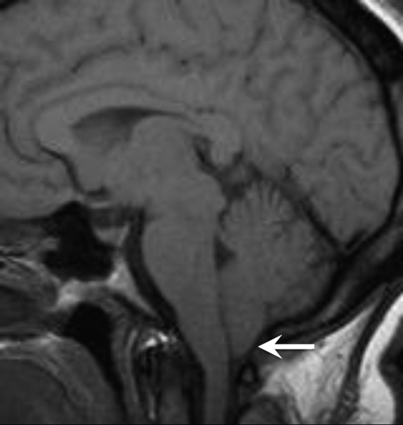
- Fig 1.
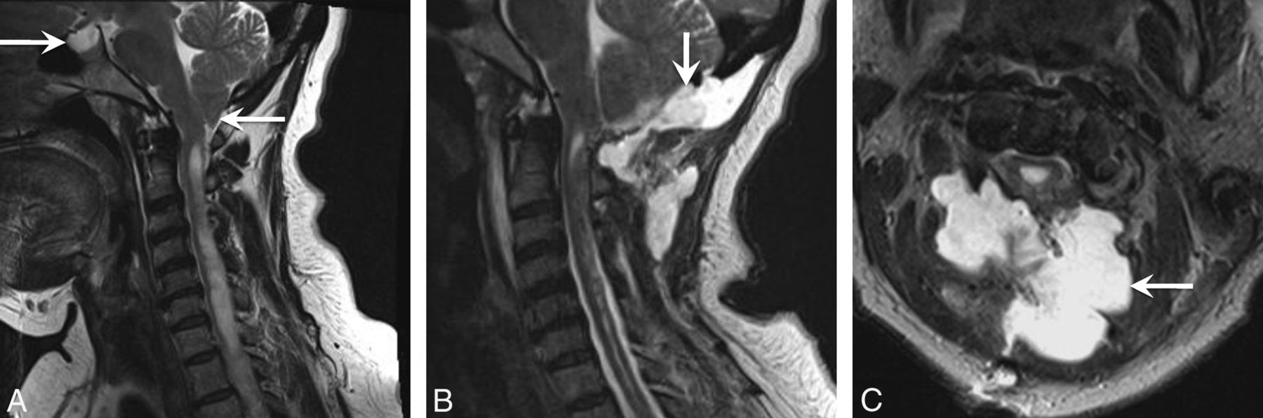
A 43-year-old woman initially diagnosed with Chiari I and treated with surgical decompression. The patient had persistent headaches and recurrent pseudomeningoceles at the surgical site. The patient was ultimately diagnosed with IIH and underwent ventriculoperitoneal shunt surgery with symptomatic relief. A, Sagittal T2-weighted image shows a peglike herniation of the cerebellar tonsils (arrow). However, a partially empty sella is also noted (arrow), which could have been a clue to the underlying or coexistent IIH. B and C, Sagittal and axial T2WI shows the postsurgical changes from suboccipital craniectomy and a complex extracranial fluid collection, compatible with pseudomeningocele (arrow).
- Fig 2.
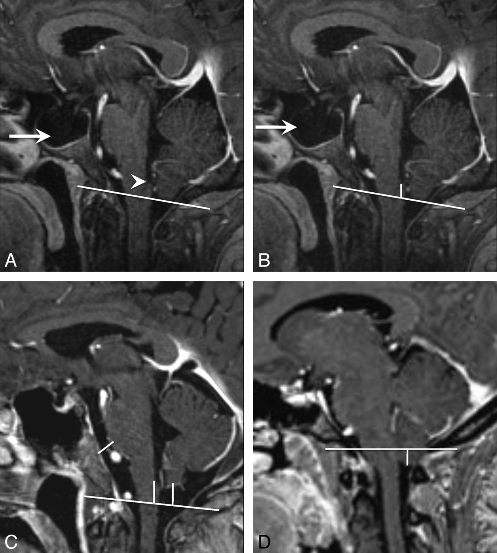
Cerebellar tonsil, obex position, and prepontine cistern width measurements. A, Sagittal T1-weighted image shows the foramen magnum reference line from the opisthion to the basion. Note the appearance of the obex (arrowhead) and the large empty sella in this patient with IIH (arrow). B, Sagittal T1WI shows the measurement from the obex to the foramen magnum reference line. Note that the obex was above the foramen magnum and would be assigned a negative number (−8 mm). C, Sagittal T1WI shows all 3 measurements in a healthy control. Note that the cerebellar tonsils are above the foramen magnum and would, therefore, be assigned a negative number (−9 mm for tonsil and obex positions). D, Sagittal T1WI shows the measurement from the cerebellar tonsil to the foramen magnum. Note that the cerebellar tonsils are below the foramen magnum and would therefore be assigned a positive number (5 mm).
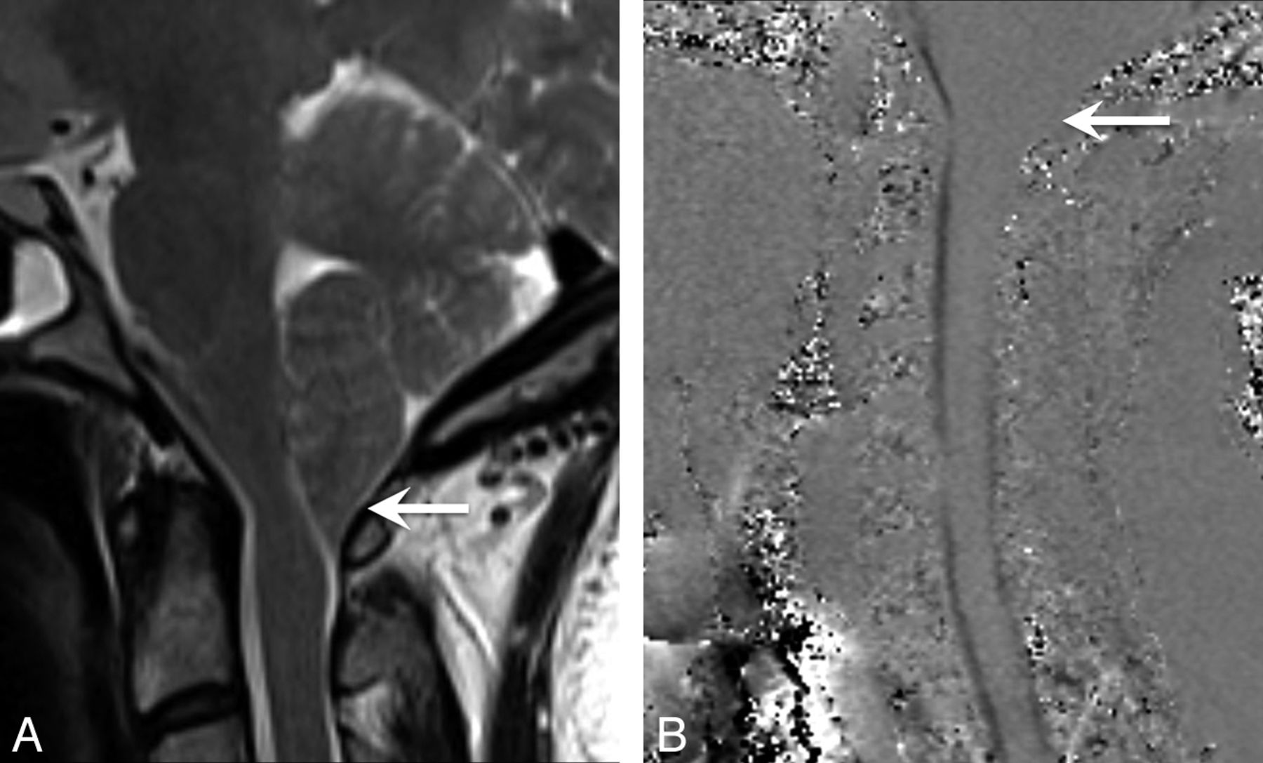
- Fig 3.
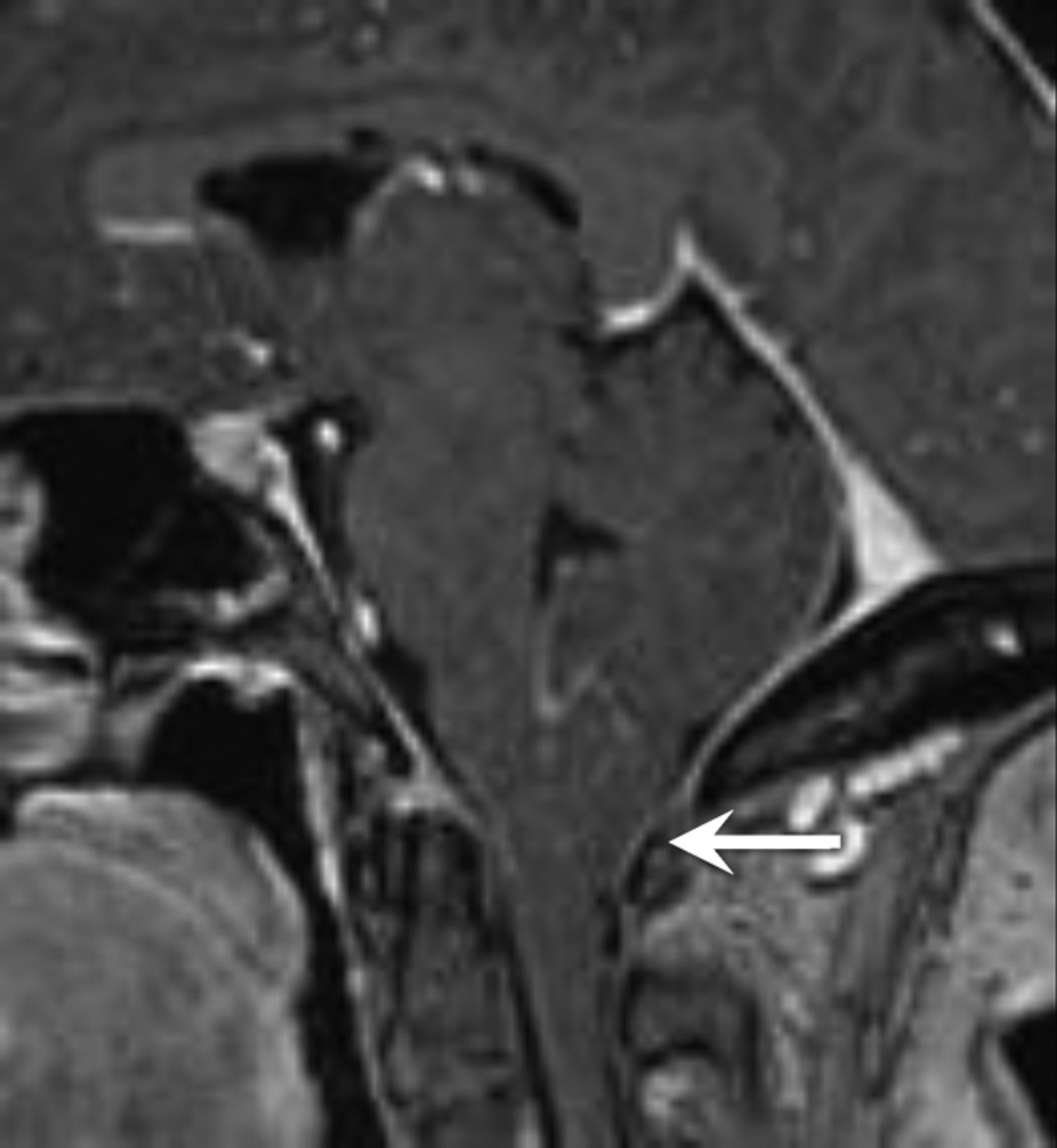
A patient with IIH with tonsillar ectopia of ≥5 mm. Sagittal T1-weighted image demonstrates herniation and a peglike configuration of the cerebellar tonsils below the foramen magnum.
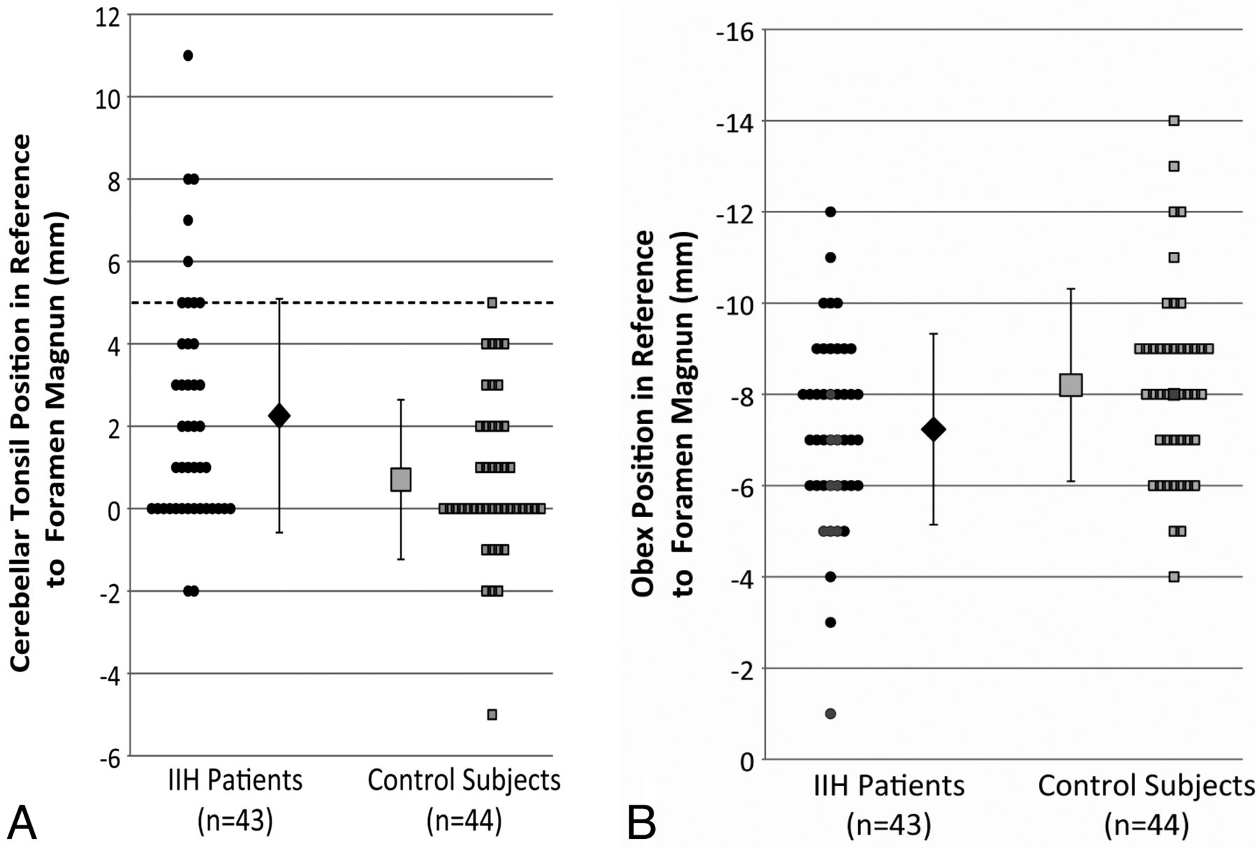
- Fig 4.
Differences in the tonsillar and obex positions between patients with IIH and healthy controls. Note that negative numbers are above the foramen magnum and positive numbers are below the foramen magnum. A, Differences in the tonsillar position between patients with IIH and control subjects. More than 20% of patients with IIH had cerebellar tonsillar ectopia of ≥5 mm (dotted line) (P < .05), the current criterion for radiologically diagnosed Chiari I malformation. B, The position of the obex relative to the foramen magnum was also significantly different (P < .01) between patients with IIH and control subjects. Black markers in B correspond to subjects with cerebellar tonsillar ectopia of ≥5 mm, indicating that it may be possible for additional discrimination between Chiari I and patients with IIH.
- Fig 5.
A patient with IIH with cerebellar tonsillar ectopia of ≥5 mm. This was the only patient with IIH who ultimately underwent surgical decompression after being managed for 12 years with CSF shunt surgery procedures alone. A, Sagittal T2-weighted image demonstrates herniation of the cerebellar tonsils with a peglike configuration and crowding at the foramen magnum. B, Cine CSF flow study shows loss of CSF flow posteriorly at the foramen magnum (arrow).
- Fig 6.
A patient with IIH with tonsillar ectopia of ≥5 mm. Sagittal T1-weighted image shows herniation and a peglike configuration of cerebellar tonsils (arrow).


Tables
In this issue


Jump to section
Related Articles
Cited By...
- Evolution of MRI Findings in Patients with Idiopathic Intracranial Hypertension after Venous Sinus Stenting
- Chiari malformations: principles of diagnosis and management
- Noninvasive Assessment of Intracranial Pressure Status in Idiopathic Intracranial Hypertension Using Displacement Encoding with Stimulated Echoes (DENSE) MRI: A Prospective Patient Study with Contemporaneous CSF Pressure Correlation
- Structural Brain Changes following Long-Term 6{degrees} Head-Down Tilt Bed Rest as an Analog for Spaceflight
- Chiari type 1 malformation in a pseudotumour cerebri patient: is it an acquired or congenital Chiari malformation?