
Article Figures & Data
Figures
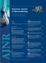
- Fig 1.
A 63-year-old man with nonspecific chronic lower back pain. A, MR imaging shows dilated radicular and perimedullary veins without myelopathy. B, Angiography reveals an SDAVF at the right L1 level. The fistula was surgically disconnected.
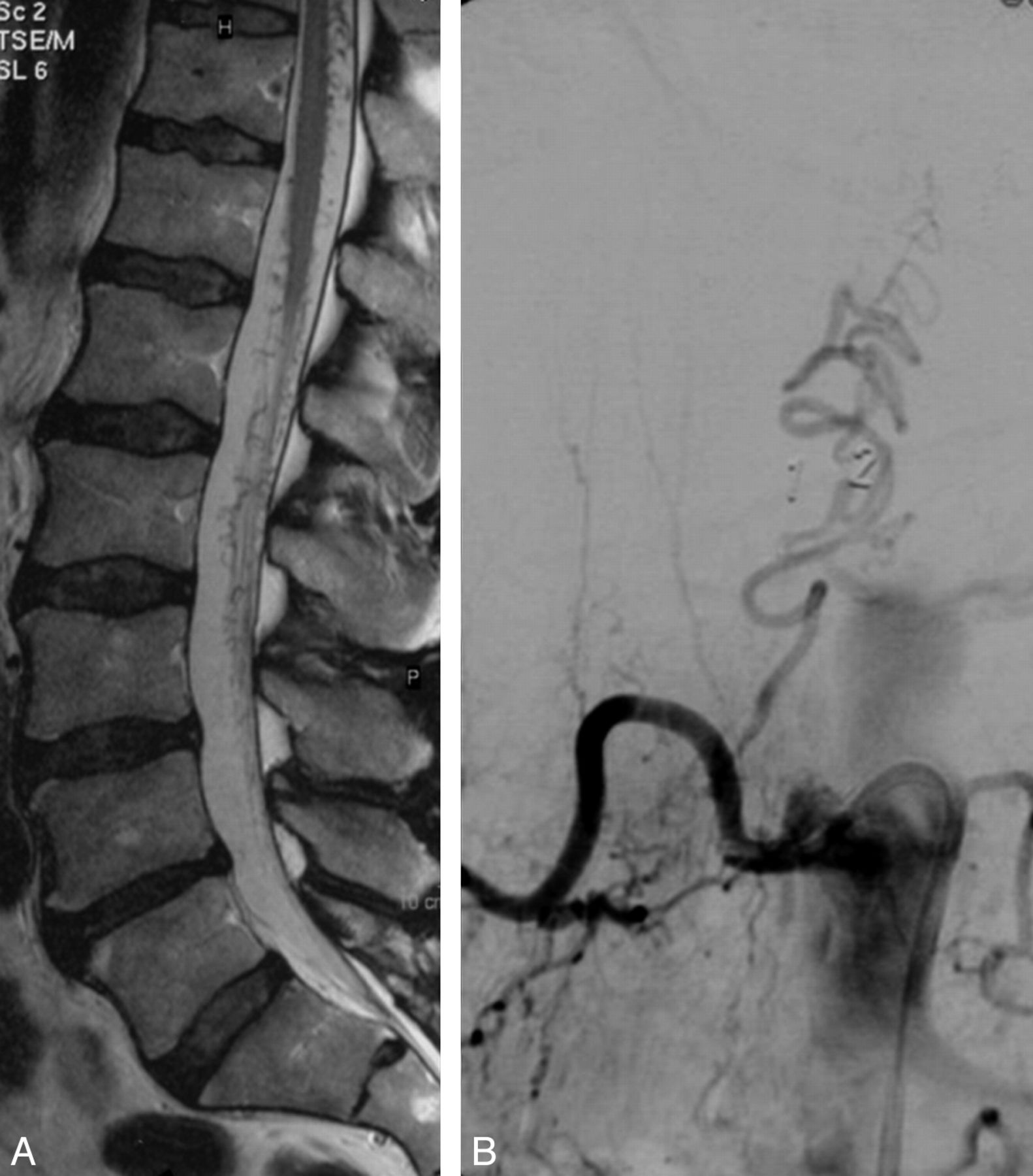
- Fig 2.
A 68-year-old man with nonspecific chronic back pain. A, T2-weighted MR imaging shows dilated radicular and perimedullary veins without swelling and edema of the conus. B, Anteroposterior view of a 3D right internal iliac angiogram confirms the presence of a high-flow SDAVF located at the right L5-S1 level. Note retrograde filling of both L5 lumbar arteries. C, MR imaging 3 months after embolization of the fistula no longer shows the dilated radicular and perimedullary veins. Note L4-L5 disk herniation.
- Fig 3.
A 41-year-old woman with Osler-Weber-Rendu disease and nonspecific back pain. A and B, T2-weighted MR imaging shows dilated perimedullary veins around the conus and lower cord without myelopathy. C, Angiography of the left L1 lumbar artery reveals an SDAVF with a high flow and perimedullary drainage in a cranial direction. Arrow indicates the transition point from artery to vein. The fistula was closed with acrylic glue. D, Follow-up MR imaging 3 months later confirms permanent closure of the SDAVF.
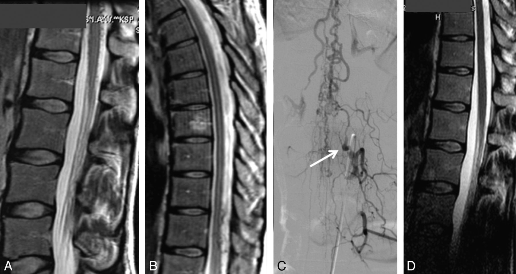
- Fig 4.
A 52-year-old woman with an incidental cervical SDAVF. A, CT angiography of the carotid arteries performed for suspected carotid stenosis shows dilated perimedullary veins at the C2-C3 level. B, MR imaging confirms the dilated perimedullary veins in the presence of a normal cervical cord. C and D, Anteroposterior view of a left vertebral angiogram in 2D (C) and 3D (B) demonstrates the SDAVF with drainage in cranial direction. E, Superselective angiogram through the microcatheter (arrow) just before glue injection under balloon protection of the vertebral artery. F, Complete obliteration of the cervical SDAVF.
- Fig 5.
A 71-year-old man with nonspecific lower back pain. A and B, T2-weighted MR imaging shows dilated perimedullary veins around the conus and lower cord without myelopathy. C, Spinal angiography shows an SDAVF at the right T8 level.


