Advertisement

Main menu

  • Home
  • Content
    • Current Issue
    • Accepted Manuscripts
    • Article Preview
    • Past Issue Archive
    • Video Articles
    • AJNR Case Collection
    • Case of the Week Archive
    • Case of the Month Archive
    • Classic Case Archive
  • Special Collections
    • AJNR Awards
    • Low-Field MRI
    • Alzheimer Disease
    • ASNR Foundation Special Collection
    • Photon-Counting CT
    • View All
  • Multimedia
    • AJNR Podcasts
    • AJNR SCANtastic
    • Trainee Corner
    • MRI Safety Corner
    • Imaging Protocols
  • For Authors
    • Submit a Manuscript
    • Submit a Video Article
    • Submit an eLetter to the Editor/Response
    • Manuscript Submission Guidelines
    • Statistical Tips
    • Fast Publishing of Accepted Manuscripts
    • Graphical Abstract Preparation
    • Imaging Protocol Submission
    • Author Policies
  • About Us
    • About AJNR
    • Editorial Board
    • Editorial Board Alumni
  • More
    • Become a Reviewer/Academy of Reviewers
    • Subscribers
    • Permissions
    • Alerts
    • Feedback
    • Advertisers
    • ASNR Home

User menu

  • Alerts
  • Log in

Search

  • Advanced search
American Journal of Neuroradiology

American Journal of Neuroradiology

ASHNR American Society of Functional Neuroradiology ASHNR American Society of Pediatric Neuroradiology ASSR
  • Alerts
  • Log in

Advanced Search

  • Home
  • Content
    • Current Issue
    • Accepted Manuscripts
    • Article Preview
    • Past Issue Archive
    • Video Articles
    • AJNR Case Collection
    • Case of the Week Archive
    • Case of the Month Archive
    • Classic Case Archive
  • Special Collections
    • AJNR Awards
    • Low-Field MRI
    • Alzheimer Disease
    • ASNR Foundation Special Collection
    • Photon-Counting CT
    • View All
  • Multimedia
    • AJNR Podcasts
    • AJNR SCANtastic
    • Trainee Corner
    • MRI Safety Corner
    • Imaging Protocols
  • For Authors
    • Submit a Manuscript
    • Submit a Video Article
    • Submit an eLetter to the Editor/Response
    • Manuscript Submission Guidelines
    • Statistical Tips
    • Fast Publishing of Accepted Manuscripts
    • Graphical Abstract Preparation
    • Imaging Protocol Submission
    • Author Policies
  • About Us
    • About AJNR
    • Editorial Board
    • Editorial Board Alumni
  • More
    • Become a Reviewer/Academy of Reviewers
    • Subscribers
    • Permissions
    • Alerts
    • Feedback
    • Advertisers
    • ASNR Home
  • Follow AJNR on Twitter
  • Visit AJNR on Facebook
  • Follow AJNR on Instagram
  • Join AJNR on LinkedIn
  • RSS Feeds

AJNR Awards, New Junior Editors, and more. Read the latest AJNR updates

Research ArticleSpine Imaging and Spine Image-Guided Interventions

Flat Panel Catheter Angiotomography of the Spinal Venous System: An Enhanced Venous Phase for Spinal Digital Subtraction Angiography

J. Chen, T. Ethiati and P. Gailloud
American Journal of Neuroradiology November 2012, 33 (10) 1875-1881; DOI: https://doi.org/10.3174/ajnr.A3111
J. Chen
aFrom the Division of Interventional Neuroradiology (J.C., T.E., P.G.), The Johns Hopkins Hospital, Baltimore, Maryland
  • Find this author on Google Scholar
  • Find this author on PubMed
  • Search for this author on this site
T. Ethiati
aFrom the Division of Interventional Neuroradiology (J.C., T.E., P.G.), The Johns Hopkins Hospital, Baltimore, Maryland
bSiemens Corporate Research (T.E.), Princeton, New Jersey.
  • Find this author on Google Scholar
  • Find this author on PubMed
  • Search for this author on this site
P. Gailloud
aFrom the Division of Interventional Neuroradiology (J.C., T.E., P.G.), The Johns Hopkins Hospital, Baltimore, Maryland
  • Find this author on Google Scholar
  • Find this author on PubMed
  • Search for this author on this site
  • Article
  • Figures & Data
  • Info & Metrics
  • Responses
  • References
  • PDF
Loading

Article Figures & Data

Figures

  • Fig 1.
    • Download figure
    • Open in new tab
    • Download powerpoint
    Fig 1.

    47-year-old woman investigated for progressive myelopathy. A, Spinal DSA, left T10 injection, arterial phase, documenting the artery of the lumbosacral enlargement (artery of Adamkiewicz). B, Spinal DSA, left T10 injection, venous phase, illustrating the typical appearance of the perimedullary venous system during spinal angiography performed under optimal conditions, that is, with a lean patient able to hold breath long enough to prevent motion artifacts. The anterior spinal venous axis is seen (arrowheads), as well as a medullary vein (short arrow) draining into the internal vertebral venous plexus (long arrow). C, FPCA, left T10 injection, sagittal MIP, voxel size = 0.4, section thickness = 1.8. The anterior and posterior perimedullary venous systems are easily differentiated. On FPCA, depending on the level of injection, overlapping may exist between arterial and venous structures related to the length of the rotational acquisition (20 seconds). D, FPCA, left T10 injection, axial MIP, voxel size = 0.1, section thickness = 4.0. The axial projection best characterizes this anastomosis, showing its course over the lateral of the spinal cord, connecting the anterior spinal vein (gray arrowhead) to a right posterior-lateral vein (white arrowhead), separate from the posterior spinal vein (black arrowhead). Because of their close topographic relationship and the thickness of the section (4.0 mm), the anterior spinal vein (gray arrowhead) and the anterior spinal artery are particularly difficult to separate in this image. The lack of dynamic information in FPCA can render the distinction between adjoining arteries and veins difficult; correct interpretation is helped by the analysis of the course and connection pattern of the vessels, the use of different reconstruction planes, or the correlation with the spinal DSA information.

  • Fig 2.
    • Download figure
    • Open in new tab
    • Download powerpoint
    Fig 2.

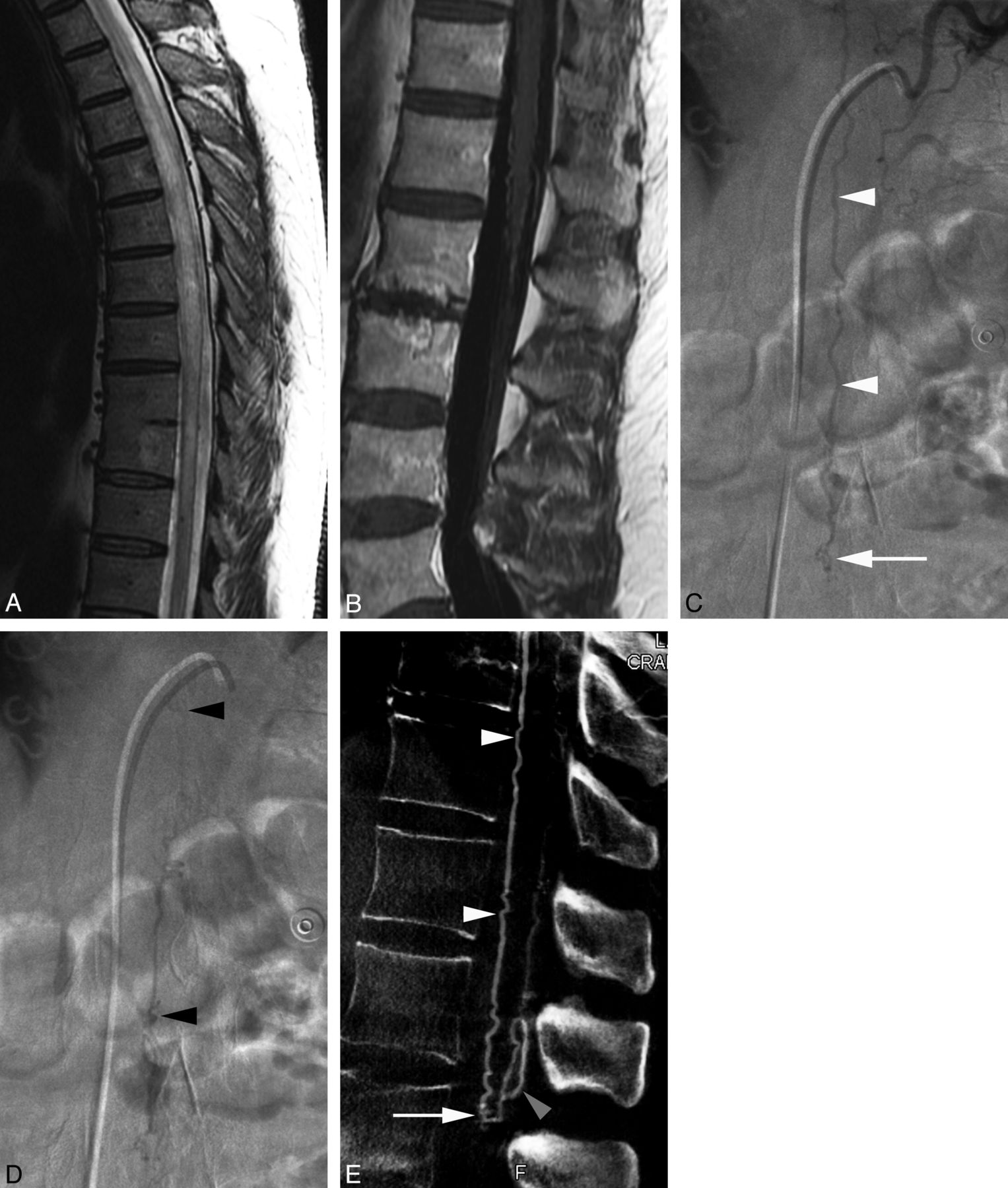
    57-year-old man with a perimedullary arteriovenous fistula of the conus medullaris. A, MR imaging, sagittal T2-weighted image of the thoracic spine, showing diffuse spinal cord swelling and hyperintensity; B, MR imaging, sagittal T1-weighted image after gadolinium administration of the lumbar spine. Note the presence of enhancing venous structures around the conus medullaris and along the filum terminale. C, Spinal DSA, left T10 injection, anteroposterior view, early arterial phase, showing a prominent descending ramus of the anterior spinal artery (white arrowheads) reaching the level of the conus medullaris, where a small tangle of blood vessels may be seen (arrow). D, Spinal DSA, left T10 injection, anteroposterior view. In the late arterial phase, while the vascular tangle is still visible, extensive opacification of the perimedullary venous system is noted (black arrowheads). E, FPCA, left T10 injection, sagittal MIP, voxel size = 0.4, section thickness = 3.0, demonstrating the anterior spinal artery (white arrowheads) ending at the tip of the conus medullaris (L1/L2) into a Type I perimedullary arteriovenous fistula (arrow). The posterior location of the enlarged draining veins detected by spinal DSA is well documented on this sagittal reconstruction (gray arrowhead).

  • Fig 3.
    • Download figure
    • Open in new tab
    • Download powerpoint
    Fig 3.

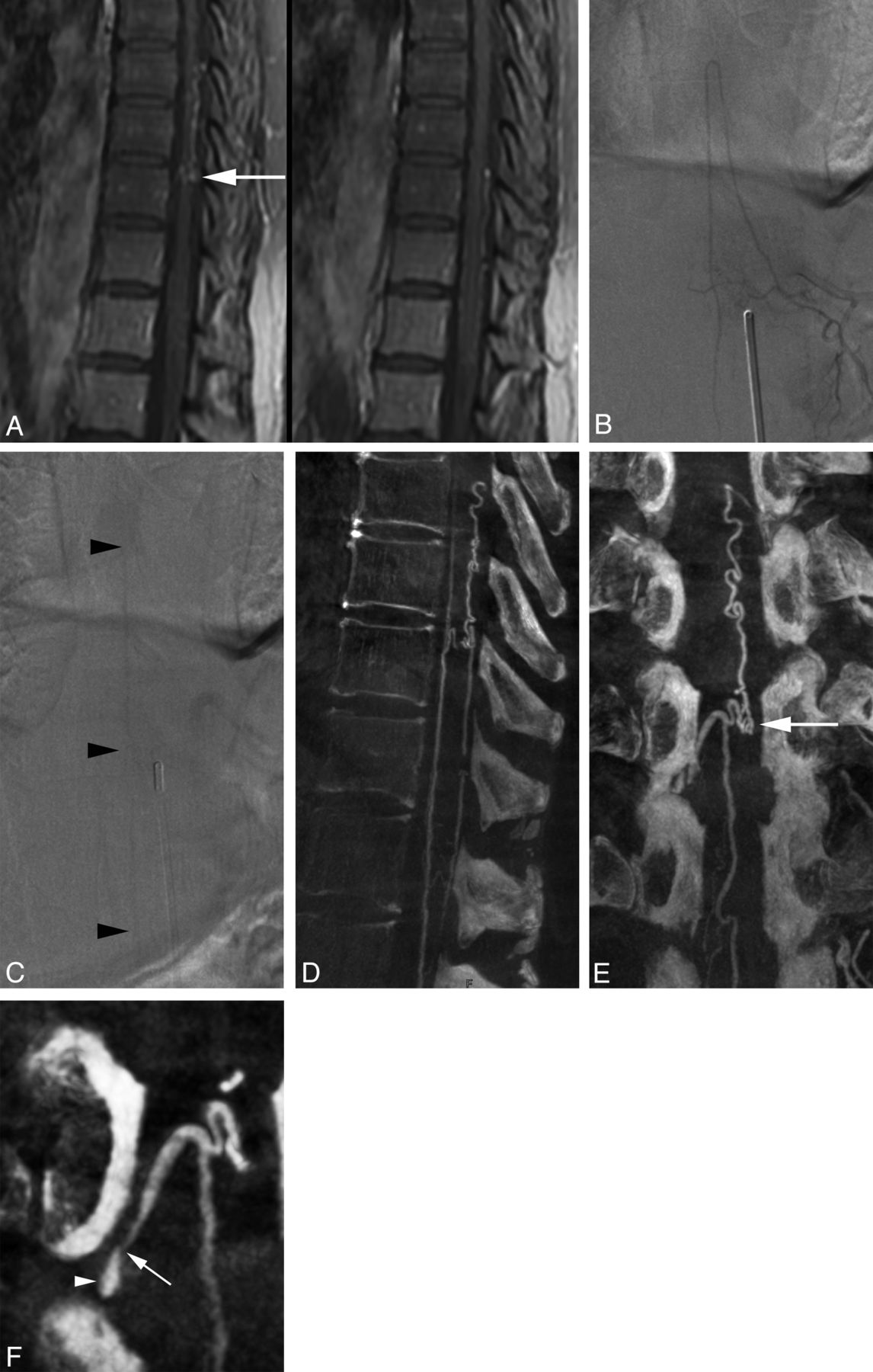
    58-year-old woman with a history of severe midscapular pain. A, MR imaging, sagittal T1-weighted images after gadolinium administration, showing prominent venous structures along the dorsal surface of the spinal cord between T8 and T12. A tangle of veins at the upper aspect of T10 was considered particularly suspicious for a vascular malformation (arrow). B, Spinal DSA, left T11 injection, anteroposterior view centered at T10/T11, arterial phase, documenting the artery of the lumbosacral enlargement (artery of Adamkiewicz). C, Spinal DSA, left T11 injection, anteroposterior view centered at T10/T11, venous phase. In this venous phase, the anterior spinal axis can be seen (arrowheads), without evidence of enlarged or abnormal veins. D, FPCA, left T11 injection, sagittal MIP, voxel size = 0.4, section thickness = 3.0, demonstrating the thoracolumbar perimedullary venous system, including the dorsal venous structures documented by MR imaging. E, FPCA, left T11 injection, coronal MIP, voxel size = 0.4, section thickness = 2.0. This coronal projection is an enlarged but otherwise unremarkable venous segment ending in a tangle of veins at the upper aspect of T10 (arrow), matching the suspicious structure seen by MR imaging (A, left). F, FPCA, left T10 injection, oblique MIP, voxel size = 0.1, section thickness = 1.0. This high-resolution reconstruction depicts the fine morphology of the right medullary vein exiting the dural sac at T10/T11. Note the narrowing of the vein as it pierces the dura (arrow) to join the internal vertebral venous plexus (epidural plexus; arrowhead).

PreviousNext
Back to top

In this issue

American Journal of Neuroradiology: 33 (10)
American Journal of Neuroradiology
Vol. 33, Issue 10
1 Nov 2012
  • Table of Contents
  • Index by author
Advertisement
Print
Download PDF
Email Article

Thank you for your interest in spreading the word on American Journal of Neuroradiology.

NOTE: We only request your email address so that the person you are recommending the page to knows that you wanted them to see it, and that it is not junk mail. We do not capture any email address.

Enter multiple addresses on separate lines or separate them with commas.
Flat Panel Catheter Angiotomography of the Spinal Venous System: An Enhanced Venous Phase for Spinal Digital Subtraction Angiography
(Your Name) has sent you a message from American Journal of Neuroradiology
(Your Name) thought you would like to see the American Journal of Neuroradiology web site.
CAPTCHA
This question is for testing whether or not you are a human visitor and to prevent automated spam submissions.
Cite this article
J. Chen, T. Ethiati, P. Gailloud
Flat Panel Catheter Angiotomography of the Spinal Venous System: An Enhanced Venous Phase for Spinal Digital Subtraction Angiography
American Journal of Neuroradiology Nov 2012, 33 (10) 1875-1881; DOI: 10.3174/ajnr.A3111

Citation Manager Formats

  • BibTeX
  • Bookends
  • EasyBib
  • EndNote (tagged)
  • EndNote 8 (xml)
  • Medlars
  • Mendeley
  • Papers
  • RefWorks Tagged
  • Ref Manager
  • RIS
  • Zotero
0 Responses
Respond to this article
Share
Bookmark this article
Flat Panel Catheter Angiotomography of the Spinal Venous System: An Enhanced Venous Phase for Spinal Digital Subtraction Angiography
J. Chen, T. Ethiati, P. Gailloud
American Journal of Neuroradiology Nov 2012, 33 (10) 1875-1881; DOI: 10.3174/ajnr.A3111
del.icio.us logo Twitter logo Facebook logo Mendeley logo
  • Tweet Widget
  • Facebook Like
  • Google Plus One
Purchase

Jump to section

  • Article
    • Abstract
    • ABBREVIATIONS:
    • Materials and Methods
    • Results
    • Discussion
    • Conclusions
    • Footnotes
    • References
  • Figures & Data
  • Info & Metrics
  • Responses
  • References
  • PDF

Related Articles

  • No related articles found.
  • PubMed
  • Google Scholar

Cited By...

  • Intersegmental artery dissection resulting in spinal infarction
  • Periconal arterial anastomotic circle and posterior lumbosacral watershed zone of the spinal cord
  • Transmedullary Venous Anastomoses: Anatomy and Angiographic Visualization Using Flat Panel Catheter Angiotomography
  • Crossref
  • Google Scholar

This article has not yet been cited by articles in journals that are participating in Crossref Cited-by Linking.

More in this TOC Section

  • Management Outcomes For VO Spine Biopsy
  • Characteristics of SIH Type I Culprit Lesions
  • Advanced Imaging of Type 2 Spinal CSF Leaks
Show more Spine Imaging and Spine Image-Guided Interventions

Similar Articles

Advertisement

Indexed Content

  • Current Issue
  • Accepted Manuscripts
  • Article Preview
  • Past Issues
  • Editorials
  • Editor's Choice
  • Fellows' Journal Club
  • Letters to the Editor
  • Video Articles

Cases

  • Case Collection
  • Archive - Case of the Week
  • Archive - Case of the Month
  • Archive - Classic Case

More from AJNR

  • Trainee Corner
  • Imaging Protocols
  • MRI Safety Corner
  • Book Reviews

Multimedia

  • AJNR Podcasts
  • AJNR Scantastics

Resources

  • Turnaround Time
  • Submit a Manuscript
  • Submit a Video Article
  • Submit an eLetter to the Editor/Response
  • Manuscript Submission Guidelines
  • Statistical Tips
  • Fast Publishing of Accepted Manuscripts
  • Graphical Abstract Preparation
  • Imaging Protocol Submission
  • Evidence-Based Medicine Level Guide
  • Publishing Checklists
  • Author Policies
  • Become a Reviewer/Academy of Reviewers
  • News and Updates

About Us

  • About AJNR
  • Editorial Board
  • Editorial Board Alumni
  • Alerts
  • Permissions
  • Not an AJNR Subscriber? Join Now
  • Advertise with Us
  • Librarian Resources
  • Feedback
  • Terms and Conditions
  • AJNR Editorial Board Alumni

American Society of Neuroradiology

  • Not an ASNR Member? Join Now

© 2025 by the American Society of Neuroradiology All rights, including for text and data mining, AI training, and similar technologies, are reserved.
Print ISSN: 0195-6108 Online ISSN: 1936-959X

Powered by HighWire