
Article Figures & Data
Figures
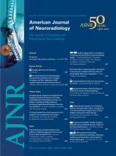
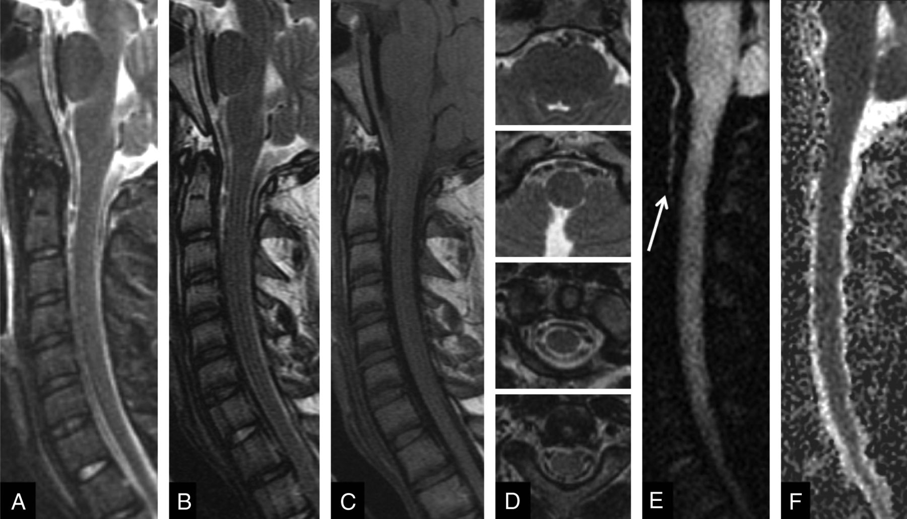
- Fig 1.
A 63-year-old man with a remote history of throat cancer and prior neck radiation, with acute onset of quadriparesis. The focal area of T2 hyperintensity (white arrows on all images) is identified at C4-C5 on the sagittal T2-weighted image (A) and STIR image (B), with no enhancement on the postgadolinium T1-weighted image (C). Increased marrow signal is present on the T2-weighted image (white arrowheads), consistent with a history of prior neck radiation. Initial diagnostic considerations might include transverse myelitis, radiation myelitis, spinal cord infarct, acute demyelination, neoplasm, and infection. On review of the sagittal rFOV DWI (D) and the corresponding ADC map (E) demonstrating reduced diffusion, readers were more confident in the diagnosis of acute cord infarct, and this was consistent with the patient's clinical course and diagnosis.
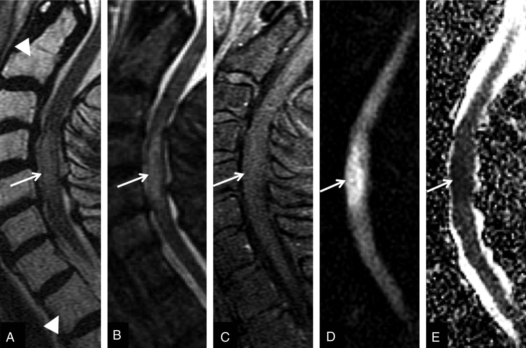
- Fig 2.
A 59-year-old woman with complex medical history including Crohn disease and pulmonary fibrosis who presented with lower extremity weakness and hyperreflexia. Three consecutive sagittal rFOV DWIs (A) demonstrate high signal in the cervical cord at the C2 level (arrowheads), confirmed centrally within the cord substance on an axial T2-weighted fast spin-echo image (C). Corresponding decreased ADC signal (not shown) and clinical presentation are most consistent with cord infarct. Additionally, an intradural extramedullary mass is noted on the last 2 sagittal rFOV DWIs (white arrows), which was difficult to appreciate on conventional sagittal (B) and axial (D) T2-weighted fast spin-echo images. While it was not biopsied, the high signal intensity of the mass on T2-weighted imaging suggests that this is likely a schwannoma.
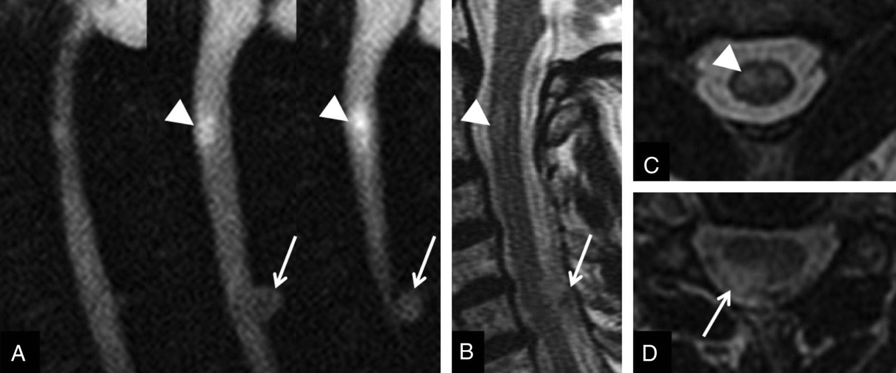
- Fig 3.
A 19-year-old woman with new-onset back pain. rFOV DWI (A), trace ADC map (B), T2 fast-spin-echo (C), and postgadolinium T1-weighted (D) sagittal image demonstrate a hypercellular process in multiple vertebral bodies (white arrows) and the epidural space (asterisks). The complete extent of vertebral body and posterior element involvement is better defined on the rFOV DWI compared with the conventional images. Biopsy demonstrated lymphoma.
- Fig 4.
A 17-year-old boy status post-assault, found to have multiple facial fractures. Sagittal anatomic images include STIR (A), T2 FSE (B), and spin-echo T1-weighted (C), and multiple axial FSE T2-weighted images (D). A retroclival and upper cervical subdural or epidural hematoma extending caudally to the C2-C3 level is far more conspicuous on the rFOV DWI (white arrow in E) than on the conventional images, where it mimics CSF flow artifact on the STIR and FSE T2-weighted images.
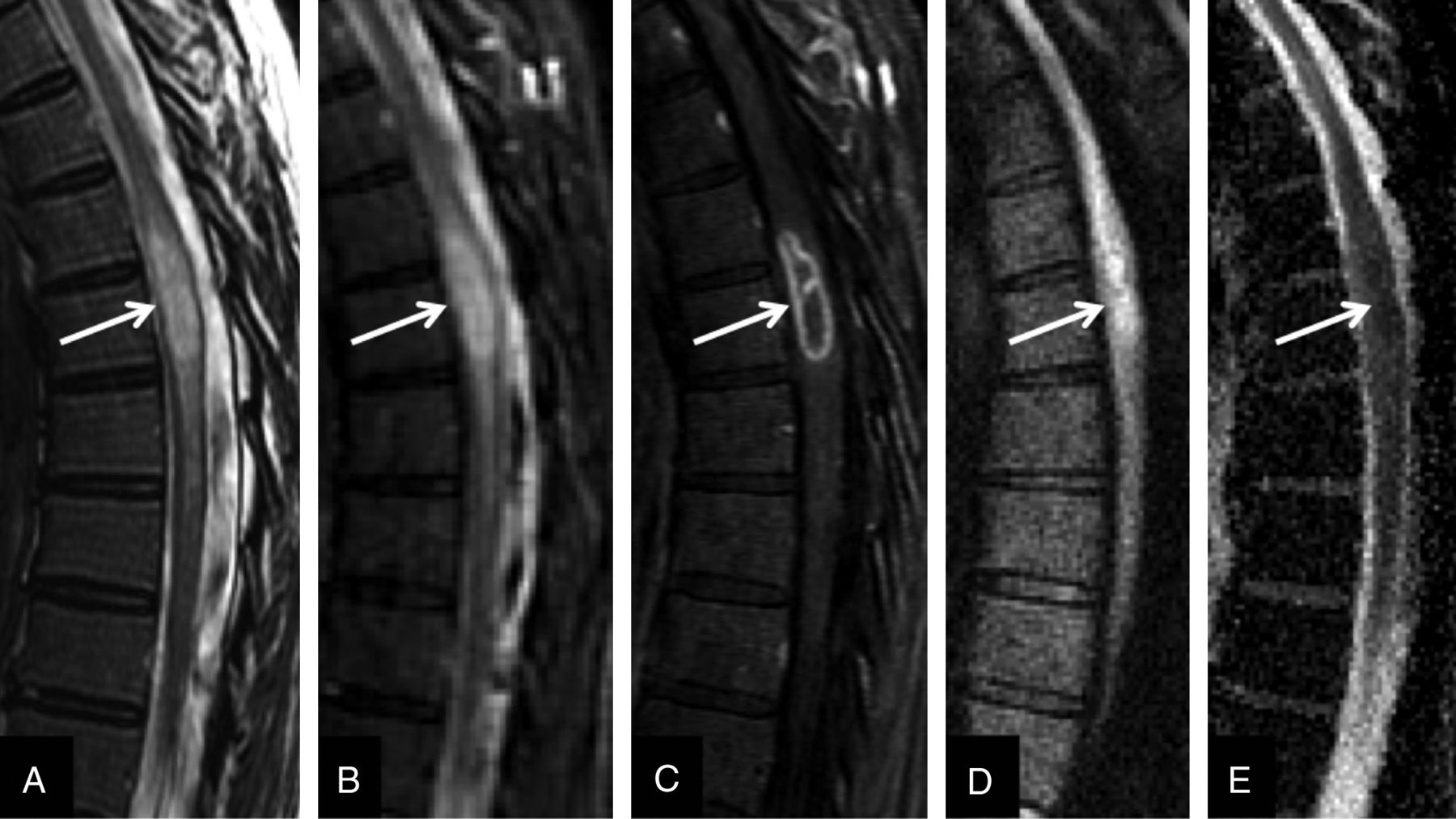
- Fig 5.
A 60-year-old woman with a history of chronic granulomatous disease and recent dental work presented to an outside hospital with left arm weakness and general malaise. An expansile focal area of T2 signal abnormality (white arrow on all images) is identified on the sagittal FSE T2-weighted image (A) and confirmed on STIR (B). Sagittal postgadolinium T1-weighted image (C) confirms a rim-enhancing intramedullary lesion, with a central nonenhancing portion. The rFOV DWI (D) demonstrates diffusion restriction centrally within this lesion, evidenced by low ADC (E), suggesting the presence of pus. This was interpreted as consistent with intramedullary abscess, and the patient responded well to antibiotics.


Tables
Spinal Pathology Total Cases Paired Wilcoxon Test Increased Confidence with Added DWI (P value)a Trauma (extradural)b 28 .0005 Neoplasm (extradural)b 16 .0047 Infection (all locations)b 14 <.0001 Disk herniation 112 .1573 Mass (intradural extramedullary)b 4 .0455 Mass (intramedullary)b 4 .0253 Hemorrhage (intradural extramedullary)b 2 .0143 Hemorrhage (intramedullary) 2 .3173 Collection (intradural extramedullaryb 4 .0003 Demyelinationb 20 <.0001 Cord infarctionb 6 <.0001 Syrinx 3 .0833 Cord contusionb 23 <.0001 Vascular malformation 1 .1573 No radiologic evidence of pathology 48 N/A Reader Agreement Unweighted κ 95% CI 95% CI (P Value) Symmetry Test (P Value) Identify pathology 0.77 0.73–0.80 <.0001 <.0001a Confidence in pathology 0.57 0.50–0.64 <.0001 <.0001a Confidence in DWI 0.33 0.21–0.45 <.0001 .2145 Added value of DWI 0.77 0.70–0.83 <.0001 .0008a -
↵a Bowker symmetry test is statistically significant (implying overall higher scores by 1 reader compared with the other).
-


