Advertisement

Main menu

  • Home
  • Content
    • Current Issue
    • Accepted Manuscripts
    • Article Preview
    • Past Issue Archive
    • Video Articles
    • AJNR Case Collection
    • Case of the Week Archive
    • Case of the Month Archive
    • Classic Case Archive
  • Special Collections
    • AJNR Awards
    • Low-Field MRI
    • Alzheimer Disease
    • ASNR Foundation Special Collection
    • Photon-Counting CT
    • View All
  • Multimedia
    • AJNR Podcasts
    • AJNR SCANtastic
    • Trainee Corner
    • MRI Safety Corner
    • Imaging Protocols
  • For Authors
    • Submit a Manuscript
    • Submit a Video Article
    • Submit an eLetter to the Editor/Response
    • Manuscript Submission Guidelines
    • Statistical Tips
    • Fast Publishing of Accepted Manuscripts
    • Graphical Abstract Preparation
    • Imaging Protocol Submission
    • Author Policies
  • About Us
    • About AJNR
    • Editorial Board
    • Editorial Board Alumni
  • More
    • Become a Reviewer/Academy of Reviewers
    • Subscribers
    • Permissions
    • Alerts
    • Feedback
    • Advertisers
    • ASNR Home

User menu

  • Alerts
  • Log in

Search

  • Advanced search
American Journal of Neuroradiology

American Journal of Neuroradiology

ASHNR American Society of Functional Neuroradiology ASHNR American Society of Pediatric Neuroradiology ASSR
  • Alerts
  • Log in

Advanced Search

  • Home
  • Content
    • Current Issue
    • Accepted Manuscripts
    • Article Preview
    • Past Issue Archive
    • Video Articles
    • AJNR Case Collection
    • Case of the Week Archive
    • Case of the Month Archive
    • Classic Case Archive
  • Special Collections
    • AJNR Awards
    • Low-Field MRI
    • Alzheimer Disease
    • ASNR Foundation Special Collection
    • Photon-Counting CT
    • View All
  • Multimedia
    • AJNR Podcasts
    • AJNR SCANtastic
    • Trainee Corner
    • MRI Safety Corner
    • Imaging Protocols
  • For Authors
    • Submit a Manuscript
    • Submit a Video Article
    • Submit an eLetter to the Editor/Response
    • Manuscript Submission Guidelines
    • Statistical Tips
    • Fast Publishing of Accepted Manuscripts
    • Graphical Abstract Preparation
    • Imaging Protocol Submission
    • Author Policies
  • About Us
    • About AJNR
    • Editorial Board
    • Editorial Board Alumni
  • More
    • Become a Reviewer/Academy of Reviewers
    • Subscribers
    • Permissions
    • Alerts
    • Feedback
    • Advertisers
    • ASNR Home
  • Follow AJNR on Twitter
  • Visit AJNR on Facebook
  • Follow AJNR on Instagram
  • Join AJNR on LinkedIn
  • RSS Feeds

AJNR Awards, New Junior Editors, and more. Read the latest AJNR updates

Case ReportCase Report

Dedifferentiated Liposarcoma of the Neck: CT Findings

Y. Wang and H. Shi
American Journal of Neuroradiology January 2012, 33 (1) E4-E6; DOI: https://doi.org/10.3174/ajnr.A2431
Y. Wang
  • Find this author on Google Scholar
  • Find this author on PubMed
  • Search for this author on this site
H. Shi
  • Find this author on Google Scholar
  • Find this author on PubMed
  • Search for this author on this site
  • Article
  • Figures & Data
  • Info & Metrics
  • Responses
  • References
  • PDF
Loading

Abstract

SUMMARY: We report a case of DDL with an osteosarcomatous component affecting the right neck. CT showed a lipomatous region with thick septa, a low-attenuation high-water-content component, and a sporadic heterogeneous high-attenuation calcified area.,

Abbreviations

DDL
dedifferentiated liposarcoma
HE
hematoxylin-eosin
WDL
well-differentiated liposarcoma

Liposarcoma is one of the most common soft-tissue sarcomas. It is currently classified into 5 subtypes: well-differentiated, dedifferentiated, myxoid, pleomorphic, and mixed.1 DDL represents a biphasic neoplasm, with 1 component being a WDL and the other a nonadipose cellular sarcoma. The most common dedifferentiated component is undifferentiated pleomorphic sarcoma or fibrosarcoma. Dedifferentiation to osteosarcoma is rare, and only 8 cases, to our knowledge, have been described in the literature.2–9

We report a case of a young woman who had DDL of the neck with an osteosarcomatous component, to illustrate the imaging and pathologic findings.

Case Report

A 20-year-old woman presented with a painless mass in the right neck, which had been progressively enlarging for approximately 2 months. On physical examination, the right neck was markedly larger than the left. An oral examination revealed a submucosal bulging of the right lateral pharyngeal wall. The overlying mucosa appeared to be normal. The patient denied dysphagia or bucking (choking cough). The other physical examination findings were fairly unremarkable.

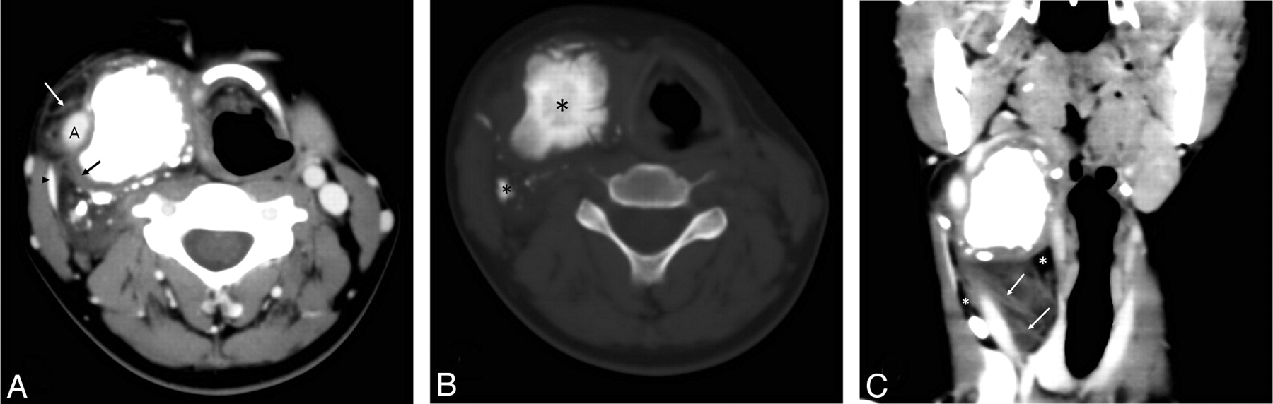
A contrast-enhanced CT scan of the head and neck revealed a large neck mass that had 3 components of differing attenuation, including a lipomatous region with thick septa, a low-attenuation high-water-content component, and a sporadic heterogeneous high-attenuation calcified area (Fig 1). After infusion of the contrast material, no enhancement was seen (Fig 1). The mass had well-defined margins between the sternocleidomastoideus muscle and the lateral pharyngeal wall (Fig 1). The mass surrounded the right common carotid artery and displaced the jugular vein laterally.

Fig 1.
  • Download figure
  • Open in new tab
  • Download powerpoint
Fig 1.

A, Contrast-enhanced axial CT scan shows lipomatous regions with thick septa (white arrow), a low-attenuation high-water-content component (black arrow), and sporadic heterogeneous high-attenuation calcified areas in the right common carotid space. The mass surrounds the right common carotid artery and displaces the jugular vein (black arrowhead) laterally. B, In this precontrast axial CT scan with a bone algorithm, the tumor ossification (black asterisks) is evident. C, A multiplanar reformatted image shows the mass with well-defined margins between the sternocleidomastoideus muscle and the lateral pharyngeal wall, displacing the right jugular vein. Thick septa (white arrows) and fat (white asterisks) are noted.

Fig 2.
  • Download figure
  • Open in new tab
  • Download powerpoint
Fig 2.

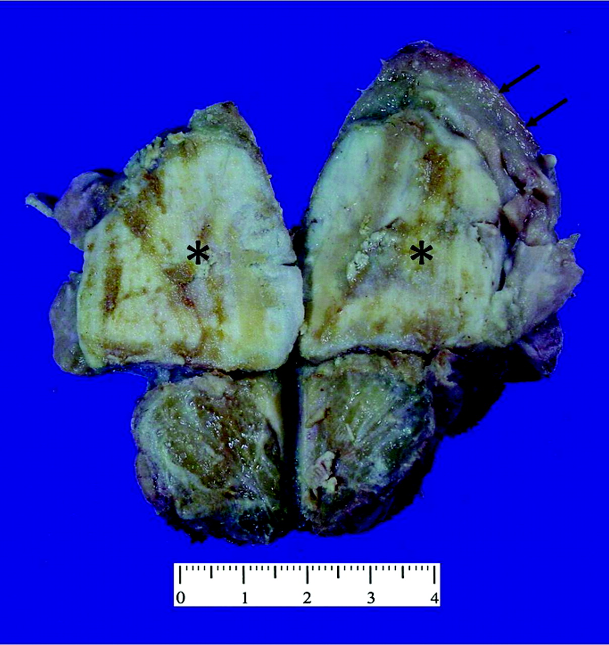
Photograph of the gross specimen shows the intramuscular sclerotic bone-forming lesion (black asterisks) with sharply delineated lipomatous caps (black arrows).

Fig 3.
  • Download figure
  • Open in new tab
  • Download powerpoint
Fig 3.

A, Photomicrographs of a biopsy specimen section obtained from the gritty nodular portion of the mass. Mineralized immature tumor bone trabeculae are present, and spindle-shaped tumor cells are seen adjacent to the tumor bone. The overall findings are consistent with osteosarcoma (HE, original magnification ×40). B, Photomicrographs of the lipoid portion. Mature-appearing adipose tissue combined with scattered lipoblasts (insert) (HE, original magnification ×100; insert, original magnification ×400).

The patient underwent a tumor resection via cervical approach. On gross inspection, a portion of the resected mass, measuring 5 × 5 × 4 cm, was gritty and nodular, with variegated tan-yellow to red-brown cut surfaces; another portion was lipoid tissue with approximately 8 cm of thickness, well-demarcated from the gritty nodular portion within the mass. Neither hemorrhage nor necrosis was identified. Microscopically, the nodular and gritty portion was composed of proliferating spindle-shaped cells that directly produced osteoid or immature tumor bone trabeculae. The lipoid area was characterized by mature-appearing adipose tissue, with only scattered lipoblasts. A diagnosis of a WDL juxtaposed to a high-grade osteosarcoma (ie, the DDL) was made. Five months after the initial surgery, the patient is disease-free without any signs of local recurrence or distant metastases.

Discussion

DDL occurs most frequently in patients in their seventh decade. Men and women are affected approximately equally.10 The areas of dedifferentiation are usually larger than 3 cm at imaging.10 The Armed Forces Institute of Pathology reported that 8.9% of all liposarcomas are dedifferentiated lesions, with 28.5% in the extremities, 66% in the retroperitoneum, and 6% in other locations.11

To the best of our knowledge, this is the ninth case of a DDL with osteosarcoma and the first case located in the neck reported in the literature.2–9

Radiographically, CT appearances of lipomatous tumors are based on the distribution of the fat and nonfat components, and patterns are classified into 3 types12: Type A tumors, homogeneous fatty tumors, are low-grade lipomatous neoplasms, including lipoma and lipomalike liposarcoma. Type B tumors, fat-attenuation tumors intermingled with higher attenuation components, are higher grade liposarcomas, including myxoid and mixed liposarcoma with pleomorphic or small cell types. Type C tumors, complex tumors containing both a fat component and a well-defined nonfatty component, are the typical pattern of DDL.12

In our case, osteosarcomatous components were approximately 5 cm. Calcification or ossification is a distinct imaging pattern of DDL. Osteosarcomatous and chondrosarcomatous dedifferentiation is the cause of such mineralization.

In the neck, the differential diagnosis for DDL with osteosarcomatous components and teratoma may be troublesome radiographically. CT of a teratoma demonstrates heterogeneous solid, fatty tissue, and cystic mass with specks of calcification.13 Other head and neck tumors that have fatty tissue include lipomas and lipoblastomas.

The clinical behavior of DDL, which reflects the high-grade histologic characteristics of most of these lesions, is more aggressive than well-differentiated liposarcomas. DDLs are treated with wide surgical excision and frequently with radiation therapy; chemotherapy may also be used as an adjunct. The disease is characterized by a tendency to recur locally in at least 40% of cases.1,14–19 Distant metastases are observed in 15%–20% of cases, with an overall mortality rate of 28%–30% at 5-year follow-up.3,20,21

Conclusions

We describe a case of DDL with an osteosarcomatous component in the neck. Radiologists should be familiar with the different location, and this case emphasizes the wide spectrum of DDL.

References

  1. 1.↵
    1. Fletcher CDM,
    2. Unni KK,
    3. Mertens F
    , for the International Agency for Research. Pathology and Genetics of Tumours of Soft Tissue and Bone: IARC WHO Classification of Tumours. Lyon, France: IARC Press; 2002:38–39
  2. 2.↵
    1. Toshiyasu T,
    2. Ehara S,
    3. Yamaguchi T,
    4. et al
    . Dedifferentiated liposarcoma of the retroperitoneum with osteosarcomatous component: report of two cases. Clin Imaging 2009;33:70–74
    CrossRefPubMed
  3. 3.↵
    1. Henricks WH,
    2. Chu YC,
    3. Goldblum JR,
    4. et al
    . Dedifferentiated liposarcoma: a clinicopathological analysis of 155 cases with a proposal for an expanded definition of dedifferentiation. Am J Surg Pathol 1997;21:271–81
    CrossRefPubMed
  4. 4.↵
    1. Toms AP,
    2. White LM,
    3. Kandle R,
    4. et al
    . Low-grade liposarcoma with osteosarcomatous dedifferentiation: radiological and histological features. Skeletal Radiol 2003;32:286–89
    PubMed
  5. 5.↵
    1. Evans HL
    . Liposarcoma: a study of 55 cases with a reassessment of its classification. Am J Surg Pathol 1979;3:507–23
    CrossRefPubMed
  6. 6.↵
    1. Ippolito V,
    2. Brien EW,
    3. Menedez LR,
    4. et al
    . Case report 797. Skeletal Radiol 1993;22:604–08
    PubMed
  7. 7.↵
    1. Yamamoto T,
    2. Matsushima T,
    3. Marui T,
    4. et al
    . Dedifferentiated liposarcoma with chondroblastic osteosarcomatous dedifferentiation. Pathol Int 2000;50:558–61
    CrossRefPubMed
  8. 8.↵
    1. Yu L,
    2. Jung S,
    3. Hojnowski L,
    4. et al
    . Dedifferentiated liposarcoma of soft tissue with high-grade osteosarcomatous dedifferentiation. Radiographics 2005;25:1082–86
    CrossRefPubMed
  9. 9.↵
    1. Orui H,
    2. Ishikawa A,
    3. Tsuchiya T,
    4. et al
    . Chondro-osseous differentiation in fat tissue tumors: magnetic resonance imaging with pathologic correlation. Skeletal Radiol 2000;29:459–65
    CrossRefPubMed
  10. 10.↵
    1. Murphey MD,
    2. Arcara LK,
    3. Fanburg-Smith J
    . Imaging of musculoskeletal liposarcoma with radiologic-pathologic correction. Radiographics 2005;25:1371–95
    CrossRefPubMed
  11. 11.↵
    1. Nascimento AG,
    2. Kurtin PJ,
    3. Guillou L,
    4. et al
    . Dedifferentiated liposarcoma: a report of nine cases with a peculiar neurallike whirling pattern associated with metaplastic bone formation. Am J Surg Pathol 1998;22:945–55
    CrossRefPubMed
  12. 12.↵
    1. Ehara S,
    2. Rosenberg AE,
    3. Kattapuram SV
    . Atypical lipomas, liposarcomas, and other fat-containing sarcomas: CT analysis of fat element. Clin Imaging 1995;19:50–53
    CrossRefPubMed
  13. 13.↵
    1. Robson CD
    . Imaging of head and neck neoplasms in children. Pediatr Radiol 2010;40:499–509
    CrossRefPubMed
  14. 14.↵
    1. Kransdorf MJ,
    2. Murphey M
    1. Kransdorf MJ,
    2. Murphey M
    . Liposarcoma. In: Kransdorf MJ, Murphey M. Imaging of Soft Tissue Tumors. Philadelphia: Saunders; 1997:79–98
  15. 15.↵
    1. Miettinen M
    . Liposarcoma. In: Diagnostic Soft Tissue Pathology. New York: Churchill Livingstone; 2003:227–40
  16. 16.↵
    1. Resnick D
    . Tumors and tumor-like lesions of bone: imaging and pathology of specific lesions. In: Diagnosis of Bone and Joint Disorders. 4th ed. Philadelphia: Saunders; 2002:4152–55
  17. 17.↵
    1. Kransdorf MJ
    . Malignant soft-tissue tumors in a large referral population: distribution of diagnoses by age, sex, and location. AJR Am J Roentgenol 1995;164:129–34
    CrossRefPubMed
  18. 18.↵
    1. Weiss S,
    2. Goldblum J
    . Liposarcoma. In: Enzinger and Weiss's Soft Tissue Tumors. 4th ed. St Louis: Mosby; 2001:641–93
  19. 19.↵
    1. Peterson JJ,
    2. Kransdorf MJ,
    3. Bancroft LW,
    4. et al
    . Malignant fatty tumors: classification, clinical course, imaging appearance and treatment. Skeletal Radiol 2003;32:493–503
    CrossRefPubMed
  20. 20.↵
    1. Weiss SW,
    2. Rao VK
    . Well-differentiated liposarcoma (atypical lipoma) of deep soft tissue of the extremities, retroperitoneum, and miscellaneous sites: a follow-up study of 92 cases with analysis of the incidence of “dedifferentiation.” Am J Surg Pathol 1992;16:1051–58
    CrossRefPubMed
  21. 21.↵
    1. McCormick D,
    2. Mentzel T,
    3. Beham A,
    4. et al
    . Dedifferentiated liposarcoma: clinicopathologic analysis of 32 cases suggesting a better prognostic subgroup among pleomorphic sarcomas. Am J Surg Pathol 1994;18:1213–23
    CrossRefPubMed
  • Received September 22, 2010.
  • Accepted after revision October 20, 2010.
  • © 2012 by American Journal of Neuroradiology
View Abstract
PreviousNext
Back to top

In this issue

American Journal of Neuroradiology: 33 (1)
American Journal of Neuroradiology
Vol. 33, Issue 1
1 Jan 2012
  • Table of Contents
  • Index by author
Advertisement
Print
Download PDF
Email Article

Thank you for your interest in spreading the word on American Journal of Neuroradiology.

NOTE: We only request your email address so that the person you are recommending the page to knows that you wanted them to see it, and that it is not junk mail. We do not capture any email address.

Enter multiple addresses on separate lines or separate them with commas.
Dedifferentiated Liposarcoma of the Neck: CT Findings
(Your Name) has sent you a message from American Journal of Neuroradiology
(Your Name) thought you would like to see the American Journal of Neuroradiology web site.
CAPTCHA
This question is for testing whether or not you are a human visitor and to prevent automated spam submissions.
Cite this article
Y. Wang, H. Shi
Dedifferentiated Liposarcoma of the Neck: CT Findings
American Journal of Neuroradiology Jan 2012, 33 (1) E4-E6; DOI: 10.3174/ajnr.A2431

Citation Manager Formats

  • BibTeX
  • Bookends
  • EasyBib
  • EndNote (tagged)
  • EndNote 8 (xml)
  • Medlars
  • Mendeley
  • Papers
  • RefWorks Tagged
  • Ref Manager
  • RIS
  • Zotero
0 Responses
Respond to this article
Share
Bookmark this article
Dedifferentiated Liposarcoma of the Neck: CT Findings
Y. Wang, H. Shi
American Journal of Neuroradiology Jan 2012, 33 (1) E4-E6; DOI: 10.3174/ajnr.A2431
del.icio.us logo Twitter logo Facebook logo Mendeley logo
  • Tweet Widget
  • Facebook Like
  • Google Plus One
Purchase

Jump to section

  • Article
    • Abstract
    • Abbreviations
    • Case Report
    • Discussion
    • Conclusions
    • References
  • Figures & Data
  • Info & Metrics
  • Responses
  • References
  • PDF

Related Articles

  • No related articles found.
  • PubMed
  • Google Scholar

Cited By...

  • No citing articles found.
  • Crossref (7)
  • Google Scholar

This article has been cited by the following articles in journals that are participating in Crossref Cited-by Linking.

  • From head and neck lipoma to liposarcoma: a wide spectrum of differential diagnoses and their therapeutic implications
    Marta Barisella, Lorenzo Giannini, Cesare Piazza
    Current Opinion in Otolaryngology & Head & Neck Surgery 2020 28 2
  • Fat: friend or foe? A review of fat-containing masses within the head and neck
    Hrishikesh A Kale, Arpan V Prabhu, Andrey Sinelnikov, Barton Branstetter
    The British Journal of Radiology 2016 89 1067
  • Liposarcoma of the retropharyngeal space with rapidly worsening dyspnea: A case report and review of the literature
    JIAN-GUO HE, HUA JIANG, BEI-BEI YANG, PENG-FANG LIN
    Oncology Letters 2013 5 6
  • Head and Neck Sarcoma Tumor Board Survival Guide for Neuroradiologists: Imaging Findings, History, and Pathology
    Daniel Warren, Cameron Koch, Matthew S. Parsons, Gloria J. Guzmán Pérez-Carrillo, Rami W. Eldaya
    Current Problems in Diagnostic Radiology 2023 52 4
  • Dedifferentiated Liposarcoma of the Deep (Paralaryngeal) Soft Tissue: Lessons Learnt from a Case with a Partly Deceptively Benign Appearing Dedifferentiated Component
    Fredrik Petersson, Euan Murugasu
    Head and Neck Pathology 2014 8 2
  • A case of dedifferentiated liposarcoma arising in the neck
    Junki Kobai, Kensuke Suzuki, Masao Yagi, Takuo Fujisawa, Tomofumi Sakagami, Minaki Shimizu, Yuri Noda, Hiroshi Iwai
    JOURNAL OF JAPAN SOCIETY FOR HEAD AND NECK SURGERY 2023 33 1
  • Dedifferentiated Liposarcoma of the Parotid Gland: A Rare Presentation
    Siddharth Arora, Kriti Grover, Mansi Dey, Bibhu Prasad Mishra
    Indian Journal of Otolaryngology and Head & Neck Surgery 2025 77 2

More in this TOC Section

  • Atypical Diffusion-Restricted Lesion in 5-Fluorouracil Encephalopathy
  • Dural Infantile Hemangioma Masquerading as a Skull Vault Lesion
  • Multimodal Imaging of Spike Propagation: A Technical Case Report
Show more Case Reports

Similar Articles

Advertisement

Indexed Content

  • Current Issue
  • Accepted Manuscripts
  • Article Preview
  • Past Issues
  • Editorials
  • Editor's Choice
  • Fellows' Journal Club
  • Letters to the Editor
  • Video Articles

Cases

  • Case Collection
  • Archive - Case of the Week
  • Archive - Case of the Month
  • Archive - Classic Case

More from AJNR

  • Trainee Corner
  • Imaging Protocols
  • MRI Safety Corner
  • Book Reviews

Multimedia

  • AJNR Podcasts
  • AJNR Scantastics

Resources

  • Turnaround Time
  • Submit a Manuscript
  • Submit a Video Article
  • Submit an eLetter to the Editor/Response
  • Manuscript Submission Guidelines
  • Statistical Tips
  • Fast Publishing of Accepted Manuscripts
  • Graphical Abstract Preparation
  • Imaging Protocol Submission
  • Evidence-Based Medicine Level Guide
  • Publishing Checklists
  • Author Policies
  • Become a Reviewer/Academy of Reviewers
  • News and Updates

About Us

  • About AJNR
  • Editorial Board
  • Editorial Board Alumni
  • Alerts
  • Permissions
  • Not an AJNR Subscriber? Join Now
  • Advertise with Us
  • Librarian Resources
  • Feedback
  • Terms and Conditions
  • AJNR Editorial Board Alumni

American Society of Neuroradiology

  • Not an ASNR Member? Join Now

© 2025 by the American Society of Neuroradiology All rights, including for text and data mining, AI training, and similar technologies, are reserved.
Print ISSN: 0195-6108 Online ISSN: 1936-959X

Powered by HighWire