
Article Figures & Data
Figures
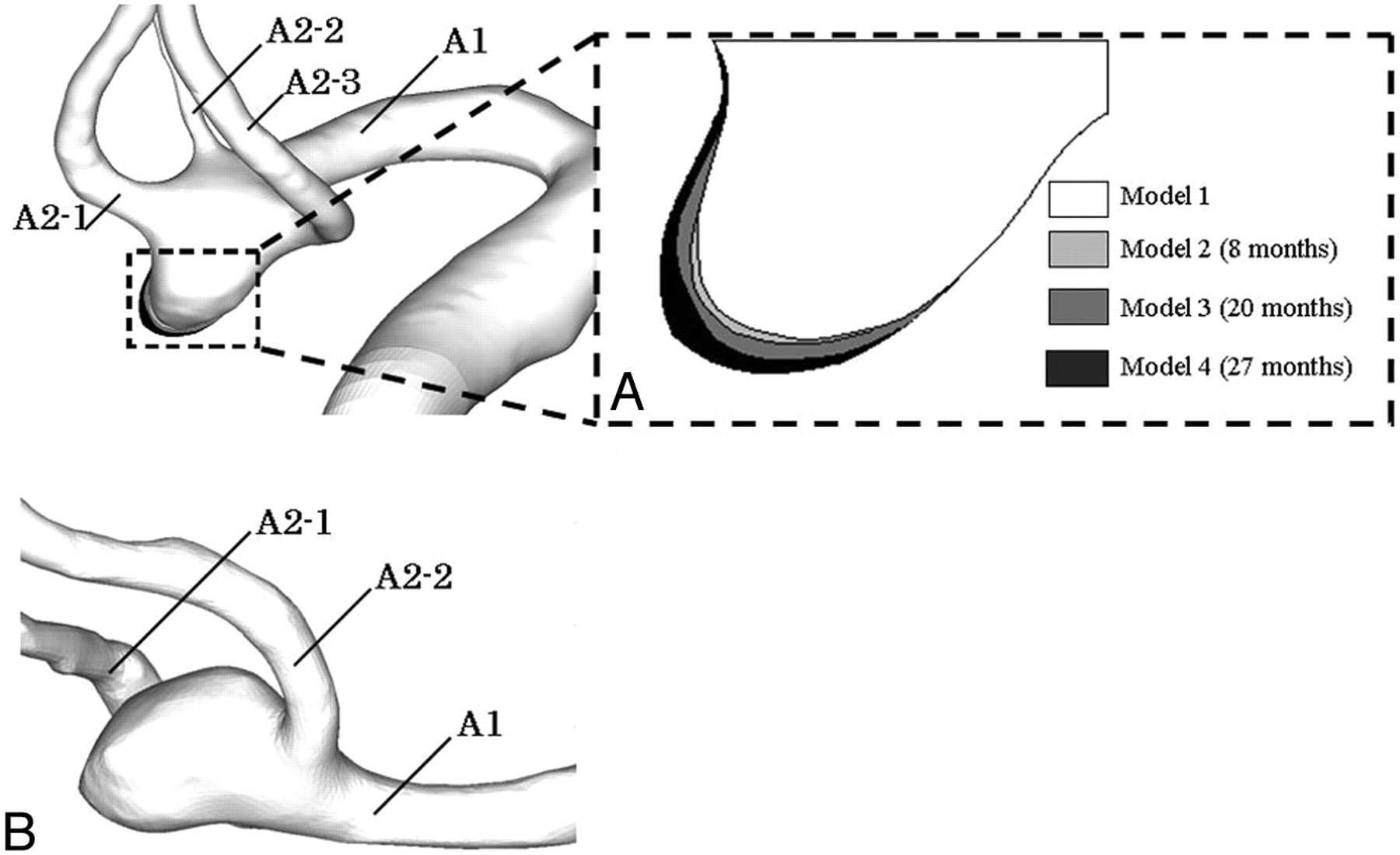
- Fig 1.
A, STL view of the growing model and an enlarged view of the growing area. The growing area was detected by alignment of the aneurysm geometry. The alignment was assessed with the matching of surfaces in nonaneurysmal vessel segments between each model. B, STL view of the nongrowth model.
- Fig 2.
Calculated volume and the surface area of each growing model.
- Fig 3.
Inflow waveform, measured by LDV. The inflow condition was set by referencing the typical blood flow waveform at the ACA.
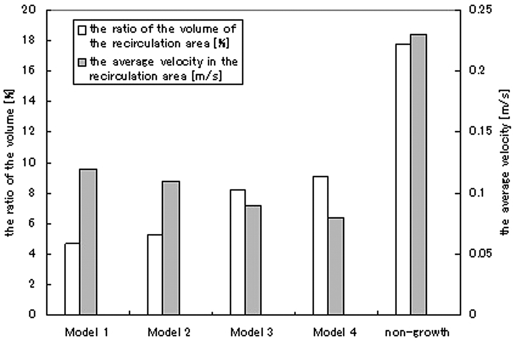
- Fig 4.
Ratio of recirculating volume to that of aneurysms and the average velocity in the recirculation area in each model.
- Fig 5.
WSS distribution on the planes where the enlarging region was observed in model 1. The enlarging region on each plane is shown.


In this issue


Jump to section
Related Articles
Cited By...
- Hemodynamic Characteristics in Ruptured and Unruptured Intracranial Aneurysms: A Prospective Cohort Study Utilizing the AneurysmFlow Tool
- Aneurysm pressure measurement before and after placement of a Pipeline stent: feasibility study using a 0.014 inch pressure wire for coronary intervention
- Novel High-Throughput In Vitro Model for Identifying Hemodynamic-Induced Inflammatory Mediators of Cerebral Aneurysm Formation
- Wall Shear Stress Distribution of Small Aneurysms Prone to Rupture: A Case-Control Study