Advertisement

Main menu

  • Home
  • Content
    • Current Issue
    • Accepted Manuscripts
    • Article Preview
    • Past Issue Archive
    • Video Articles
    • AJNR Case Collection
    • Case of the Week Archive
    • Case of the Month Archive
    • Classic Case Archive
  • Special Collections
    • Advancing NeuroMRI with High-Relaxivity Contrast Agents
    • AJNR Awards
    • Low-Field MRI
    • Alzheimer Disease
    • ASNR Foundation Special Collection
    • Photon-Counting CT
    • View All
  • Multimedia
    • AJNR Podcasts
    • AJNR SCANtastic
    • Trainee Corner
    • MRI Safety Corner
    • Imaging Protocols
  • For Authors
    • Submit a Manuscript
    • Submit a Video Article
    • Submit an eLetter to the Editor/Response
    • Manuscript Submission Guidelines
    • Statistical Tips
    • Fast Publishing of Accepted Manuscripts
    • Graphical Abstract Preparation
    • Imaging Protocol Submission
    • Author Policies
  • About Us
    • About AJNR
    • Editorial Board
    • Editorial Board Alumni
  • More
    • Become a Reviewer/Academy of Reviewers
    • Subscribers
    • Permissions
    • Alerts
    • Feedback
    • Advertisers
    • ASNR Home

User menu

  • Alerts
  • Log in

Search

  • Advanced search
American Journal of Neuroradiology

American Journal of Neuroradiology

ASHNR American Society of Functional Neuroradiology ASHNR American Society of Pediatric Neuroradiology ASSR
  • Alerts
  • Log in

Advanced Search

  • Home
  • Content
    • Current Issue
    • Accepted Manuscripts
    • Article Preview
    • Past Issue Archive
    • Video Articles
    • AJNR Case Collection
    • Case of the Week Archive
    • Case of the Month Archive
    • Classic Case Archive
  • Special Collections
    • Advancing NeuroMRI with High-Relaxivity Contrast Agents
    • AJNR Awards
    • Low-Field MRI
    • Alzheimer Disease
    • ASNR Foundation Special Collection
    • Photon-Counting CT
    • View All
  • Multimedia
    • AJNR Podcasts
    • AJNR SCANtastic
    • Trainee Corner
    • MRI Safety Corner
    • Imaging Protocols
  • For Authors
    • Submit a Manuscript
    • Submit a Video Article
    • Submit an eLetter to the Editor/Response
    • Manuscript Submission Guidelines
    • Statistical Tips
    • Fast Publishing of Accepted Manuscripts
    • Graphical Abstract Preparation
    • Imaging Protocol Submission
    • Author Policies
  • About Us
    • About AJNR
    • Editorial Board
    • Editorial Board Alumni
  • More
    • Become a Reviewer/Academy of Reviewers
    • Subscribers
    • Permissions
    • Alerts
    • Feedback
    • Advertisers
    • ASNR Home
  • Follow AJNR on Twitter
  • Visit AJNR on Facebook
  • Follow AJNR on Instagram
  • Join AJNR on LinkedIn
  • RSS Feeds

AJNR Awards, New Junior Editors, and more. Read the latest AJNR updates


Improved Turnaround Times | Median time to first decision: 12 days

Research ArticleBrain

Comparison of MR and Contrast Venography of the Cervical Venous System in Multiple Sclerosis

G. Zaharchuk, N.J. Fischbein, J. Rosenberg, R.J. Herfkens and M.D. Dake
American Journal of Neuroradiology September 2011, 32 (8) 1482-1489; DOI: https://doi.org/10.3174/ajnr.A2549
G. Zaharchuk
  • Find this author on Google Scholar
  • Find this author on PubMed
  • Search for this author on this site
N.J. Fischbein
  • Find this author on Google Scholar
  • Find this author on PubMed
  • Search for this author on this site
J. Rosenberg
  • Find this author on Google Scholar
  • Find this author on PubMed
  • Search for this author on this site
R.J. Herfkens
  • Find this author on Google Scholar
  • Find this author on PubMed
  • Search for this author on this site
M.D. Dake
  • Find this author on Google Scholar
  • Find this author on PubMed
  • Search for this author on this site
  • Article
  • Figures & Data
  • Supplemental
  • Info & Metrics
  • Responses
  • References
  • PDF
Loading

Article Figures & Data

Figures

  • Tables
  • Fig 1.
    • Download figure
    • Open in new tab
    • Download powerpoint
    Fig 1.

    Qualitative grading of venous stenosis between 0 (none) and 3 (severe). Examples at the high IJ vein level demonstrate either no stenosis (A) or bilateral mild (B), moderate (C), and severe (D) stenosis. In some severe cases, the IJ was not visualized, as on the right in this example. The cases shown are bilateral for the purposes of example, but in general, left-right asymmetry in the degree of stenosis was more common.

  • Fig 2.
    • Download figure
    • Open in new tab
    • Download powerpoint
    Fig 2.

    Grading scale for collateral venous flow. Images shown are sagittal MIPs of either the left or right side of the neck from TRICKS MRV from the phase with the most venous opacification. Collateral scorings shown are: (A) none, 0, (B) mild, 1, (C) moderate, 2, and (D) severe, 3. In severe cases, prominent collaterals were sometimes seen over the posterior neck regions, as in this example.

  • Fig 3.
    • Download figure
    • Open in new tab
    • Download powerpoint
    Fig 3.

    A 35-year-old woman with relapsing-remitting MS diagnosed 5 years before imaging showing characteristic locations of venous abnormalities on 2D-TOF MRV. In the upper neck (A–C), flattening was often seen near C1 (arrows, B). In the mid- and lower neck (D–F), flattening can be seen between the sternocleidomastoid and anterior scalene muscles on the right (arrows, E).
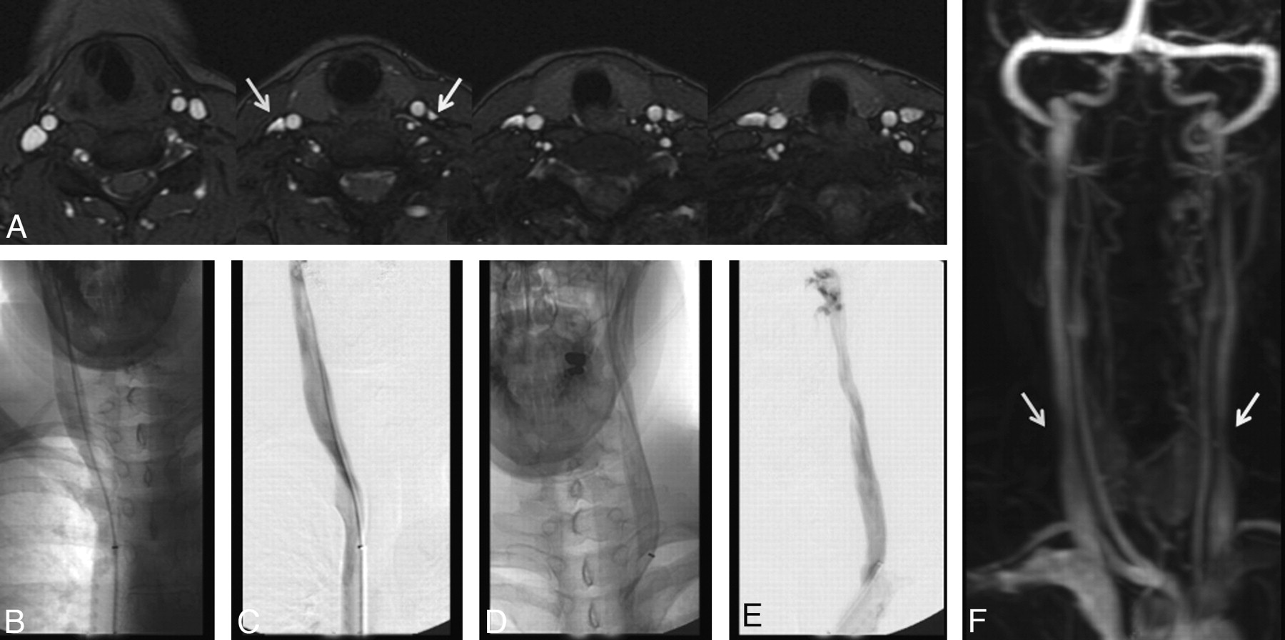
  • Fig 4.
    • Download figure
    • Open in new tab
    • Download powerpoint
    Fig 4.

    A 38-year-old woman with relapsing-remitting MS for 3 years. A, High IJ stenosis adjacent to the right C1 lateral mass (arrow). B, CV shows the focal stenosis, whereas a delayed image (C) shows associated non-IJ collateral venous vessels. The left IJ seems normal on CV. The right high IJ was graded severe on both MRV and CV; the left IJ was graded by MRV as mild stenosis, due to the slightly flattened appearance, whereas it was called normal on CV (not shown).

  • Fig 5.
    • Download figure
    • Open in new tab
    • Download powerpoint
    Fig 5.

    A 36-year-old man with relapsing-remitting MS for 2 years, showing severe stenosis of the left mid-IJ on MRV (A) and early (B) and late (C) phases of CV (arrows). Enlarged posterior paraspinal venous collaterals are also noted on the venogram. In certain individuals, the course of the carotid artery swings across the IJ vein at this level, possibly causing compression.

  • Fig 6.
    • Download figure
    • Open in new tab
    • Download powerpoint
    Fig 6.

    A 41-year-old woman with relapsing-remitting MS for 6 years. A, MRV demonstrates moderate right (arrow) and severe left (arrow) lower IJ stenosis. CV demonstrates no appreciable stenosis in the low IJ segment on either side. Unsubtracted (B) and subtracted (C) CV after right IJ injection; unsubtracted (D) and subtracted (E) CV after left IJ injection. The cause of this lack of agreement between modalities remains unclear, though the degree of stenosis was less severe on the TRICKS images (F).
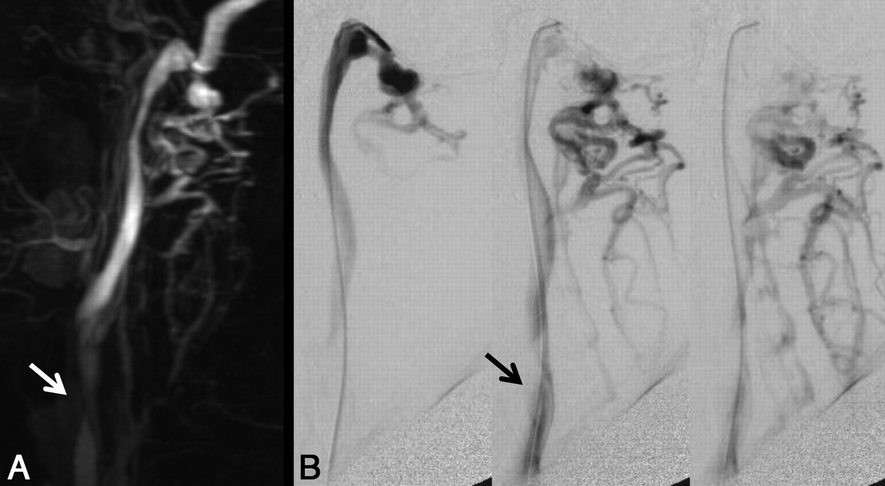
  • Fig 7.
    • Download figure
    • Open in new tab
    • Download powerpoint
    Fig 7.

    A 47-year-old man with 2 years of relapsing-remitting MS. A, Lateral view of MIP image of the left neck from TRICKS MRV. B, Multiple frames from CV with injection of the high left IJ. Both modalities rated the posterior paraspinal collaterals in this patient as moderate. An arrow points to a location of stenosis noted in the mid-IJ.

  • Fig 8.
    • Download figure
    • Open in new tab
    • Download powerpoint
    Fig 8.

    Relationship between collateral rating versus composite IJ vein stenosis score. There was a significant relationship between more non-IJ collaterals and increasing stenosis for the CV examination (ρ = 0.49) but not for the MRV examination (ρ = 0.18), based on rank correlation. Line is a locally weighted regression smoother. Points have been jittered for clarity.

Tables

  • Figures
    • View popup
    Table 1:

    Frequency of stenosis ratings by location and modality

    Scan Type and LocationSide and IJ Stenosis Scorea
    LeftRight
    01230123
    CV
        High108912811155
        Mid2618434320
        Low111288191082
    MRV
        High951411871410
        Mid2782234500
        Low16968147612
    • a Scores: 0, none; 1, mild; 2, moderate; and 3, severe stenosis.

    • View popup
    Table 2:

    Agreement of MRV and CV for IJ stenosis

    Nκa95% CIP
    Full scale (0–3)
        Overall2340.540.44–0.64<.001
        Left1170.510.36–0.65<.001
        Right1170.570.43–0.71<.001
        Upper780.650.48–0.78<.001
        Mid780.580.34–0.76<.001
        Lower780.17−0.05–0.38.062
    Dichotomized (normal [0] versus abnormal [1–3])b
        Overall2340.550.44–0.66<.001
        Left1170.490.32–0.65<.001
        Right1170.610.45–0.74<.001
        Upper780.670.44–0.85<.001
        Mid780.600.36–0.80<.001
        Lower780.13−0.09–0.35.120
    • a Weighted κ using squared discrepancies.

    • b For the dichotomized scores, considering abnormal CV as a standard, sensitivity, 0.79 (95% CI, 0.71%–0.86%); specificity, 0.76 (95% CI, 0.67%–0.83%); PPV, 0.79 (95% CI, 0.71%–0.86%); and NPV, 0.76 (95% CI, 0.67%–0.83%).

    • View popup
    Table 3:

    Severe stenosis (grade = 3) in any IJ segment on a per-patient basis

    MRVCVa
    NoYesTotal
    No14317
    Yes71522
    Total211839
    • a Considering CV with severe stenosis as a standard, sensitivity, 0.83 (95% CI, 0.58%–0.96%); specificity, 0.67 (95% CI, 0.43%–0.85%); PPV, 0.68 (95% CI, 0.45%–0.85%); and NPV, 0.82 (95% CI, 0.56%–0.95%).

    • View popup
    Table 4:

    Frequency of collateral scores

    Scan TypeCollateral ScoreaTotal
    0123
    CV2317271178
    MRV623331678
    Total29406027156
    P = .002 by paired Wilcoxon test, MRV scores > CV scores
    • a Scores: 0, none; 1, mild; 2, moderate; and 3, severe.

    • View popup
    Table 5:

    Agreement of collateral scores

    Nκa95% CIP
    Full scale (0–3)
        Overall780.300.09–0.50.002
        Left390.420.10–0.66.003
        Right390.17−0.08 to 0.44.096
    Dichotomized (normal [0] versus abnormal [1–3])b
        Overall780.18−0.05 to 0.40.012
        Left390.28−0.08 to 0.65.020
        Right390.11−0.10 to 0.35.148
    • a Weighted κ using squared discrepancies.

    • b Scores: 0, none; 1, mild; 2, moderate; and 3, severe.

PreviousNext
Back to top

In this issue

American Journal of Neuroradiology: 32 (8)
American Journal of Neuroradiology
Vol. 32, Issue 8
1 Sep 2011
  • Table of Contents
  • Index by author
Advertisement
Print
Download PDF
Email Article

Thank you for your interest in spreading the word on American Journal of Neuroradiology.

NOTE: We only request your email address so that the person you are recommending the page to knows that you wanted them to see it, and that it is not junk mail. We do not capture any email address.

Enter multiple addresses on separate lines or separate them with commas.
Comparison of MR and Contrast Venography of the Cervical Venous System in Multiple Sclerosis
(Your Name) has sent you a message from American Journal of Neuroradiology
(Your Name) thought you would like to see the American Journal of Neuroradiology web site.
CAPTCHA
This question is for testing whether or not you are a human visitor and to prevent automated spam submissions.
Cite this article
G. Zaharchuk, N.J. Fischbein, J. Rosenberg, R.J. Herfkens, M.D. Dake
Comparison of MR and Contrast Venography of the Cervical Venous System in Multiple Sclerosis
American Journal of Neuroradiology Sep 2011, 32 (8) 1482-1489; DOI: 10.3174/ajnr.A2549

Citation Manager Formats

  • BibTeX
  • Bookends
  • EasyBib
  • EndNote (tagged)
  • EndNote 8 (xml)
  • Medlars
  • Mendeley
  • Papers
  • RefWorks Tagged
  • Ref Manager
  • RIS
  • Zotero
0 Responses
Respond to this article
Share
Bookmark this article
Comparison of MR and Contrast Venography of the Cervical Venous System in Multiple Sclerosis
G. Zaharchuk, N.J. Fischbein, J. Rosenberg, R.J. Herfkens, M.D. Dake
American Journal of Neuroradiology Sep 2011, 32 (8) 1482-1489; DOI: 10.3174/ajnr.A2549
del.icio.us logo Twitter logo Facebook logo Mendeley logo
  • Tweet Widget
  • Facebook Like
  • Google Plus One
Purchase

Jump to section

  • Article
    • Abstract
    • Abbreviations
    • Materials and Methods
    • Results
    • Discussion
    • Conclusions
    • Footnotes
    • References
  • Figures & Data
  • Supplemental
  • Info & Metrics
  • Responses
  • References
  • PDF

Related Articles

  • PubMed
  • Google Scholar

Cited By...

  • Lower Arterial Cross-Sectional Area of Carotid and Vertebral Arteries and Higher Frequency of Secondary Neck Vessels Are Associated with Multiple Sclerosis
  • Jugular Anomalies in Multiple Sclerosis Are Associated with Increased Collateral Venous Flow
  • Phlebographic Study Does Not Show Differences Between Patients with MS and Control Subjects
  • Reproducibility of Cerebrospinal Venous Blood Flow and Vessel Anatomy with the Use of Phase Contrast-Vastly Undersampled Isotropic Projection Reconstruction and Contrast-Enhanced MRA
  • No Association Between Conventional Brain MR Imaging and Chronic Cerebrospinal Venous Insufficiency in Multiple Sclerosis
  • Extracranial Venous Drainage Patterns in Patients with Multiple Sclerosis and Healthy Controls
  • Crossref
  • Google Scholar

This article has not yet been cited by articles in journals that are participating in Crossref Cited-by Linking.

More in this TOC Section

  • Progression of Microstructural Damage in Spinocerebellar Ataxia Type 2: A Longitudinal DTI Study
  • SWI or T2*: Which MRI Sequence to Use in the Detection of Cerebral Microbleeds? The Karolinska Imaging Dementia Study
  • Statin Therapy Does Not Affect the Radiographic and Clinical Profile of Patients with TIA and Minor Stroke
Show more Brain

Similar Articles

Advertisement

Indexed Content

  • Current Issue
  • Accepted Manuscripts
  • Article Preview
  • Past Issues
  • Editorials
  • Editor's Choice
  • Fellows' Journal Club
  • Letters to the Editor
  • Video Articles

Cases

  • Case Collection
  • Archive - Case of the Week
  • Archive - Case of the Month
  • Archive - Classic Case

More from AJNR

  • Trainee Corner
  • Imaging Protocols
  • MRI Safety Corner
  • Book Reviews

Multimedia

  • AJNR Podcasts
  • AJNR Scantastics

Resources

  • Turnaround Time
  • Submit a Manuscript
  • Submit a Video Article
  • Submit an eLetter to the Editor/Response
  • Manuscript Submission Guidelines
  • Statistical Tips
  • Fast Publishing of Accepted Manuscripts
  • Graphical Abstract Preparation
  • Imaging Protocol Submission
  • Evidence-Based Medicine Level Guide
  • Publishing Checklists
  • Author Policies
  • Become a Reviewer/Academy of Reviewers
  • News and Updates

About Us

  • About AJNR
  • Editorial Board
  • Editorial Board Alumni
  • Alerts
  • Permissions
  • Not an AJNR Subscriber? Join Now
  • Advertise with Us
  • Librarian Resources
  • Feedback
  • Terms and Conditions
  • AJNR Editorial Board Alumni

American Society of Neuroradiology

  • Not an ASNR Member? Join Now

© 2025 by the American Society of Neuroradiology All rights, including for text and data mining, AI training, and similar technologies, are reserved.
Print ISSN: 0195-6108 Online ISSN: 1936-959X

Powered by HighWire