Advertisement

Main menu

  • Home
  • Content
    • Current Issue
    • Accepted Manuscripts
    • Article Preview
    • Past Issue Archive
    • Video Articles
    • AJNR Case Collection
    • Case of the Week Archive
    • Case of the Month Archive
    • Classic Case Archive
  • Special Collections
    • AJNR Awards
    • Low-Field MRI
    • Alzheimer Disease
    • ASNR Foundation Special Collection
    • Photon-Counting CT
    • View All
  • Multimedia
    • AJNR Podcasts
    • AJNR SCANtastic
    • Trainee Corner
    • MRI Safety Corner
    • Imaging Protocols
  • For Authors
    • Submit a Manuscript
    • Submit a Video Article
    • Submit an eLetter to the Editor/Response
    • Manuscript Submission Guidelines
    • Statistical Tips
    • Fast Publishing of Accepted Manuscripts
    • Graphical Abstract Preparation
    • Imaging Protocol Submission
    • Author Policies
  • About Us
    • About AJNR
    • Editorial Board
    • Editorial Board Alumni
  • More
    • Become a Reviewer/Academy of Reviewers
    • Subscribers
    • Permissions
    • Alerts
    • Feedback
    • Advertisers
    • ASNR Home

User menu

  • Alerts
  • Log in

Search

  • Advanced search
American Journal of Neuroradiology

American Journal of Neuroradiology

ASHNR American Society of Functional Neuroradiology ASHNR American Society of Pediatric Neuroradiology ASSR
  • Alerts
  • Log in

Advanced Search

  • Home
  • Content
    • Current Issue
    • Accepted Manuscripts
    • Article Preview
    • Past Issue Archive
    • Video Articles
    • AJNR Case Collection
    • Case of the Week Archive
    • Case of the Month Archive
    • Classic Case Archive
  • Special Collections
    • AJNR Awards
    • Low-Field MRI
    • Alzheimer Disease
    • ASNR Foundation Special Collection
    • Photon-Counting CT
    • View All
  • Multimedia
    • AJNR Podcasts
    • AJNR SCANtastic
    • Trainee Corner
    • MRI Safety Corner
    • Imaging Protocols
  • For Authors
    • Submit a Manuscript
    • Submit a Video Article
    • Submit an eLetter to the Editor/Response
    • Manuscript Submission Guidelines
    • Statistical Tips
    • Fast Publishing of Accepted Manuscripts
    • Graphical Abstract Preparation
    • Imaging Protocol Submission
    • Author Policies
  • About Us
    • About AJNR
    • Editorial Board
    • Editorial Board Alumni
  • More
    • Become a Reviewer/Academy of Reviewers
    • Subscribers
    • Permissions
    • Alerts
    • Feedback
    • Advertisers
    • ASNR Home
  • Follow AJNR on Twitter
  • Visit AJNR on Facebook
  • Follow AJNR on Instagram
  • Join AJNR on LinkedIn
  • RSS Feeds

AJNR Awards, New Junior Editors, and more. Read the latest AJNR updates

Research ArticleNeurointervention

3D Quantitative Evaluation of Atherosclerotic Plaque Based on Rotational Angiography

A. Chien, J. Sayre, B. Dong, J. Ye and F. Viñuela
American Journal of Neuroradiology August 2011, 32 (7) 1249-1254; DOI: https://doi.org/10.3174/ajnr.A2483
A. Chien
  • Find this author on Google Scholar
  • Find this author on PubMed
  • Search for this author on this site
J. Sayre
  • Find this author on Google Scholar
  • Find this author on PubMed
  • Search for this author on this site
B. Dong
  • Find this author on Google Scholar
  • Find this author on PubMed
  • Search for this author on this site
J. Ye
  • Find this author on Google Scholar
  • Find this author on PubMed
  • Search for this author on this site
F. Viñuela
  • Find this author on Google Scholar
  • Find this author on PubMed
  • Search for this author on this site
  • Article
  • Figures & Data
  • Info & Metrics
  • Responses
  • References
  • PDF
Loading

Article Figures & Data

Figures

  • Fig 1.
    • Download figure
    • Open in new tab
    • Download powerpoint
    Fig 1.

    Representative angiographic images indicating the location of atherosclerotic plaque (yellow arrow, A) and 3D reconstruction of the same blood vessel (B) in mathematic indices ready for the plaque-estimation algorithm vector field (C), which encodes surface curvature.
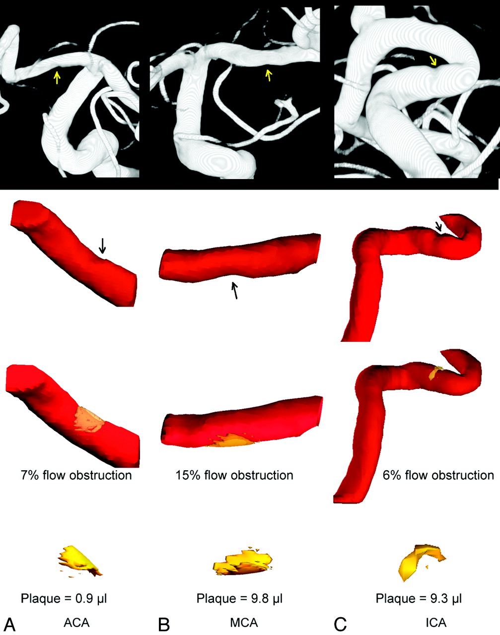
  • Fig 2.
    • Download figure
    • Open in new tab
    • Download powerpoint
    Fig 2.

    Plaque quantification for cases I the ACA (A), II the MCA (B), and III the ICA (C). The top row is the diagnostic images with arrows indicating stenosis. The second row shows the 3D geometry of the vessel built with 3DRA images with arrows indicating stenosis. The third row shows the reconstructed plaque (yellow) and the percentage of flow obstruction computed by the program. The bottom row shows the morphology and volume of the plaque in units of microliters.
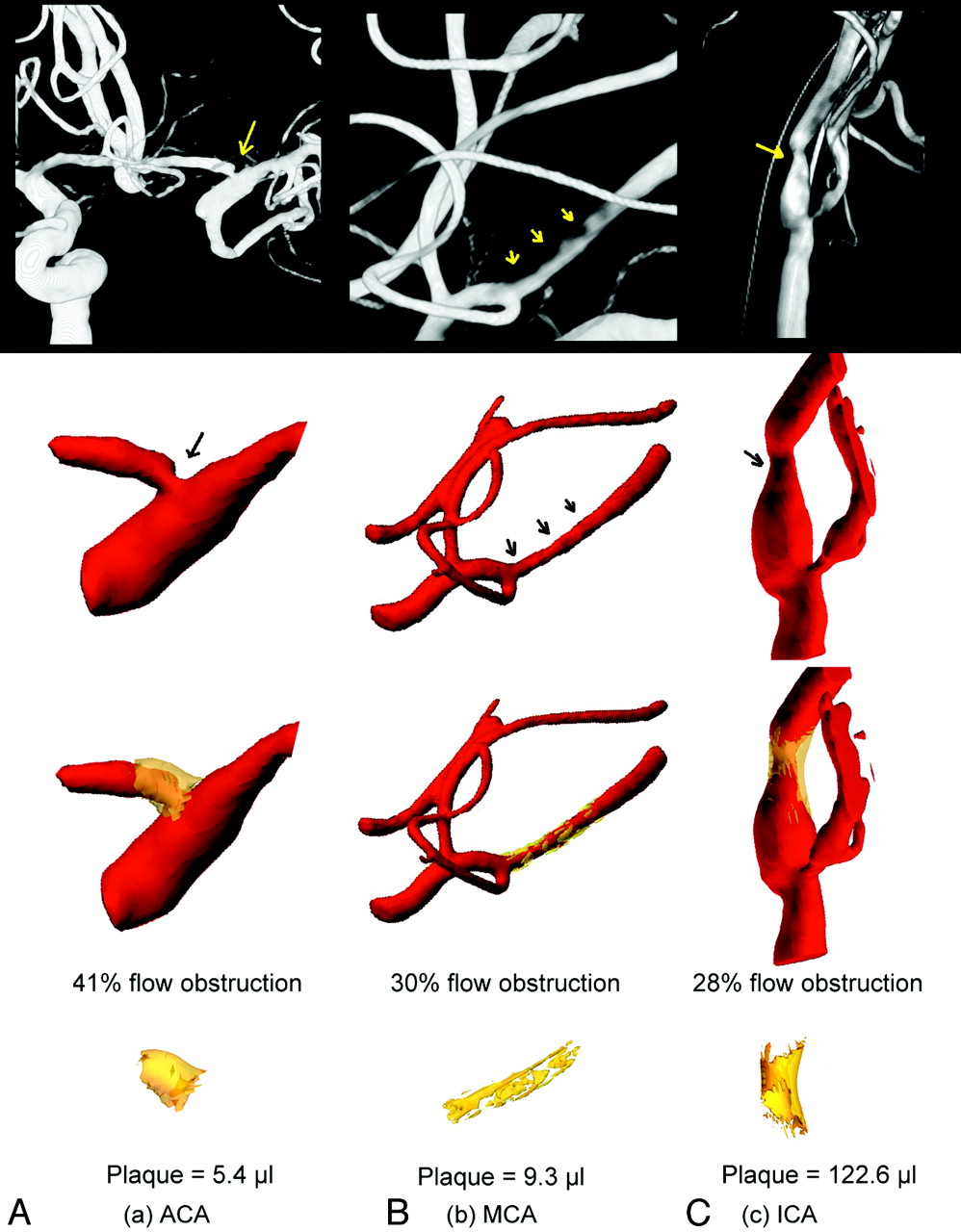
  • Fig 3.
    • Download figure
    • Open in new tab
    • Download powerpoint
    Fig 3.

    Plaque quantification for cases IV the ACA (A), V the MCA (B), and VI the ICA (C). The top row is the diagnostic images with arrows indicating stenosis. The second row shows the 3D geometry of the vessel built with 3DRA images and arrows indicating stenosis. The third row shows the reconstructed plaque (yellow) and the percentage of flow obstruction. The bottom row shows the morphology and volume of the plaque in units of microliters.

  • Fig 4.
    • Download figure
    • Open in new tab
    • Download powerpoint
    Fig 4.

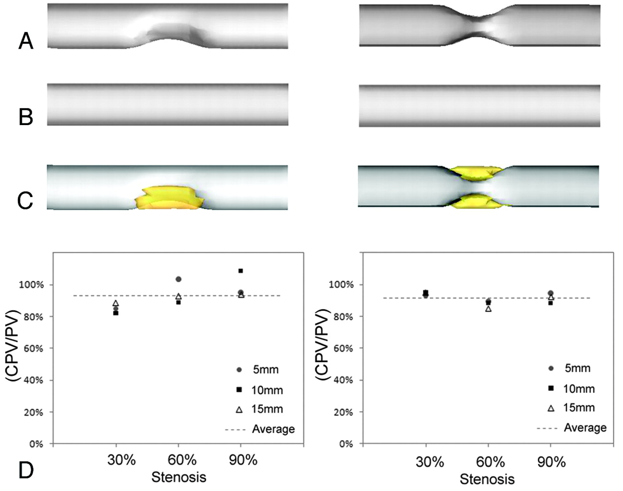
    A, Representative unilateral (left) and bilateral (right) stenosis phantoms. B, The ideal vessel without stenosis. C, The reconstructed plaque for the unilateral (left) and bilateral (right) models. D, The percentage of the volume estimation (CPV/PV) for lesion sizes 5, 10, and 15 mm in the direction of the vessel long axis.

PreviousNext
Back to top

In this issue

American Journal of Neuroradiology: 32 (7)
American Journal of Neuroradiology
Vol. 32, Issue 7
1 Aug 2011
  • Table of Contents
  • Index by author
Advertisement
Print
Download PDF
Email Article

Thank you for your interest in spreading the word on American Journal of Neuroradiology.

NOTE: We only request your email address so that the person you are recommending the page to knows that you wanted them to see it, and that it is not junk mail. We do not capture any email address.

Enter multiple addresses on separate lines or separate them with commas.
3D Quantitative Evaluation of Atherosclerotic Plaque Based on Rotational Angiography
(Your Name) has sent you a message from American Journal of Neuroradiology
(Your Name) thought you would like to see the American Journal of Neuroradiology web site.
CAPTCHA
This question is for testing whether or not you are a human visitor and to prevent automated spam submissions.
Cite this article
A. Chien, J. Sayre, B. Dong, J. Ye, F. Viñuela
3D Quantitative Evaluation of Atherosclerotic Plaque Based on Rotational Angiography
American Journal of Neuroradiology Aug 2011, 32 (7) 1249-1254; DOI: 10.3174/ajnr.A2483

Citation Manager Formats

  • BibTeX
  • Bookends
  • EasyBib
  • EndNote (tagged)
  • EndNote 8 (xml)
  • Medlars
  • Mendeley
  • Papers
  • RefWorks Tagged
  • Ref Manager
  • RIS
  • Zotero
0 Responses
Respond to this article
Share
Bookmark this article
3D Quantitative Evaluation of Atherosclerotic Plaque Based on Rotational Angiography
A. Chien, J. Sayre, B. Dong, J. Ye, F. Viñuela
American Journal of Neuroradiology Aug 2011, 32 (7) 1249-1254; DOI: 10.3174/ajnr.A2483
del.icio.us logo Twitter logo Facebook logo Mendeley logo
  • Tweet Widget
  • Facebook Like
  • Google Plus One
Purchase

Jump to section

  • Article
    • Abstract
    • Abbreviations
    • Materials and Methods
    • Results
    • Discussion
    • Conclusions
    • Appendix
    • Footnotes
    • References
  • Figures & Data
  • Info & Metrics
  • Responses
  • References
  • PDF

Related Articles

  • No related articles found.
  • PubMed
  • Google Scholar

Cited By...

  • IS FlowMap, a novel tool to examine blood flow changes induced by flow diverter stent treatment: initial experiences with pipeline cases
  • Crossref
  • Google Scholar

This article has not yet been cited by articles in journals that are participating in Crossref Cited-by Linking.

More in this TOC Section

  • Cavernous dural AVF treated by transfacial route
  • A Retrospective Study in Tentorial DAVFs
  • Proximal Protection Devices for Carotid Stenting
Show more Neurointervention

Similar Articles

Advertisement

Indexed Content

  • Current Issue
  • Accepted Manuscripts
  • Article Preview
  • Past Issues
  • Editorials
  • Editor's Choice
  • Fellows' Journal Club
  • Letters to the Editor
  • Video Articles

Cases

  • Case Collection
  • Archive - Case of the Week
  • Archive - Case of the Month
  • Archive - Classic Case

More from AJNR

  • Trainee Corner
  • Imaging Protocols
  • MRI Safety Corner
  • Book Reviews

Multimedia

  • AJNR Podcasts
  • AJNR Scantastics

Resources

  • Turnaround Time
  • Submit a Manuscript
  • Submit a Video Article
  • Submit an eLetter to the Editor/Response
  • Manuscript Submission Guidelines
  • Statistical Tips
  • Fast Publishing of Accepted Manuscripts
  • Graphical Abstract Preparation
  • Imaging Protocol Submission
  • Evidence-Based Medicine Level Guide
  • Publishing Checklists
  • Author Policies
  • Become a Reviewer/Academy of Reviewers
  • News and Updates

About Us

  • About AJNR
  • Editorial Board
  • Editorial Board Alumni
  • Alerts
  • Permissions
  • Not an AJNR Subscriber? Join Now
  • Advertise with Us
  • Librarian Resources
  • Feedback
  • Terms and Conditions
  • AJNR Editorial Board Alumni

American Society of Neuroradiology

  • Not an ASNR Member? Join Now

© 2025 by the American Society of Neuroradiology All rights, including for text and data mining, AI training, and similar technologies, are reserved.
Print ISSN: 0195-6108 Online ISSN: 1936-959X

Powered by HighWire