
Article Figures & Data
Figures
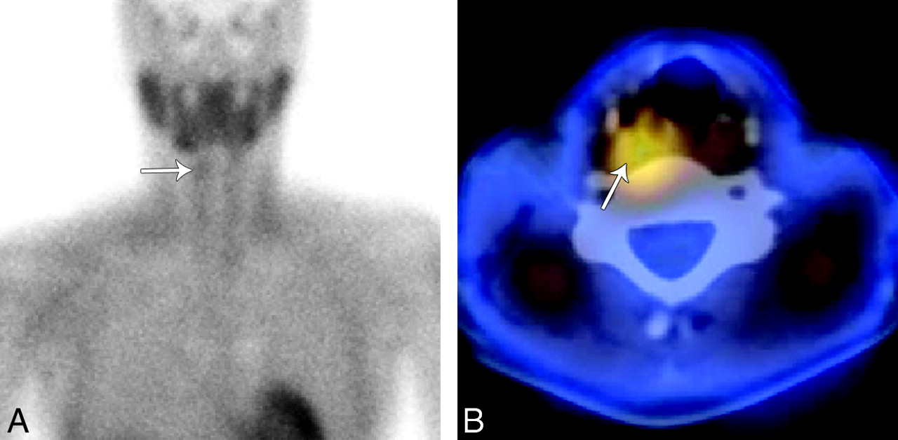
- Fig 1.
Technetium Tc99m sestamibi imaging with SPECT. A, A 2-hour planar sestamibi image shows minimal residual uptake in the right neck (arrow) inferior to the submandibular gland. B, A fused axial SPECT image demonstrates tracer activity (arrow) superior and posterior to the right thyroid lobe in the region of the right retropharyngeal space.
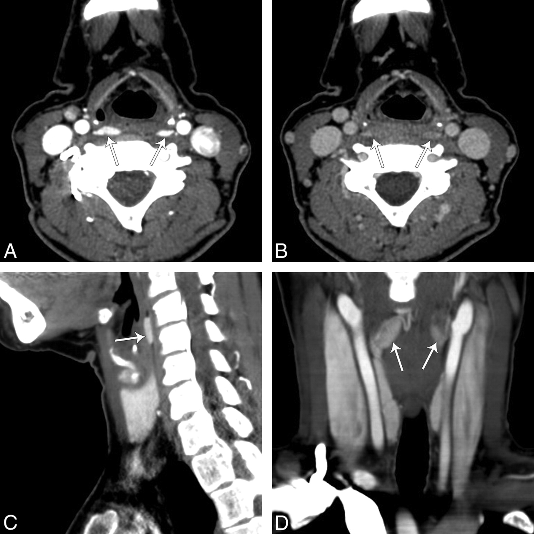
- Fig 2.
4D-CT of the neck showing bilateral retropharyngeal masses. A, An arterial phase axial image of the neck at the level of the pyriform sinus demonstrates 2 hyperenhancing lesions (arrows), right larger than left, in the retropharyngeal space. B, On delayed images at the same level there was rapid washout of contrast in the lesions. Reformatted right sagittal (C) and coronal arterial phase (D) images demonstrate that hyperenhancing lesions (arrows) in the retropharyngeal space have an oval shape.


In this issue


Jump to section
Related Articles
Cited By...
- 4D-CT for Preoperative Localization of Abnormal Parathyroid Glands in Patients with Hyperparathyroidism: Accuracy and Ability to Stratify Patients by Unilateral versus Bilateral Disease in Surgery-Naive and Re-Exploration Patients
- Parathyroid Lesions: Characterization with Dual-Phase Arterial and Venous Enhanced CT of the Neck