Advertisement

Main menu

  • Home
  • Content
    • Current Issue
    • Accepted Manuscripts
    • Article Preview
    • Past Issue Archive
    • Video Articles
    • AJNR Case Collection
    • Case of the Week Archive
    • Case of the Month Archive
    • Classic Case Archive
  • Special Collections
    • AJNR Awards
    • Low-Field MRI
    • Alzheimer Disease
    • ASNR Foundation Special Collection
    • Photon-Counting CT
    • View All
  • Multimedia
    • AJNR Podcasts
    • AJNR SCANtastic
    • Trainee Corner
    • MRI Safety Corner
    • Imaging Protocols
  • For Authors
    • Submit a Manuscript
    • Submit a Video Article
    • Submit an eLetter to the Editor/Response
    • Manuscript Submission Guidelines
    • Statistical Tips
    • Fast Publishing of Accepted Manuscripts
    • Graphical Abstract Preparation
    • Imaging Protocol Submission
    • Author Policies
  • About Us
    • About AJNR
    • Editorial Board
    • Editorial Board Alumni
  • More
    • Become a Reviewer/Academy of Reviewers
    • Subscribers
    • Permissions
    • Alerts
    • Feedback
    • Advertisers
    • ASNR Home

User menu

  • Alerts
  • Log in

Search

  • Advanced search
American Journal of Neuroradiology

American Journal of Neuroradiology

ASHNR American Society of Functional Neuroradiology ASHNR American Society of Pediatric Neuroradiology ASSR
  • Alerts
  • Log in

Advanced Search

  • Home
  • Content
    • Current Issue
    • Accepted Manuscripts
    • Article Preview
    • Past Issue Archive
    • Video Articles
    • AJNR Case Collection
    • Case of the Week Archive
    • Case of the Month Archive
    • Classic Case Archive
  • Special Collections
    • AJNR Awards
    • Low-Field MRI
    • Alzheimer Disease
    • ASNR Foundation Special Collection
    • Photon-Counting CT
    • View All
  • Multimedia
    • AJNR Podcasts
    • AJNR SCANtastic
    • Trainee Corner
    • MRI Safety Corner
    • Imaging Protocols
  • For Authors
    • Submit a Manuscript
    • Submit a Video Article
    • Submit an eLetter to the Editor/Response
    • Manuscript Submission Guidelines
    • Statistical Tips
    • Fast Publishing of Accepted Manuscripts
    • Graphical Abstract Preparation
    • Imaging Protocol Submission
    • Author Policies
  • About Us
    • About AJNR
    • Editorial Board
    • Editorial Board Alumni
  • More
    • Become a Reviewer/Academy of Reviewers
    • Subscribers
    • Permissions
    • Alerts
    • Feedback
    • Advertisers
    • ASNR Home
  • Follow AJNR on Twitter
  • Visit AJNR on Facebook
  • Follow AJNR on Instagram
  • Join AJNR on LinkedIn
  • RSS Feeds

AJNR Awards, New Junior Editors, and more. Read the latest AJNR updates

Case ReportCase Report

Bilateral Retropharyngeal Parathyroid Hyperplasia Detected with 4D Multidetector Row CT

R.D. Welling, J.A. Olson, P.G. Kranz, J.D. Eastwood and J.K. Hoang
American Journal of Neuroradiology May 2011, 32 (5) E80-E82; DOI: https://doi.org/10.3174/ajnr.A2104
R.D. Welling
  • Find this author on Google Scholar
  • Find this author on PubMed
  • Search for this author on this site
J.A. Olson Jr.
  • Find this author on Google Scholar
  • Find this author on PubMed
  • Search for this author on this site
P.G. Kranz
  • Find this author on Google Scholar
  • Find this author on PubMed
  • Search for this author on this site
J.D. Eastwood
  • Find this author on Google Scholar
  • Find this author on PubMed
  • Search for this author on this site
J.K. Hoang
  • Find this author on Google Scholar
  • Find this author on PubMed
  • Search for this author on this site
  • Article
  • Figures & Data
  • Info & Metrics
  • Responses
  • References
  • PDF
Loading

Abstract

SUMMARY: We present a case of bilateral retropharyngeal parathyroid hyperplasia detected with 4D-CT in a patient with persistent primary hyperparathyroidism and failed neck exploration. We discuss the embryologic basis of ectopic retropharyngeal parathyroid adenomas and hyperplasia and the utility of 4D-CT in their localization for surgical planning.

Abbreviations

PTH
parathyroid hormone
SPECT
single-photon emission computed tomography

Primary hyperparathyroidism is caused by a single-gland adenoma in most patients (≤90%) or multiglandular hyperplasia (10%–15%).1 Ectopic parathyroid adenomas and hyperplasia, defined by their location in sites other than the juxtathyroid position, are a well-documented cause of primary hyperparathyroidism and are reported in ≤22% of cases.2,3 When an adenoma or glandular hyperplasia is ectopic, findings of the initial work-up with sonography and sestamibi scintigraphy may be negative. In such cases, MR imaging and CT are often performed. These second-line investigations have become increasingly important since the surgical treatment of parathyroid masses has transitioned from bilateral 4-gland exploration to unilateral minimally invasive parathyroidectomy.4,5 The need to accurately localize parathyroid lesions and advances in CT technology have led to new protocols such as 4D-CT.6–8

We present a case of ectopic bilateral retropharyngeal parathyroid hyperplasia detected on 4D-CT and review its role and technique.

Case Report

A 47-year-old woman was referred to our institution for evaluation of persistent primary hyperparathyroidism after bilateral neck exploration 4 years prior. At that time, she had undergone right inferior parathyroidectomy and biopsy of the inferior left parathyroid gland, which revealed hyperplasia. The superior parathyroid glands were not identified.

Postoperatively, the patient continued to experience hypercalcemia and was treated with the calcimimetic, cinacalcet (Sensipar). Despite medical therapy, the patient's serum PTH and serum and urine calcium levels remained elevated. She continued to have symptoms of fatigue, memory loss, and muscle aches and had osteopenia detected by a dual energy x-ray absorptiometry scan. Given her biochemical abnormalities, symptoms, and bone loss, she again presented for surgical evaluation.

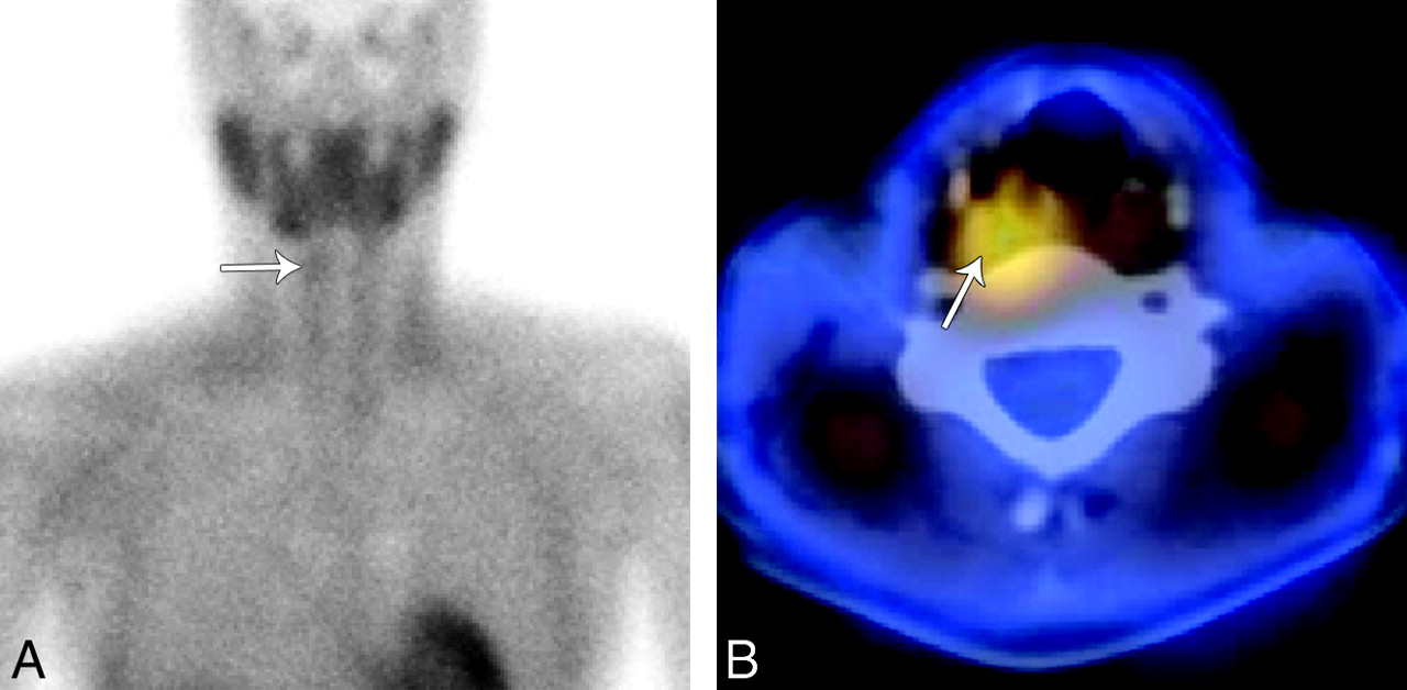
The imaging work-up began with sonography, which did not demonstrate a parathyroid adenoma in the usual juxtathyroid locations. Next she underwent sestamibi imaging with SPECT correlation (Fig 1). Two-hour planar sestamibi imaging demonstrated minimal residual uptake in the right neck inferior to the submandibular gland. Fused SPECT imaging demonstrated a region of increased radiotracer activity in the right retropharyngeal space, superior to the upper pole of the right thyroid. No activity was present in the left neck on delayed sestamibi images.

Fig 1.
  • Download figure
  • Open in new tab
  • Download powerpoint
Fig 1.

Technetium Tc99m sestamibi imaging with SPECT. A, A 2-hour planar sestamibi image shows minimal residual uptake in the right neck (arrow) inferior to the submandibular gland. B, A fused axial SPECT image demonstrates tracer activity (arrow) superior and posterior to the right thyroid lobe in the region of the right retropharyngeal space.

Finally, 4D-CT of the neck and mediastinum was performed (Fig 2). Our protocol involves imaging with 64-section multidetector row CT after 120 mL of iopamidol (Isovue-300; Bristol-Meyers Squibb, Princeton, New Jersey) is administered intravenously via an 18-ga intravenous cannula in the right antecubital fossa at a rate of 3 mL/s, followed by a 50-mL saline flush. We acquire contiguous axial images from the lower margin of the mandible to the level of the inferior aortic arch in arterial (25-second delay) and venous phases (80-second delay) with the following settings: 0.625 section thickness; gantry rotation time, 0.8 seconds; 20-cm FOV; 120 kV(peak); and automatic tube current modulation (Smart mA; GE Healthcare, Milwaukee, Wisconsin). Reformatted images in the 2 phases are sent to a PACS as 2.5-mm-thick contiguous images in the axial, coronal, and sagittal planes.

Fig 2.
  • Download figure
  • Open in new tab
  • Download powerpoint
Fig 2.

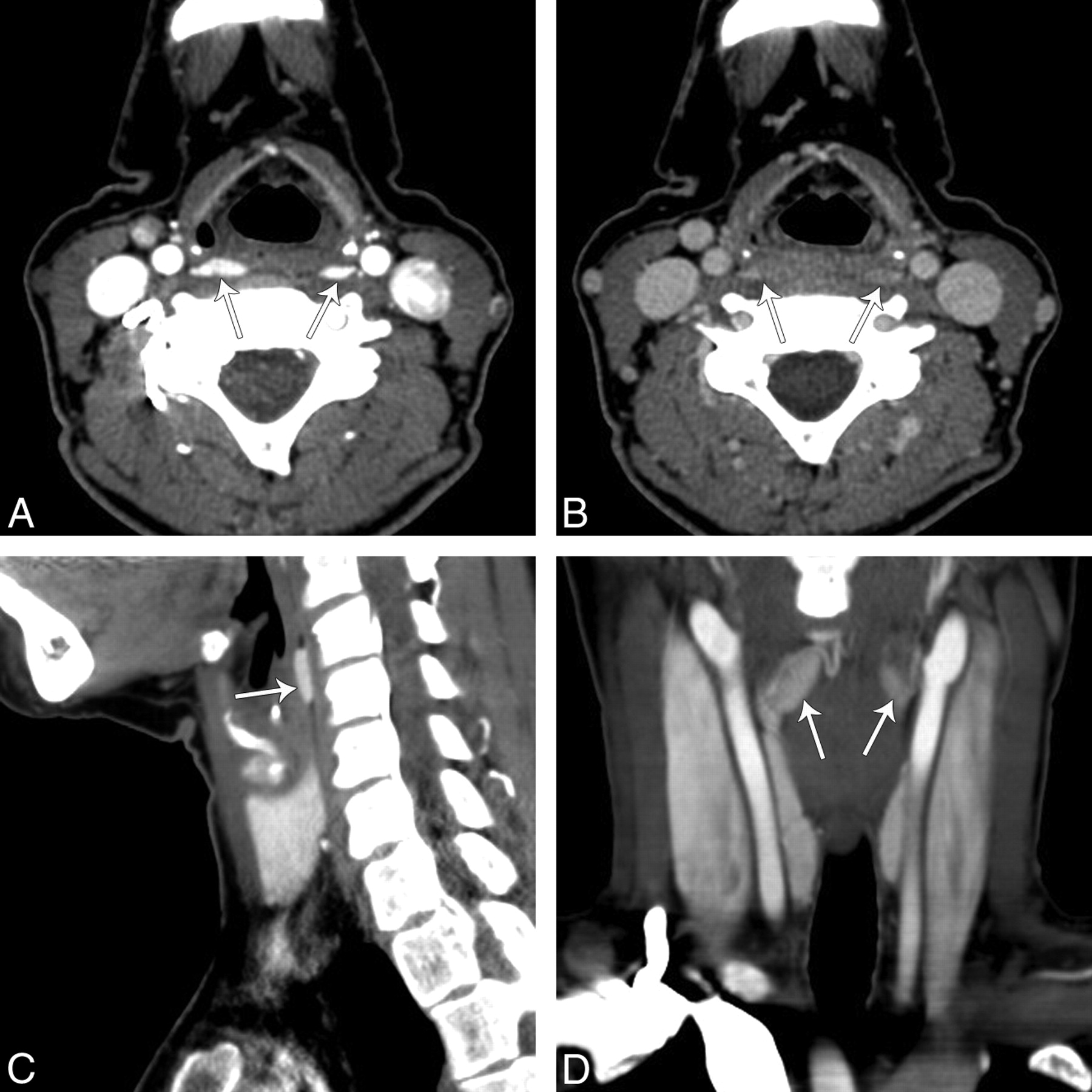
4D-CT of the neck showing bilateral retropharyngeal masses. A, An arterial phase axial image of the neck at the level of the pyriform sinus demonstrates 2 hyperenhancing lesions (arrows), right larger than left, in the retropharyngeal space. B, On delayed images at the same level there was rapid washout of contrast in the lesions. Reformatted right sagittal (C) and coronal arterial phase (D) images demonstrate that hyperenhancing lesions (arrows) in the retropharyngeal space have an oval shape.

On arterial phase images, 2 hyperenhancing lesions, right larger than left, were seen in the retropharyngeal space at the level of the pyriform sinus. On delayed images, there was rapid washout of contrast in the lesions, which were both oval on reformatted sagittal and coronal images.

On the basis of these findings, the surgeon performed a targeted neck exploration in the right retropharyngeal space through a 3-cm incision and identified a 600-mg parathyroid mass. After its removal, intraoperative serum PTH dropped from its highest baseline level but remained elevated. Because of this plateau of the intraoperative PTH level and a probable second abnormality on 4D-CT, the surgeon explored the left retropharyngeal space and found a second 360-mg mass. Following resection of the second mass, the intraoperative serum PTH level normalized and the procedure was concluded.

Pathology showed that both parathyroid masses were hypercellular, consistent with multiple gland parathyroid hyperplasia. At 2-week and 1-month follow-ups, the total calcium and PTH levels were normal.

Discussion

Parathyroid adenomas and hyperplasia are most commonly found posterior to the thyroid between the hyoid and sternal notch. Ectopic locations for parathyroid tumors include mediastinal, intrathyroid, intracarotid sheath, and intraneural sites.3,9 Retropharyngeal ectopic parathyroid lesions are rare and, when present, are a diagnostic challenge for the radiologist and surgeon. A case of bilateral ectopic retropharyngeal parathyroid hyperplasia has not previously been reported.

The retropharyngeal location of the parathyroid gland is due to the common embryologic origin of the superior parathyroid gland and the apex of the pyriform sinus from the fourth branchial pouch of the fetal pharyngeal wall.10 If the parathyroid gland fails to separate from the pyriform sinus, the structures may migrate together to a retropharyngeal location. Hence, retropharyngeal parathyroid tumors are typically found at the level of the pyriform sinus, as shown in our patient.

Bilateral retropharyngeal parathyroid tumors could be mistaken for venous plexuses on a routine axial enhanced CT scan (Fig 2B). 4D-CT helped us make a correct diagnosis. The original description of 4D-CT includes image sets in 3 planes (axial, coronal, sagittal).6 The “fourth” dimension of 4D-CT is the perfusion information derived from noncontrast, arterial, and delayed (venous) phase imaging. In this reported case, the arterial phase imaging was useful for detection of discrete hyperenhancing tumors, and delayed images showed rapid washout of contrast, distinguishing the lesions from surrounding venous structures (Fig 2). The noncontrast phase has been removed from our protocol because we did not find that it helped with diagnosis.

Rodgers et al6 reported that 4D-CT had improved sensitivity (88%) over sestamibi imaging (65%) and sonography (57%) when the studies were used to lateralize hyperfunctioning parathyroid glands to 1 side of the neck. Furthermore, when used to localize parathyroid tumors to the correct quadrant of the neck, the sensitivity of 4D-CT (70%) was significantly higher than that of sestamibi imaging (33%) and sonography (29%). In another study of patients with repeat neck surgery, 4D-CT more often localized and lateralized hyperfunctioning parathyroid tissue than sestamibi imaging.8 4D-CT has also been shown to have better sensitivity for the detection of multiglandular disease, which is a common cause for repeat surgery.6 The advantage of 4D-CT was illustrated in our case because the sestamibi study did not demonstrate the surgically proved left retropharyngeal hyperplastic gland, a phenomenon thought to be due to rapid washout of radiotracer from the left parathyroid tumor,11,12 a potential pitfall of sestamibi imaging.

A disadvantage of 4D-CT is its increased radiation exposure to the patient. To reduce the radiation dose, we modified our current protocol to exclude the precontrast imaging described in the original studies of 4D-CT. We also use dose-reduction techniques of automatic tube current modulation, and we limit the superior level of imaging to the lower mandible to exclude the lens of the orbit and the brain. Despite these attempts, the dose-length product for arterial and venous phase in this study was 1300 mGy cm, which is equivalent to just <2 CT neck scans with an optimized dose-reduction technique.13 Hence, 4D-CT should be reserved for cases of primary hyperparathyroidism with indeterminate or negative findings on sonography and sestamibi scintigraphy.

MR imaging with contrast is another imaging option, depending on the experience of the institution.14,15 MR imaging has a sensitivity similar to that of sonography and sestamibi imaging.16 On MR imaging, parathyroid adenomas may have variable T1 and T2 signal-intensity patterns. Contrast imaging increases the sensitivity for detection of lesions that are T1 and T2 isointense, but false-negative results can still arise from motion artifacts or because adenomas may have signal-intensity and enhancement characteristics similar to those of cervical lymph nodes.17

This case of bilateral retropharyngeal parathyroid hyperplasia shows the value of 4D-CT in defining a discrete parathyroid lesion and in determining the enhancement characteristics of the lesion. Accurate localization of parathyroid tumors is important for repeat parathyroid surgery and for minimally invasive directed parathyroidectomy.

Acknowledgment

We thank Michael Hanson, MD, for his clinical input.

References

  1. 1.↵
    1. Schachter PP,
    2. Ayesh S,
    3. Schneider T,
    4. et al
    . Expression of kinase genes in primary hyperparathyroidism: adenoma versus hyperplastic parathyroid tissue. Surgery 2002; 132: 1094–98, discussion 1098–99
    CrossRefPubMed
  2. 2.↵
    1. Thompson NW,
    2. Eckhauser FE,
    3. Harness JK
    . The anatomy of primary hyperparathyroidism. Surgery 1982; 92: 814–21
    PubMed
  3. 3.↵
    1. Lumachi F,
    2. Zucchetta P,
    3. Varotto S,
    4. et al
    . Noninvasive localization procedures in ectopic hyperfunctioning parathyroid tumors. Endocr Relat Cancer 1999; 6: 123–25
    Abstract
  4. 4.↵
    1. Ruda JM,
    2. Hollenbeak CS,
    3. Stack BC Jr..
    A systematic review of the diagnosis and treatment of primary hyperparathyroidism from 1995 to 2003. Otolaryngol Head Neck Surg 2005; 132: 359–72
    Abstract/FREE Full Text
  5. 5.↵
    1. Adil E,
    2. Adil T,
    3. Fedok F,
    4. et al
    . Minimally invasive radioguided parathyroidectomy performed for primary hyperparathyroidism. Otolaryngol Head Neck Surg 2009; 141: 34–38
    Abstract/FREE Full Text
  6. 6.↵
    1. Rodgers SE,
    2. Hunter GJ,
    3. Hamberg LM,
    4. et al
    . Improved preoperative planning for directed parathyroidectomy with 4-dimensional computed tomography. Surgery 2006; 140: 932–40, discussion 940–41
    CrossRefPubMed
  7. 7.↵
    1. Philip M,
    2. Guerrero MA,
    3. Evans DB,
    4. et al
    . Efficacy of 4D-CT preoperative localization in 2 patients with MEN 2A. J Surg Educ 2008; 65: 182–85
    CrossRefPubMed
  8. 8.↵
    1. Mortenson MM,
    2. Evans DB,
    3. Lee JE,
    4. et al
    . Parathyroid exploration in the reoperative neck: improved preoperative localization with 4D-computed tomography. J Am Coll Surg 2008; 206: 888–95, discussion 895–96
    CrossRefPubMed
  9. 9.↵
    1. Chan TJ,
    2. Libutti SK,
    3. McCart JA,
    4. et al
    . Persistent primary hyperparathyroidism caused by adenomas identified in pharyngeal or adjacent structures. World J Surg 2003; 27: 675–79
    CrossRefPubMed
  10. 10.↵
    1. Benson MT,
    2. Dalen K,
    3. Mancuso AA,
    4. et al
    . Congenital anomalies of the branchial apparatus: embryology and pathologic anatomy. Radiographics 1992; 12: 943–60
    CrossRefPubMed
  11. 11.↵
    1. Benard F,
    2. Lefebvre B,
    3. Beuvon F,
    4. et al
    . Rapid washout of technetium-99m-MIBI from a large parathyroid adenoma. J Nucl Med 1995; 36: 241–43
    Abstract/FREE Full Text
  12. 12.↵
    1. Nguyen BD
    . Parathyroid imaging with Tc-99m sestamibi planar and SPECT scintigraphy. Radiographics 1999; 19: 601–14, discussion 615–16
    PubMed
  13. 13.↵
    1. Russell MT,
    2. Fink JR,
    3. Rebeles F,
    4. et al
    . Balancing radiation dose and image quality: clinical applications of neck volume CT. AJNR Am J Neuroradiol 2008; 29: 727–31
    Abstract/FREE Full Text
  14. 14.↵
    1. Fakhran S,
    2. Branstetter BF 4th.,
    3. Pryma DA
    . Parathyroid imaging. Neuroimaging Clin N Am 2008; 18: 537–49, ix
    CrossRefPubMed
  15. 15.↵
    1. Lee VS,
    2. Spritzer CE,
    3. Coleman RE,
    4. et al.
    The complementary roles of fast spin-echo MR imaging and double-phase 99m Tc-sestamibi scintigraphy for localization of hyperfunctioning parathyroid glands. AJR Am J Roentgenol 1996; 167: 1555–62
    PubMed
  16. 16.↵
    1. Lopez Hanninen E,
    2. Vogl TJ,
    3. Steinmuller T,
    4. et al
    . Preoperative contrast-enhanced MRI of the parathyroid glands in hyperparathyroidism. Invest Radiol 2000; 35: 426–30
    CrossRefPubMed
  17. 17.↵
    1. Johnson NA,
    2. Tublin ME,
    3. Ogilvie JB
    . Parathyroid imaging: technique and role in the preoperative evaluation of primary hyperparathyroidism. AJR Am J Roentgenol 2007; 188: 1706–15
    CrossRefPubMed
  • Received February 5, 2010.
  • Accepted after revision February 20, 2010.
  • Copyright © American Society of Neuroradiology
View Abstract
PreviousNext
Back to top

In this issue

American Journal of Neuroradiology: 32 (5)
American Journal of Neuroradiology
Vol. 32, Issue 5
1 May 2011
  • Table of Contents
  • Index by author
Advertisement
Print
Download PDF
Email Article

Thank you for your interest in spreading the word on American Journal of Neuroradiology.

NOTE: We only request your email address so that the person you are recommending the page to knows that you wanted them to see it, and that it is not junk mail. We do not capture any email address.

Enter multiple addresses on separate lines or separate them with commas.
Bilateral Retropharyngeal Parathyroid Hyperplasia Detected with 4D Multidetector Row CT
(Your Name) has sent you a message from American Journal of Neuroradiology
(Your Name) thought you would like to see the American Journal of Neuroradiology web site.
CAPTCHA
This question is for testing whether or not you are a human visitor and to prevent automated spam submissions.
Cite this article
R.D. Welling, J.A. Olson, P.G. Kranz, J.D. Eastwood, J.K. Hoang
Bilateral Retropharyngeal Parathyroid Hyperplasia Detected with 4D Multidetector Row CT
American Journal of Neuroradiology May 2011, 32 (5) E80-E82; DOI: 10.3174/ajnr.A2104

Citation Manager Formats

  • BibTeX
  • Bookends
  • EasyBib
  • EndNote (tagged)
  • EndNote 8 (xml)
  • Medlars
  • Mendeley
  • Papers
  • RefWorks Tagged
  • Ref Manager
  • RIS
  • Zotero
0 Responses
Respond to this article
Share
Bookmark this article
Bilateral Retropharyngeal Parathyroid Hyperplasia Detected with 4D Multidetector Row CT
R.D. Welling, J.A. Olson, P.G. Kranz, J.D. Eastwood, J.K. Hoang
American Journal of Neuroradiology May 2011, 32 (5) E80-E82; DOI: 10.3174/ajnr.A2104
del.icio.us logo Twitter logo Facebook logo Mendeley logo
  • Tweet Widget
  • Facebook Like
  • Google Plus One
Purchase

Jump to section

  • Article
    • Abstract
    • Abbreviations
    • Case Report
    • Discussion
    • Acknowledgment
    • References
  • Figures & Data
  • Info & Metrics
  • Responses
  • References
  • PDF

Related Articles

  • No related articles found.
  • PubMed
  • Google Scholar

Cited By...

  • 4D-CT for Preoperative Localization of Abnormal Parathyroid Glands in Patients with Hyperparathyroidism: Accuracy and Ability to Stratify Patients by Unilateral versus Bilateral Disease in Surgery-Naive and Re-Exploration Patients
  • Parathyroid Lesions: Characterization with Dual-Phase Arterial and Venous Enhanced CT of the Neck
  • Crossref (22)
  • Google Scholar

This article has been cited by the following articles in journals that are participating in Crossref Cited-by Linking.

  • How to Perform Parathyroid 4D CT: Tips and Traps for Technique and Interpretation
    Jenny K. Hoang, Won-kyung Sung, Manisha Bahl, C. Douglas Phillips
    Radiology 2014 270 1
  • Diagnostic Accuracy of 4D-CT for Parathyroid Adenomas and Hyperplasia
    J.L. Chazen, A. Gupta, A. Dunning, C.D. Phillips
    American Journal of Neuroradiology 2012 33 3
  • 4D-CT for Preoperative Localization of Abnormal Parathyroid Glands in Patients with Hyperparathyroidism: Accuracy and Ability to Stratify Patients by Unilateral versus Bilateral Disease in Surgery-Naïve and Re-Exploration Patients
    H.R. Kelly, L.M. Hamberg, G.J. Hunter
    American Journal of Neuroradiology 2014 35 1
  • Lifetime Attributable Risk of Cancer From Radiation Exposure During Parathyroid Imaging: Comparison of 4D CT and Parathyroid Scintigraphy
    Jenny K. Hoang, Robert E. Reiman, Giao B. Nguyen, Natalie Januzis, Bennett B. Chin, Carolyn Lowry, Terry T. Yoshizumi
    American Journal of Roentgenology 2015 204 5
  • Parathyroid Lesions: Characterization with Dual-Phase Arterial and Venous Enhanced CT of the Neck
    A.R. Gafton, C.M. Glastonbury, J.D. Eastwood, J.K. Hoang
    American Journal of Neuroradiology 2012 33 5
  • Imaging of the Parathyroid Glands
    C. Douglas Phillips, Deborah R. Shatzkes
    Seminars in Ultrasound, CT and MRI 2012 33 2
  • Discriminating Parathyroid Adenoma from Local Mimics by Using Inherent Tissue Attenuation and Vascular Information Obtained with Four-Dimensional CT: Formulation of a Multinomial Logistic Regression Model
    George J. Hunter, Daniel T. Ginat, Hillary R. Kelly, Elkan F. Halpern, Leena M. Hamberg
    Radiology 2014 270 1
  • 3-phase dual-energy CT scan as a feasible salvage imaging modality for the identification of non-localizing parathyroid adenomas: A prospective study
    Michael Roskies, Xiaoyang Liu, Michael P. Hier, Richard J. Payne, Alex Mlynarek, Veronique Forest, Mark Levental, Reza Forghani
    Journal of Otolaryngology - Head & Neck Surgery 2015 44 1
  • Update on Preoperative Parathyroid Localization in Primary Hyperparathyroidism
    Hye-Sun Park, Namki Hong, Jong Ju Jeong, Mijin Yun, Yumie Rhee
    Endocrinology and Metabolism 2022 37 5

More in this TOC Section

  • Atypical Diffusion-Restricted Lesion in 5-Fluorouracil Encephalopathy
  • Dural Infantile Hemangioma Masquerading as a Skull Vault Lesion
  • Multimodal Imaging of Spike Propagation: A Technical Case Report
Show more Case Reports

Similar Articles

Advertisement

Indexed Content

  • Current Issue
  • Accepted Manuscripts
  • Article Preview
  • Past Issues
  • Editorials
  • Editor's Choice
  • Fellows' Journal Club
  • Letters to the Editor
  • Video Articles

Cases

  • Case Collection
  • Archive - Case of the Week
  • Archive - Case of the Month
  • Archive - Classic Case

More from AJNR

  • Trainee Corner
  • Imaging Protocols
  • MRI Safety Corner
  • Book Reviews

Multimedia

  • AJNR Podcasts
  • AJNR Scantastics

Resources

  • Turnaround Time
  • Submit a Manuscript
  • Submit a Video Article
  • Submit an eLetter to the Editor/Response
  • Manuscript Submission Guidelines
  • Statistical Tips
  • Fast Publishing of Accepted Manuscripts
  • Graphical Abstract Preparation
  • Imaging Protocol Submission
  • Evidence-Based Medicine Level Guide
  • Publishing Checklists
  • Author Policies
  • Become a Reviewer/Academy of Reviewers
  • News and Updates

About Us

  • About AJNR
  • Editorial Board
  • Editorial Board Alumni
  • Alerts
  • Permissions
  • Not an AJNR Subscriber? Join Now
  • Advertise with Us
  • Librarian Resources
  • Feedback
  • Terms and Conditions
  • AJNR Editorial Board Alumni

American Society of Neuroradiology

  • Not an ASNR Member? Join Now

© 2025 by the American Society of Neuroradiology All rights, including for text and data mining, AI training, and similar technologies, are reserved.
Print ISSN: 0195-6108 Online ISSN: 1936-959X

Powered by HighWire