Advertisement

Main menu

  • Home
  • Content
    • Current Issue
    • Accepted Manuscripts
    • Article Preview
    • Past Issue Archive
    • Video Articles
    • AJNR Case Collection
    • Case of the Week Archive
    • Case of the Month Archive
    • Classic Case Archive
  • Special Collections
    • Advancing NeuroMRI with High-Relaxivity Contrast Agents
    • AJNR Awards
    • Low-Field MRI
    • Alzheimer Disease
    • ASNR Foundation Special Collection
    • Photon-Counting CT
    • View All
  • Multimedia
    • AJNR Podcasts
    • AJNR SCANtastic
    • Trainee Corner
    • MRI Safety Corner
    • Imaging Protocols
  • For Authors
    • Submit a Manuscript
    • Submit a Video Article
    • Submit an eLetter to the Editor/Response
    • Manuscript Submission Guidelines
    • Statistical Tips
    • Fast Publishing of Accepted Manuscripts
    • Graphical Abstract Preparation
    • Imaging Protocol Submission
    • Author Policies
  • About Us
    • About AJNR
    • Editorial Board
    • Editorial Board Alumni
  • More
    • Become a Reviewer/Academy of Reviewers
    • Subscribers
    • Permissions
    • Alerts
    • Feedback
    • Advertisers
    • ASNR Home

User menu

  • Alerts
  • Log in

Search

  • Advanced search
American Journal of Neuroradiology

American Journal of Neuroradiology

ASHNR American Society of Functional Neuroradiology ASHNR American Society of Pediatric Neuroradiology ASSR
  • Alerts
  • Log in

Advanced Search

  • Home
  • Content
    • Current Issue
    • Accepted Manuscripts
    • Article Preview
    • Past Issue Archive
    • Video Articles
    • AJNR Case Collection
    • Case of the Week Archive
    • Case of the Month Archive
    • Classic Case Archive
  • Special Collections
    • Advancing NeuroMRI with High-Relaxivity Contrast Agents
    • AJNR Awards
    • Low-Field MRI
    • Alzheimer Disease
    • ASNR Foundation Special Collection
    • Photon-Counting CT
    • View All
  • Multimedia
    • AJNR Podcasts
    • AJNR SCANtastic
    • Trainee Corner
    • MRI Safety Corner
    • Imaging Protocols
  • For Authors
    • Submit a Manuscript
    • Submit a Video Article
    • Submit an eLetter to the Editor/Response
    • Manuscript Submission Guidelines
    • Statistical Tips
    • Fast Publishing of Accepted Manuscripts
    • Graphical Abstract Preparation
    • Imaging Protocol Submission
    • Author Policies
  • About Us
    • About AJNR
    • Editorial Board
    • Editorial Board Alumni
  • More
    • Become a Reviewer/Academy of Reviewers
    • Subscribers
    • Permissions
    • Alerts
    • Feedback
    • Advertisers
    • ASNR Home
  • Follow AJNR on Twitter
  • Visit AJNR on Facebook
  • Follow AJNR on Instagram
  • Join AJNR on LinkedIn
  • RSS Feeds

AJNR Awards, New Junior Editors, and more. Read the latest AJNR updates


Improved Turnaround Times | Median time to first decision: 12 days

Case ReportCase Report

High T2 Signal in Primary Lateral Sclerosis Supports the Topographic Distribution of Fibers in the Corpus Callosum: Assessing Disease in the Primary Motor Segment

S.M. Riad, H. Hathout and J.C. Huang
American Journal of Neuroradiology April 2011, 32 (4) E61-E64; DOI: https://doi.org/10.3174/ajnr.A2067
S.M. Riad
  • Find this author on Google Scholar
  • Find this author on PubMed
  • Search for this author on this site
H. Hathout
  • Find this author on Google Scholar
  • Find this author on PubMed
  • Search for this author on this site
J.C. Huang
  • Find this author on Google Scholar
  • Find this author on PubMed
  • Search for this author on this site
  • Article
  • Figures & Data
  • Info & Metrics
  • Responses
  • References
  • PDF
Loading

Article Figures & Data

Figures

  • Tables
  • Fig 1.
    • Download figure
    • Open in new tab
    • Download powerpoint
    Fig 1.

    Case 1. Axial FLAIR image.

  • Fig 2.
    • Download figure
    • Open in new tab
    • Download powerpoint
    Fig 2.

    Case 1. Midsagittal T1-weighted MR image.
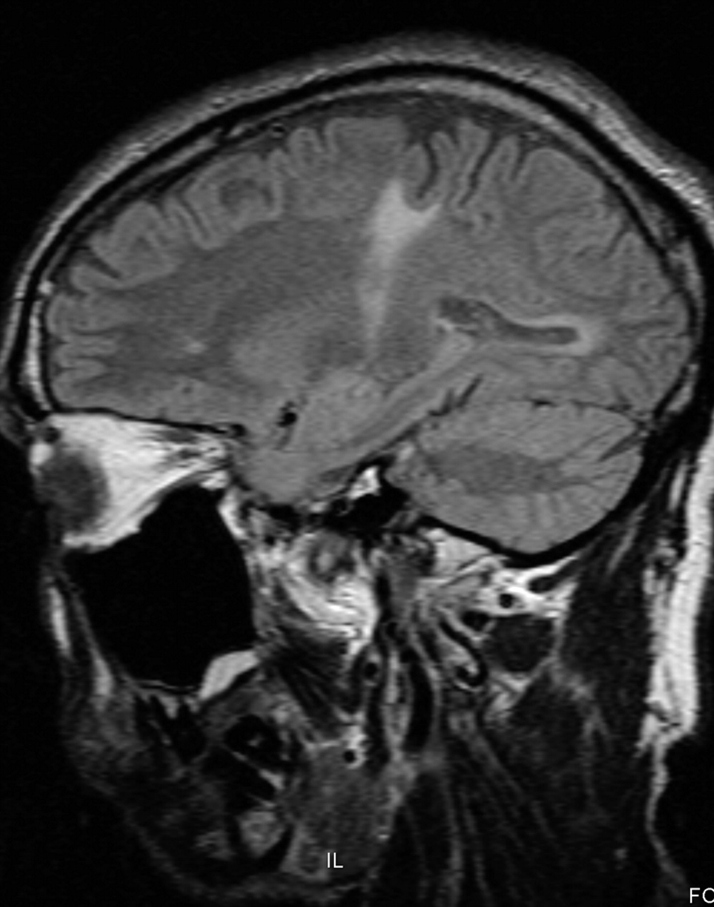
  • Fig 3.
    • Download figure
    • Open in new tab
    • Download powerpoint
    Fig 3.

    Case 1. Coronal T1-weighted MR image.

  • Fig 4.
    • Download figure
    • Open in new tab
    • Download powerpoint
    Fig 4.

    Case 1. Parasagittal T1-weighted MR image.

  • Fig 5.
    • Download figure
    • Open in new tab
    • Download powerpoint
    Fig 5.

    Case 2. A, Midsagittal FLAIR image. B, Primary motor segment of the CC, as defined by tractography.27 Reprinted with permission from Hofer and Frahm,27 and the Copyright Clearance Center of Elsevier. C, Graphic overlay of images from Fig 5A and B. MR imaging findings depicted in Fig 5A appear to involve the primary motor segment of the CC.

  • Fig 6.
    • Download figure
    • Open in new tab
    • Download powerpoint
    Fig 6.

    Case 2. Parasagittal FLAIR image.

  • Fig 7.
    • Download figure
    • Open in new tab
    • Download powerpoint
    Fig 7.

    Case 2. Coronal T1-weighted MR image.

  • Fig 8.
    • Download figure
    • Open in new tab
    • Download powerpoint
    Fig 8.

    Case 2. Axial FLAIR image.

  • Fig 9.
    • Download figure
    • Open in new tab
    • Download powerpoint
    Fig 9.

    Midsagittal schematic of the CC, showing Witelson scheme (top) and Hofer and Frahm scheme (bottom). Witelson scheme: I, premotor; II, motor; III, somesthetic; IV, parietal/temporal; V, temporal/occipital. Hofer and Frahm scheme: I, prefrontal; II, premotor; III, motor; IV, sensory; V, parietal/occipital/temporal. Note the posterior shift in the location of the primary motor segment from Witelson segment II to Hofer and Frahm segment III. Reprinted with permission from Hofer and Frahm,27 and the Copyright Clearance Center of Elsevier.

Tables

  • Figures
  • Region-of-interest analysis for white matter MR imaging findings in cases 1 and 2

    Area (cm2)Signal Intensity
    Case 1, Axial FLAIR section 5
        Left lesion0.08554.6
        Right lesion0.11596.3
        Left normal0.11382.0
        Right normal0.11381.6
        Background air0.1714.3
    Case 2, Axial FLAIR section 6
        Left lesion0.22588.5
        Right lesion0.16477.2
        Left normal0.19320.3
        Right normal0.19318.6
        Background air0.239.5
    Case 2, Sagittal FLAIR section 12
        CC lesion0.07540.0
        CC normal0.09263.8
        Background air0.1421.7
PreviousNext
Back to top

In this issue

American Journal of Neuroradiology: 32 (4)
American Journal of Neuroradiology
Vol. 32, Issue 4
1 Apr 2011
  • Table of Contents
  • Index by author
Advertisement
Print
Download PDF
Email Article

Thank you for your interest in spreading the word on American Journal of Neuroradiology.

NOTE: We only request your email address so that the person you are recommending the page to knows that you wanted them to see it, and that it is not junk mail. We do not capture any email address.

Enter multiple addresses on separate lines or separate them with commas.
High T2 Signal in Primary Lateral Sclerosis Supports the Topographic Distribution of Fibers in the Corpus Callosum: Assessing Disease in the Primary Motor Segment
(Your Name) has sent you a message from American Journal of Neuroradiology
(Your Name) thought you would like to see the American Journal of Neuroradiology web site.
CAPTCHA
This question is for testing whether or not you are a human visitor and to prevent automated spam submissions.
Cite this article
S.M. Riad, H. Hathout, J.C. Huang
High T2 Signal in Primary Lateral Sclerosis Supports the Topographic Distribution of Fibers in the Corpus Callosum: Assessing Disease in the Primary Motor Segment
American Journal of Neuroradiology Apr 2011, 32 (4) E61-E64; DOI: 10.3174/ajnr.A2067

Citation Manager Formats

  • BibTeX
  • Bookends
  • EasyBib
  • EndNote (tagged)
  • EndNote 8 (xml)
  • Medlars
  • Mendeley
  • Papers
  • RefWorks Tagged
  • Ref Manager
  • RIS
  • Zotero
0 Responses
Respond to this article
Share
Bookmark this article
High T2 Signal in Primary Lateral Sclerosis Supports the Topographic Distribution of Fibers in the Corpus Callosum: Assessing Disease in the Primary Motor Segment
S.M. Riad, H. Hathout, J.C. Huang
American Journal of Neuroradiology Apr 2011, 32 (4) E61-E64; DOI: 10.3174/ajnr.A2067
del.icio.us logo Twitter logo Facebook logo Mendeley logo
  • Tweet Widget
  • Facebook Like
  • Google Plus One
Purchase

Jump to section

  • Article
    • Abstract
    • Abbreviations
    • Case Reports
    • Results
    • Discussion
    • References
  • Figures & Data
  • Info & Metrics
  • Responses
  • References
  • PDF

Related Articles

  • No related articles found.
  • PubMed
  • Google Scholar

Cited By...

  • A Distinct MR Imaging Phenotype in Amyotrophic Lateral Sclerosis: Correlation between T1 Magnetization Transfer Contrast Hyperintensity along the Corticospinal Tract and Diffusion Tensor Imaging Analysis
  • Alterations of the Corpus Callosum as an MR Imaging-Based Hallmark of Motor Neuron Diseases
  • Crossref
  • Google Scholar

This article has not yet been cited by articles in journals that are participating in Crossref Cited-by Linking.

More in this TOC Section

  • Atypical Diffusion-Restricted Lesion in 5-Fluorouracil Encephalopathy
  • Dural Infantile Hemangioma Masquerading as a Skull Vault Lesion
  • Multimodal Imaging of Spike Propagation: A Technical Case Report
Show more Case Reports

Similar Articles

Advertisement

Indexed Content

  • Current Issue
  • Accepted Manuscripts
  • Article Preview
  • Past Issues
  • Editorials
  • Editor's Choice
  • Fellows' Journal Club
  • Letters to the Editor
  • Video Articles

Cases

  • Case Collection
  • Archive - Case of the Week
  • Archive - Case of the Month
  • Archive - Classic Case

More from AJNR

  • Trainee Corner
  • Imaging Protocols
  • MRI Safety Corner
  • Book Reviews

Multimedia

  • AJNR Podcasts
  • AJNR Scantastics

Resources

  • Turnaround Time
  • Submit a Manuscript
  • Submit a Video Article
  • Submit an eLetter to the Editor/Response
  • Manuscript Submission Guidelines
  • Statistical Tips
  • Fast Publishing of Accepted Manuscripts
  • Graphical Abstract Preparation
  • Imaging Protocol Submission
  • Evidence-Based Medicine Level Guide
  • Publishing Checklists
  • Author Policies
  • Become a Reviewer/Academy of Reviewers
  • News and Updates

About Us

  • About AJNR
  • Editorial Board
  • Editorial Board Alumni
  • Alerts
  • Permissions
  • Not an AJNR Subscriber? Join Now
  • Advertise with Us
  • Librarian Resources
  • Feedback
  • Terms and Conditions
  • AJNR Editorial Board Alumni

American Society of Neuroradiology

  • Not an ASNR Member? Join Now

© 2025 by the American Society of Neuroradiology All rights, including for text and data mining, AI training, and similar technologies, are reserved.
Print ISSN: 0195-6108 Online ISSN: 1936-959X

Powered by HighWire