Advertisement

Main menu

  • Home
  • Content
    • Current Issue
    • Accepted Manuscripts
    • Article Preview
    • Past Issue Archive
    • Video Articles
    • AJNR Case Collection
    • Case of the Week Archive
    • Case of the Month Archive
    • Classic Case Archive
  • Special Collections
    • Advancing NeuroMRI with High-Relaxivity Contrast Agents
    • AJNR Awards
    • Low-Field MRI
    • Alzheimer Disease
    • ASNR Foundation Special Collection
    • Photon-Counting CT
    • View All
  • Multimedia
    • AJNR Podcasts
    • AJNR SCANtastic
    • Trainee Corner
    • MRI Safety Corner
    • Imaging Protocols
  • For Authors
    • Submit a Manuscript
    • Submit a Video Article
    • Submit an eLetter to the Editor/Response
    • Manuscript Submission Guidelines
    • Statistical Tips
    • Fast Publishing of Accepted Manuscripts
    • Graphical Abstract Preparation
    • Imaging Protocol Submission
    • Author Policies
  • About Us
    • About AJNR
    • Editorial Board
    • Editorial Board Alumni
  • More
    • Become a Reviewer/Academy of Reviewers
    • Subscribers
    • Permissions
    • Alerts
    • Feedback
    • Advertisers
    • ASNR Home

User menu

  • Alerts
  • Log in

Search

  • Advanced search
American Journal of Neuroradiology

American Journal of Neuroradiology

ASHNR American Society of Functional Neuroradiology ASHNR American Society of Pediatric Neuroradiology ASSR
  • Alerts
  • Log in

Advanced Search

  • Home
  • Content
    • Current Issue
    • Accepted Manuscripts
    • Article Preview
    • Past Issue Archive
    • Video Articles
    • AJNR Case Collection
    • Case of the Week Archive
    • Case of the Month Archive
    • Classic Case Archive
  • Special Collections
    • Advancing NeuroMRI with High-Relaxivity Contrast Agents
    • AJNR Awards
    • Low-Field MRI
    • Alzheimer Disease
    • ASNR Foundation Special Collection
    • Photon-Counting CT
    • View All
  • Multimedia
    • AJNR Podcasts
    • AJNR SCANtastic
    • Trainee Corner
    • MRI Safety Corner
    • Imaging Protocols
  • For Authors
    • Submit a Manuscript
    • Submit a Video Article
    • Submit an eLetter to the Editor/Response
    • Manuscript Submission Guidelines
    • Statistical Tips
    • Fast Publishing of Accepted Manuscripts
    • Graphical Abstract Preparation
    • Imaging Protocol Submission
    • Author Policies
  • About Us
    • About AJNR
    • Editorial Board
    • Editorial Board Alumni
  • More
    • Become a Reviewer/Academy of Reviewers
    • Subscribers
    • Permissions
    • Alerts
    • Feedback
    • Advertisers
    • ASNR Home
  • Follow AJNR on Twitter
  • Visit AJNR on Facebook
  • Follow AJNR on Instagram
  • Join AJNR on LinkedIn
  • RSS Feeds

AJNR Awards, New Junior Editors, and more. Read the latest AJNR updates


Improved Turnaround Times | Median time to first decision: 12 days

Case ReportCase Report

High T2 Signal in Primary Lateral Sclerosis Supports the Topographic Distribution of Fibers in the Corpus Callosum: Assessing Disease in the Primary Motor Segment

S.M. Riad, H. Hathout and J.C. Huang
American Journal of Neuroradiology April 2011, 32 (4) E61-E64; DOI: https://doi.org/10.3174/ajnr.A2067
S.M. Riad
  • Find this author on Google Scholar
  • Find this author on PubMed
  • Search for this author on this site
H. Hathout
  • Find this author on Google Scholar
  • Find this author on PubMed
  • Search for this author on this site
J.C. Huang
  • Find this author on Google Scholar
  • Find this author on PubMed
  • Search for this author on this site
  • Article
  • Figures & Data
  • Info & Metrics
  • Responses
  • References
  • PDF
Loading

Abstract

SUMMARY: PLS is a disease of the UMN, distinguished from ALS in prognosis and absence of LMN signs. We present, to our knowledge, the first conventional MR imaging visualization of the callosal motor segment, a concept previously supported by primate models, electrophysiologic studies, and postmortem examinations. Modification of the Witelson topographic scheme of the CC is supported by MR tractography. On the basis of 2 cases of PLS, we present conventional imaging confirmation of the revised topographic scheme of fiber distribution across the CC.

Abbreviations

ALS
amyotrophic lateral sclerosis
CC
corpus callosum
CST
corticospinal tract
DTI
diffusion tensor imaging
FA
fractional anisotropy
FLAIR
fluid-attenuated inversion recovery
LMN
lower motor neuron
PLS
primary lateral sclerosis
UMN
upper motor neuron

PLS is a progressive pure UMN disorder first described in the late 1800s by Charcot and Erb; it can be distinguished from ALS by its characteristic absence of LMN signs. Both conditions arise sporadically, and both have been linked to specific mutations,1 though PLS is far less common than ALS, constituting as little as 1% of motor neuron diseases.2 Since diagnostic criteria for PLS were first proposed,3 reports have questioned its distinction from ALS, instead viewing PLS as an indolent variant with an exclusively UMN presentation.4–6 However, further case series7,8 have underscored the association between longer survival time and UMN-predominant disease, making the distinction between ALS and PLS clinically relevant and suggesting at least a separate syndromology.

ALS and PLS share MR imaging features such as T2 hyperintensity within the CST,9–11 atrophy of the motor and premotor cortex,3,12 MR imaging signal-intensity changes within the precentral gyrus,10 and decreased FA on tensor imaging of the descending motor pathway.13,14

Furthermore, PLS and ALS both manifest involvement of the CC, including loss of contralateral motor inhibition on behavioral testing,15,16 decreased FA on DTI,17,18 and an isolated report of callosal T2 hyperintensity in a case of ALS.19 We present 2 cases of PLS demonstrating increased T2/FLAIR signal intensity within the posterior body of the CC and discuss their relevance to understanding the topographic distribution of cortico-cortical interhemispheric projections.

Case Reports

The 2 patients presented herein were referred from the neurology department with a diagnosis of PLS. For both patients, clinical and laboratory findings were consistent with a diagnosis of “clinically pure PLS” according to the diagnostic criteria put forth by Gordon et al.7

Case 1

A 50-year-old man presented for adjustment of his medications. His medical history was significant for the onset of progressive right-leg weakness beginning in his late 20s, followed 3 years later by right upper extremity weakness. Within 5 years, spasticity and weakness of all 4 extremities prevented the patient from working. Electromyography was negative for lower motor neuron involvement. As the disease progressed, the patient became wheelchair-dependent. One year prior, he underwent bilateral tendon release surgeries for Achilles contractures.

His examination was significant for hyperreflexia and spasticity in all 4 limbs, minimal voluntary movement of his upper extremities, lower extremity paresis, and dysarthria.

Case 2

A 59-year-old man, diagnosed previously with multiple sclerosis presented to the neurology clinic for a second opinion. He experienced the onset of right-hand weakness at 39 years of age, and during the next decade, developed right-leg weakness, dysarthria, and chronic urinary urgency. Neurologic examination in the clinic demonstrated right-sided spasticity, right pronator drift, dysarthria, frontal release signs, and global hyperreflexia. Ankle clonus and Babinsky signs were elicited bilaterally. Lumbar puncture was negative for oligoclonal bands.

Results

MR mages were acquired on a Magnetom Sonata 1.5T scanner (Siemens, Erlangen, Germany).

Case 1

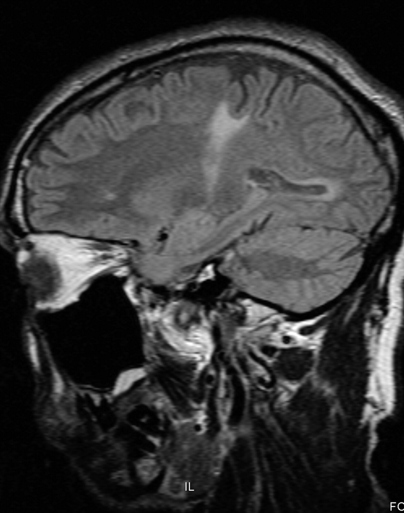
MR images included axial FLAIR (TR, 9000 ms; TE, 79 ms; TI, 2500 ms; flip angle, 15°; section thickness, 5 mm; matrix, 256 × 192; FOV, 240 × 240; NEX, 1), sagittal T1 (TR, 535 ms; TE, 15 ms; section thickness, 5 mm; matrix, 256 × 192; FOV, 240 × 240; NEX, 1), and coronal TI postcontrast sequences(TR, 687 ms; TE, 15 ms; section thickness, 5 mm; matrix, 256 × 192; FOV, 240 × 240; NEX, 1). Figure 1 is a representative axial FLAIR image (section 8) with a zone of T2 hyperintense signal in the left precentral gyrus extending across the CC toward the contralateral motor region. A midsagittal T1-weighted image from the same patient shown in Fig 2 (section 11) demonstrates focal volume loss in the posterior body of the CC. Figures 3 and 4 are coronal (section 9) and sagittal (section 14) T1 images demonstrating low signal within the CST. The bilateral CST involvement illustrated in Fig 3 was concordant with the patient's clinical presentation.

Fig 1.
  • Download figure
  • Open in new tab
  • Download powerpoint
Fig 1.

Case 1. Axial FLAIR image.

Fig 2.
  • Download figure
  • Open in new tab
  • Download powerpoint
Fig 2.

Case 1. Midsagittal T1-weighted MR image.

Fig 3.
  • Download figure
  • Open in new tab
  • Download powerpoint
Fig 3.

Case 1. Coronal T1-weighted MR image.

Fig 4.
  • Download figure
  • Open in new tab
  • Download powerpoint
Fig 4.

Case 1. Parasagittal T1-weighted MR image.

Case 2

Axial FLAIR (TR, 8000 ms; TE, 110 ms; TI, 2500 ms; flip angle, 15°; section thickness, 3 mm; matrix, 320 × 240; FOV, 240 × 240; NEX, 2), sagittal FLAIR (TR, 8710 ms; TE, 79 ms; TI, 2500 ms; flip angle, 15°; section thickness, 3 mm; matrix, 320 × 240; FOV, 240 × 240; NEX, 2), and coronal T1 postcontrast (TR, 450 ms; TE, 14 ms; section thickness, 3 mm; matrix, 256 × 192; FOV, 240 × 240; NEX, 2) sequences were performed. Figure 5A is a midsagittal FLAIR image (section 12) revealing T2 signal-intensity hyperintensity and volume loss in the posterior body of the CC. The parasagittal FLAIR image (section 19) in Fig 6 demonstrates T2 hyperintense signal within the expected course of the CST and corresponds to the distribution of T1 hypointensity in the coronal image shown in Fig 7. Figure 8is an axial FLAIR image (section 33); increased T2 signal intensity is demonstrated in the bilateral CSTs as they travel within the posterior limbs of the internal capsules. Bilateral CST involvement was concordant with the patient's clinical presentation.

Fig 5.
  • Download figure
  • Open in new tab
  • Download powerpoint
Fig 5.

Case 2. A, Midsagittal FLAIR image. B, Primary motor segment of the CC, as defined by tractography.27 Reprinted with permission from Hofer and Frahm,27 and the Copyright Clearance Center of Elsevier. C, Graphic overlay of images from Fig 5A and B. MR imaging findings depicted in Fig 5A appear to involve the primary motor segment of the CC.

Fig 6.
  • Download figure
  • Open in new tab
  • Download powerpoint
Fig 6.

Case 2. Parasagittal FLAIR image.

Fig 7.
  • Download figure
  • Open in new tab
  • Download powerpoint
Fig 7.

Case 2. Coronal T1-weighted MR image.

Fig 8.
  • Download figure
  • Open in new tab
  • Download powerpoint
Fig 8.

Case 2. Axial FLAIR image.

Region-of-Interest Analysis

Representative axial FLAIR images from patients 1 and 2 were selected, and regions of interest were generated within pathologic subcortical white matter (increased T2 signal) and normal-appearing ipsilateral white matter. A midsagittal FLAIR image from patient 2 (Fig 5A) was used for region-of-interest analysis of the CC: Regions of interest were generated within the pathologic white matter in the posterior body and the normal-appearing white matter in the anterior body. Measurements of region-of-interest area and signal intensity are displayed in the Table

View this table:
  • View inline
  • View popup

Region-of-interest analysis for white matter MR imaging findings in cases 1 and 2

Discussion

The CC is the largest white matter tract in the human brain. Knowledge of its functional anatomy derives from several lines of inquiry. Aspects of callosal function are revealed through observation of individuals who have undergone callosotomy for intractable seizures, patients with so-called “split-brain.” Comparison of these patients with those with focal callosal lesions has led to inferences about the organization of transcallosal fibers. Autoradiographic techniques in primates have visualized transcallosal projections of the primary motor cortex to homotopic and heterotopic areas of the contralateral precentral gyrus.20 Postmortem studies in humans have revealed a topographic distribution of fibers in the CC21–23 and have demonstrated heterotopic and homotopic transcallosal projections.24

Transcranial magnetic stimulation, initially a technique used to assess CST integrity,25 has been applied to the study of cortico-cortical interactions. Meyer et al15 demonstrated that stimulation of the primary motor cortex inhibits the contralateral motor cortex, a finding diminished or absent in patients with callosal lesions. By comparing patients with a range of focal callosal lesions, the investigators deduced that motor inhibitory pathways occupy the posterior body of the CC.26 Loss of motor inhibition explains the finding of increased involuntary contralateral comovements in patients with ALS 16 and is consistent with decreased FA values in the CC of patients with ALS and PLS.17,18

The Witelson scheme for partitioning the midsagittal CC had been widely accepted for more than a decade on the basis of data derived from humans and nonhuman primates. The scheme has been challenged by data from DTI-based tractography,27 which modifies the standard scheme (Fig 9), relocating the Witelson motor region (II, anterior midbody) more posteriorly within the CC (III, posterior body). Figure 5B depicts the primary motor segment of the CC, as defined by the tractography data of Hofer and Frahm.27 Figure 5C is a simple overlay of the images in 5A and 5B, localizing the midsagittal MR imaging findings from patient 2 to the callosal motor segment.

Fig 9.
  • Download figure
  • Open in new tab
  • Download powerpoint
Fig 9.

Midsagittal schematic of the CC, showing Witelson scheme (top) and Hofer and Frahm scheme (bottom). Witelson scheme: I, premotor; II, motor; III, somesthetic; IV, parietal/temporal; V, temporal/occipital. Hofer and Frahm scheme: I, prefrontal; II, premotor; III, motor; IV, sensory; V, parietal/occipital/temporal. Note the posterior shift in the location of the primary motor segment from Witelson segment II to Hofer and Frahm segment III. Reprinted with permission from Hofer and Frahm,27 and the Copyright Clearance Center of Elsevier.

We presented 2 cases of PLS, a degenerative disease of the UMN, which demonstrate volume loss and high T2 signal within the posterior body of the CC, corresponding to the primary motor segment in the Hofer and Frahm revised topographic scheme of fiber distribution within the midsagittal CC.27

References

  1. 1.↵
    1. Valdmanis PN,
    2. Dupré N,
    3. Rouleau GA
    . A locus for primary lateral sclerosis on chromosome 4ptel-4p16.1. Arch Neurol 2008;65:383–86
    CrossRefPubMed
  2. 2.↵
    1. Mitsumoto H,
    2. Chad D,
    3. Pioro EP
    1. Mitsumoto H,
    2. Chad D,
    3. Pioro EP
    History, terminology and classification of ALS. In: Mitsumoto H, Chad D, Pioro EP eds. Amyotrophic Lateral Sclerosis. Philadelphia:FA Davis;1998:3–1128
  3. 3.↵
    1. Pringle CE,
    2. Hudson AJ,
    3. Munoz DG,
    4. et al
    . Primary lateral sclerosis: clinical features, neuropathology and diagnostic criteria. Brain 1992;115:495–520
    Abstract/FREE Full Text
  4. 4.↵
    1. Le Forestier N,
    2. Maisonobe T,
    3. Piquard A,
    4. et al
    . Does primary lateral sclerosis exist? A study of 20 patients and a review of the literature. Brain 2001;124:1989–99
  5. 5.↵
    1. Bruyn RP,
    2. Koelman JH,
    3. Troost D,
    4. et al
    . Motor neuron disease (amyotrophic lateral sclerosis) arising from longstanding primary lateral sclerosis. J Neurol Neurosurg Psychiatry 1995;58:742–44
    Abstract/FREE Full Text
  6. 6.↵
    1. Kuipers-Upmeijer J,
    2. de Jager AE,
    3. Hew JM,
    4. et al
    . Primary lateral sclerosis: clinical, neurophysiological, and magnetic resonance findings. J Neurol Neurosurg Psychiatry 2001;71:615–20
    Abstract/FREE Full Text
  7. 7.↵
    1. Gordon PH,
    2. Cheng B,
    3. Katz IB,
    4. et al
    . The natural history of primary lateral sclerosis. Neurology 2006;66:647–53
    Abstract/FREE Full Text
  8. 8.↵
    1. Tartaglia MC,
    2. Rowe A,
    3. Findlater K,
    4. et al
    . Differentiation between primary lateral sclerosis and amyotrophic lateral sclerosis. Arch Neurol 2007;64:232–36
    CrossRefPubMed
  9. 9.↵
    1. Yagishita A,
    2. Nakano I,
    3. Oda M,
    4. et al
    . Location of the corticospinal tract in the internal capsule at MR imaging. Radiology 1994;191:455–60
    PubMed
  10. 10.↵
    1. Cheung G,
    2. Gawel MJ,
    3. Cooper PW,
    4. et al
    . Amyotrophic lateral sclerosis: correlation of clinical and MR imaging findings. Radiology 1995;194:263–70
    PubMed
  11. 11.↵
    1. Peretti-Viton P,
    2. Azulay JP,
    3. Trefouret S,
    4. et al
    . MRI of the intracranial corticospinal tracts in amyotrophic and primary lateral sclerosis. Neuroradiology 1999;41:744–49
    CrossRefPubMed
  12. 12.↵
    1. Butman JA,
    2. Floeter MK
    . Decreased thickness of primary motor cortex in primary lateral sclerosis. AJNR Am J Neuroradiol 2007;28:87–91
    Abstract/FREE Full Text
  13. 13.↵
    1. Ulug AM,
    2. Grunewald T,
    3. Lin MT,
    4. et al
    . Diffusion tensor imaging in the diagnosis of primary lateral sclerosis. J Magn Reson Imaging 2004;19:34–39
    CrossRefPubMed
  14. 14.↵
    1. Nelles M,
    2. Block W,
    3. Traber F,
    4. et al
    . Combined 3T diffusion tensor tractography and 1H-MR spectroscopy in motor neuron disease. AJNR Am J Neuroradiol 2008;29:1708–14
    Abstract/FREE Full Text
  15. 15.↵
    1. Meyer B,
    2. Röricht S,
    3. Gräfin von Einsiedel H,
    4. et al
    . Inhibitory and excitatory interhemispheric transfers between motor cortical areas in normal humans and patients with abnormalities of the corpus callosum. Brain 1995;118:429–40
    Abstract/FREE Full Text
  16. 16.↵
    1. Bartels C,
    2. Mertens N,
    3. Hofer S,
    4. et al
    . Callosal dysfunction in amyotrophic lateral sclerosis correlates with diffusion tensor imaging of the central motor system. Neuromuscul Disord 2008;18:398–407Epub 2008 May 5
    CrossRefPubMed
  17. 17.↵
    1. Senda J,
    2. Ito M,
    3. Watanabe H,
    4. et al
    . Correlation between pyramidal tract degeneration and widespread white matter involvement in amyotrophic lateral sclerosis: a study with tractography and diffusion-tensor imaging. Amyotroph Lateral Scler 2009;29:1–8
    CrossRef
  18. 18.↵
    1. Ciccarelli O,
    2. Behrens TE,
    3. Johansen-Berg H,
    4. et al
    . Investigation of white matter pathology in ALS and PLS using tract-based spatial statistics. Hum Brain Mapp 2009;30:615–24
    CrossRefPubMed
  19. 19.↵
    1. Zandijcke MV,
    2. Casselman J
    . Involvement of corpus callosum in amyotrophic lateral sclerosis shown by MRI. Neuroradiology 1995;37:287–88
    PubMed
  20. 20.↵
    1. Jenny AB
    . Commissural projections of the cortical hand motor area in monkeys. J Comp Neurol 1979;188:137–45
    CrossRefPubMed
  21. 21.↵
    1. De Lacoste MC,
    2. Kirkpatrick JB,
    3. Ross ED
    . Topography of the human corpus callosum. J Neuropathol Exp Neurol 1985;44:578–91
    CrossRefPubMed
  22. 22.↵
    1. Witelson SF
    . Hand and sex differences in the isthmus and genu of the human corpus callosum: a postmortem morphological study. Brain 1999;112:799–835
  23. 23.↵
    1. Aboitiz F,
    2. Scheibel AB,
    3. Fisher RS,
    4. et al
    . Fiber composition of the human corpus callosum. Brain Res 1992;598:143–53
    CrossRefPubMed
  24. 24.↵
    1. Clarke JM,
    2. Zaidel E
    . Anatomical-behavioral relationships: corpus callosum morphometry and hemispheric specialization. Brain Res 1994;2064:185–202
  25. 25.↵
    1. Reis J,
    2. Swayne OB,
    3. Vandermeeren Y,
    4. et al
    . Contribution of transcranial magnetic stimulation to the understanding of cortical mechanisms involved in motor control. J Physiol 2008;589:325–51
  26. 26.↵
    1. Meyer B,
    2. Roricht S,
    3. Woiciechowsky C
    . Topography of fibers in the human corpus callosum mediating interhemispheric inhibition between the motor cortices. Ann Neurol 2004;43:360–69
  27. 27.↵
    1. Hofer S,
    2. Frahm J
    . Topography of the human corpus callosum revisited: comprehensive fiber tractography using diffusion tensor magnetic resonance imaging. Neuroimage 2006;32:989–94
    CrossRefPubMed
  • Received November 30, 2009.
  • Accepted after revision January 20, 2010.
  • Copyright © American Society of Neuroradiology
View Abstract
PreviousNext
Back to top

In this issue

American Journal of Neuroradiology: 32 (4)
American Journal of Neuroradiology
Vol. 32, Issue 4
1 Apr 2011
  • Table of Contents
  • Index by author
Advertisement
Print
Download PDF
Email Article

Thank you for your interest in spreading the word on American Journal of Neuroradiology.

NOTE: We only request your email address so that the person you are recommending the page to knows that you wanted them to see it, and that it is not junk mail. We do not capture any email address.

Enter multiple addresses on separate lines or separate them with commas.
High T2 Signal in Primary Lateral Sclerosis Supports the Topographic Distribution of Fibers in the Corpus Callosum: Assessing Disease in the Primary Motor Segment
(Your Name) has sent you a message from American Journal of Neuroradiology
(Your Name) thought you would like to see the American Journal of Neuroradiology web site.
CAPTCHA
This question is for testing whether or not you are a human visitor and to prevent automated spam submissions.
Cite this article
S.M. Riad, H. Hathout, J.C. Huang
High T2 Signal in Primary Lateral Sclerosis Supports the Topographic Distribution of Fibers in the Corpus Callosum: Assessing Disease in the Primary Motor Segment
American Journal of Neuroradiology Apr 2011, 32 (4) E61-E64; DOI: 10.3174/ajnr.A2067

Citation Manager Formats

  • BibTeX
  • Bookends
  • EasyBib
  • EndNote (tagged)
  • EndNote 8 (xml)
  • Medlars
  • Mendeley
  • Papers
  • RefWorks Tagged
  • Ref Manager
  • RIS
  • Zotero
0 Responses
Respond to this article
Share
Bookmark this article
High T2 Signal in Primary Lateral Sclerosis Supports the Topographic Distribution of Fibers in the Corpus Callosum: Assessing Disease in the Primary Motor Segment
S.M. Riad, H. Hathout, J.C. Huang
American Journal of Neuroradiology Apr 2011, 32 (4) E61-E64; DOI: 10.3174/ajnr.A2067
del.icio.us logo Twitter logo Facebook logo Mendeley logo
  • Tweet Widget
  • Facebook Like
  • Google Plus One
Purchase

Jump to section

  • Article
    • Abstract
    • Abbreviations
    • Case Reports
    • Results
    • Discussion
    • References
  • Figures & Data
  • Info & Metrics
  • Responses
  • References
  • PDF

Related Articles

  • No related articles found.
  • PubMed
  • Google Scholar

Cited By...

  • A Distinct MR Imaging Phenotype in Amyotrophic Lateral Sclerosis: Correlation between T1 Magnetization Transfer Contrast Hyperintensity along the Corticospinal Tract and Diffusion Tensor Imaging Analysis
  • Alterations of the Corpus Callosum as an MR Imaging-Based Hallmark of Motor Neuron Diseases
  • Crossref
  • Google Scholar

This article has not yet been cited by articles in journals that are participating in Crossref Cited-by Linking.

More in this TOC Section

  • Atypical Diffusion-Restricted Lesion in 5-Fluorouracil Encephalopathy
  • Dural Infantile Hemangioma Masquerading as a Skull Vault Lesion
  • Multimodal Imaging of Spike Propagation: A Technical Case Report
Show more Case Reports

Similar Articles

Advertisement

Indexed Content

  • Current Issue
  • Accepted Manuscripts
  • Article Preview
  • Past Issues
  • Editorials
  • Editor's Choice
  • Fellows' Journal Club
  • Letters to the Editor
  • Video Articles

Cases

  • Case Collection
  • Archive - Case of the Week
  • Archive - Case of the Month
  • Archive - Classic Case

More from AJNR

  • Trainee Corner
  • Imaging Protocols
  • MRI Safety Corner
  • Book Reviews

Multimedia

  • AJNR Podcasts
  • AJNR Scantastics

Resources

  • Turnaround Time
  • Submit a Manuscript
  • Submit a Video Article
  • Submit an eLetter to the Editor/Response
  • Manuscript Submission Guidelines
  • Statistical Tips
  • Fast Publishing of Accepted Manuscripts
  • Graphical Abstract Preparation
  • Imaging Protocol Submission
  • Evidence-Based Medicine Level Guide
  • Publishing Checklists
  • Author Policies
  • Become a Reviewer/Academy of Reviewers
  • News and Updates

About Us

  • About AJNR
  • Editorial Board
  • Editorial Board Alumni
  • Alerts
  • Permissions
  • Not an AJNR Subscriber? Join Now
  • Advertise with Us
  • Librarian Resources
  • Feedback
  • Terms and Conditions
  • AJNR Editorial Board Alumni

American Society of Neuroradiology

  • Not an ASNR Member? Join Now

© 2025 by the American Society of Neuroradiology All rights, including for text and data mining, AI training, and similar technologies, are reserved.
Print ISSN: 0195-6108 Online ISSN: 1936-959X

Powered by HighWire