Advertisement

Main menu

  • Home
  • Content
    • Current Issue
    • Accepted Manuscripts
    • Article Preview
    • Past Issue Archive
    • Video Articles
    • AJNR Case Collection
    • Case of the Week Archive
    • Case of the Month Archive
    • Classic Case Archive
  • Special Collections
    • AJNR Awards
    • Low-Field MRI
    • Alzheimer Disease
    • ASNR Foundation Special Collection
    • Photon-Counting CT
    • View All
  • Multimedia
    • AJNR Podcasts
    • AJNR SCANtastic
    • Trainee Corner
    • MRI Safety Corner
    • Imaging Protocols
  • For Authors
    • Submit a Manuscript
    • Submit a Video Article
    • Submit an eLetter to the Editor/Response
    • Manuscript Submission Guidelines
    • Statistical Tips
    • Fast Publishing of Accepted Manuscripts
    • Graphical Abstract Preparation
    • Imaging Protocol Submission
    • Author Policies
  • About Us
    • About AJNR
    • Editorial Board
    • Editorial Board Alumni
  • More
    • Become a Reviewer/Academy of Reviewers
    • Subscribers
    • Permissions
    • Alerts
    • Feedback
    • Advertisers
    • ASNR Home

User menu

  • Alerts
  • Log in

Search

  • Advanced search
American Journal of Neuroradiology

American Journal of Neuroradiology

ASHNR American Society of Functional Neuroradiology ASHNR American Society of Pediatric Neuroradiology ASSR
  • Alerts
  • Log in

Advanced Search

  • Home
  • Content
    • Current Issue
    • Accepted Manuscripts
    • Article Preview
    • Past Issue Archive
    • Video Articles
    • AJNR Case Collection
    • Case of the Week Archive
    • Case of the Month Archive
    • Classic Case Archive
  • Special Collections
    • AJNR Awards
    • Low-Field MRI
    • Alzheimer Disease
    • ASNR Foundation Special Collection
    • Photon-Counting CT
    • View All
  • Multimedia
    • AJNR Podcasts
    • AJNR SCANtastic
    • Trainee Corner
    • MRI Safety Corner
    • Imaging Protocols
  • For Authors
    • Submit a Manuscript
    • Submit a Video Article
    • Submit an eLetter to the Editor/Response
    • Manuscript Submission Guidelines
    • Statistical Tips
    • Fast Publishing of Accepted Manuscripts
    • Graphical Abstract Preparation
    • Imaging Protocol Submission
    • Author Policies
  • About Us
    • About AJNR
    • Editorial Board
    • Editorial Board Alumni
  • More
    • Become a Reviewer/Academy of Reviewers
    • Subscribers
    • Permissions
    • Alerts
    • Feedback
    • Advertisers
    • ASNR Home
  • Follow AJNR on Twitter
  • Visit AJNR on Facebook
  • Follow AJNR on Instagram
  • Join AJNR on LinkedIn
  • RSS Feeds

AJNR Awards, New Junior Editors, and more. Read the latest AJNR updates

Research ArticleNeurointervention
Open Access

Hemodynamics and Anatomy of Elastase-Induced Rabbit Aneurysm Models: Similarity to Human Cerebral Aneurysms?

Z. Zeng, D.F. Kallmes, M.J. Durka, Y. Ding, D. Lewis, R. Kadirvel and A.M. Robertson
American Journal of Neuroradiology March 2011, 32 (3) 595-601; DOI: https://doi.org/10.3174/ajnr.A2324
Z. Zeng
  • Find this author on Google Scholar
  • Find this author on PubMed
  • Search for this author on this site
D.F. Kallmes
  • Find this author on Google Scholar
  • Find this author on PubMed
  • Search for this author on this site
M.J. Durka
  • Find this author on Google Scholar
  • Find this author on PubMed
  • Search for this author on this site
Y. Ding
  • Find this author on Google Scholar
  • Find this author on PubMed
  • Search for this author on this site
D. Lewis
  • Find this author on Google Scholar
  • Find this author on PubMed
  • Search for this author on this site
R. Kadirvel
  • Find this author on Google Scholar
  • Find this author on PubMed
  • Search for this author on this site
A.M. Robertson
  • Find this author on Google Scholar
  • Find this author on PubMed
  • Search for this author on this site
  • Article
  • Figures & Data
  • Info & Metrics
  • Responses
  • References
  • PDF
Loading

Article Figures & Data

Figures

  • Tables
  • Fig 1.
    • Download figure
    • Open in new tab
    • Download powerpoint
    Fig 1.

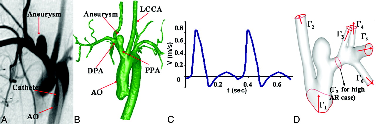
    A, Digital subtraction angiogram of an elastase-induced aneurysm. B, Reconstructed 3D geometry from 3DRA images. C, Idealized maximum velocity waveform applied at inlet. D, Computational domain for CFD analysis with flow directions (red arrows), inlet surfaces (Γ1), and outflow surface (Γ2 and Γ3 for high AR models and Γ2-Γ6 for low AR models).

  • Fig 2.
    • Download figure
    • Open in new tab
    • Download powerpoint
    Fig 2.

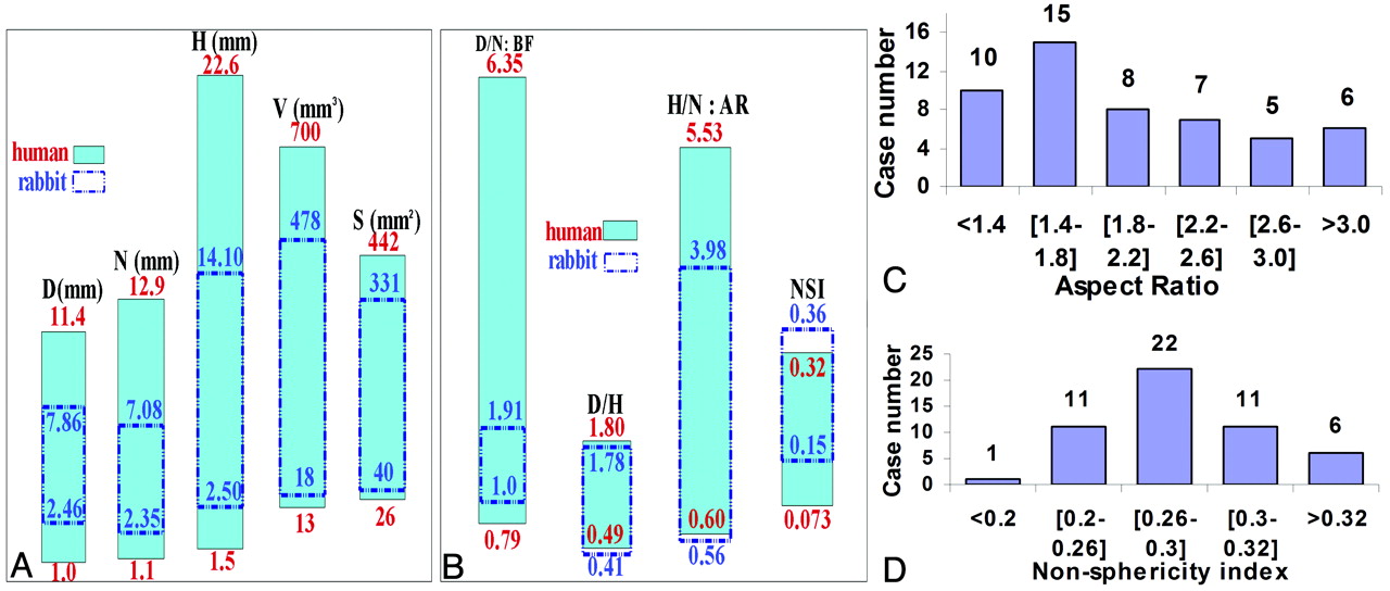
    A, Range in geometric features for human aneurysms and rabbit model aneurysms: maximum diameter (millimeters), neck diameter (millimeters), height (millimeters), volume (cubic millimeters), and surface area (square millimeters). B, Range in geometric indices: D/N, D/H, H/N, and NSI. C, Distribution of AR for rabbit aneurysms. D, Distribution of NSI for rabbit aneurysms.

  • Fig 3.
    • Download figure
    • Open in new tab
    • Download powerpoint
    Fig 3.

    Hemodynamic results for representative low and high AR cases. A, Top row: Streamlines at peak systole displaying 2 types of flow structures. Color designates velocity magnitude (millimeters per second). Bottom row: Corresponding normal component of velocity (millimeters per second) at the aneurysm neck are shown below. Heights are not drawn to scale. Color reflects magnitude. B, Left column: Pressure (left column, pascals) at peak systole relative to zero reference pressure at the inlet surface Γ1 (1 mm Hg = 133.3 Pa). Center and right columns: Two views of OSI distribution. Arrows indicate regions of elevated OSI. C, TAWSS (pascals) and WSS (pascals) at peak systole (1 PA = 10 dynes/cm2). WSS contours in logarithmic scale. Red and black arrows indicate the maximum/minimum value in aneurysm sac.

Tables

  • Figures
    • View popup
    Table 1:

    Previously reported geometric and hemodynamic features of human aneurysms

    Human Aneurysm Study ComparedNo. of AneurysmsParameters Reported Corresponding to Rabbit Aneurysms
    GeometryParlea et al (1999)987 CasesD, H, N, D/h, AR, BF
    Raghavan et al (2005)1027 Cases: 9 ruptured; 18 unrupturedD, H, N, V, S, AR, BF, NSI
    Jou et al (2008)426 Cases: 8 ruptured; 18 unrupturedDPA, N, AR, S
    HemodynamicsCebral et al (2005)262 CasesTAWSS, flow structure, impingement region, pressure, OSI
    Hassan et al (2005)368 CasesWSS, flow structure
    Jou et al (2008)426 CasesWSS (end diastole)
    Karmonik et al (2010)86 CasesOSI
    Sirenman et al (2003)72 CasesTAWSS, velocity field, flow structure, OSI
    Shojima et al (2004)520 CasesWSS (peak systole)
    Shojima et al (2005)629 CasesPressure
    • View popup
    Table 2:

    Comparison of geometric features in rabbit aneurysms (N = 51) and human aneurysms

    Geometric FeaturesRabbit AneurysmsHuman Aneurysmsa
    MeanRangeMeanRange
    D (mm)5.27 ± 1.022.46–7.86Ruptured 4.87 ± 1.86; unruptured 4.81 ± 1.37101–11.49
    N (mm)4.12 ± 1.012.35–7.08Ruptured 3.79 ± 1.58; unruptured 4.25 ± 1.3810; ruptured 4.1 ± 1.1; unruptured 4.1 ± 1.241.1–12.99
    H (mm)7.98 ± 2.792.50–14.10Ruptured 6.55 ± 3.80; unruptured 5.13 ± 1.54101.5–22.69
    DPPA (mm)4.01 ± 0.553.10–5.25Internal carotid: 3.56 mm; basilar: 3.23 mm9; internal carotid: 4.7 mm4–
    DDPA (mm)3.26 ± 0.512.5–4.23
    V (mm3)146 ± 9818–478Ruptured 146 ± 219; unruptured 84 ± 591013–70010
    S (mm2)142 ± 6640–331Ruptured 122 ± 128; unruptured 83 ± 4110; ruptured 256 ± 351; unruptured 124 ± 110426–44210
    BF (D/N)1.31 ± 0.241.00–1.91Ruptured 1.387 ± 0.492; unruptured 1.156 ± 0.17110; 1.91 ± 0.8690.79–6.359
    D/h0.72 ± 0.250.41–1.781.11 ± 0.2990.49–1.809
    AR (H/N)2.01 ± 0.760.56–3.98Ruptured 1.85 ± 0.79; unruptured 1.27 ± 0.4010; 1.86 ± 0.869; ruptured 2.0 ± 0. 9; unruptured 1.5 ± 0.840.60–5.539
    NSI0.28 ± 0.030.15–0.36Ruptured 0.233 ± 0.061; unruptured 0.156 ± 0.058100.073–0.3210
    • a From published results.

    • View popup
    Table 3:

    Hemodynamic features from rabbit aneurysms (N = 51) and human aneurysms

    Hemodynamic FeaturesRabbit AneurysmsHuman Aneurysmsa
    ReRange, 200–400Range, 200–50033,34
    αRange, 2.5–5.0Range, 1.5–5.035
    Flow patternType A: single stationary recirculation; type B: single stationary recirculation in lower sac and secondary transient recirculation in the dome4 Flow categories including types A and B2
    Pressure difference between parent artery and sac (mm Hg)0.36 ± 0.19; Range, 0.1–0.50.89 ± 0.686; ∼0.52
    OSIElevated 0.3–0.4 at neck (N = 51), sidewalls of sac (n = 15), dome (n = 18)Elevated OSI, 0.24–0.47 at neck, sidewall, and dome7,8
    Spatially averaged WSS at peak systole (aneurysm sac, Pa)0.49 ± 0.62; Range, 0.02–3.21.64 ± 1.165; Range, 0.1–3.45
    Spatially averaged TAWSS (aneurysm sac, Pa)0.13 ± 0.20; Range, 0.002–1.23–
    Spatially averaged WSS at peak systole (PPA, Pa)2.16 ± 0.93; Range, 1.02–4.893.64 ± 1.255
    Spatially averaged TAWSS (PPA, Pa)0.81 ± 0.33; Range; 0.23–1.48–
    Location of maximum WSSDistal neck (n = 51)Neck3,5; often at distal neck4
    Location of minimum WSSDome (<0.1 Pa)Dome3–5
    • a From published results.

PreviousNext
Back to top

In this issue

American Journal of Neuroradiology: 32 (3)
American Journal of Neuroradiology
Vol. 32, Issue 3
1 Mar 2011
  • Table of Contents
  • Index by author
Advertisement
Print
Download PDF
Email Article

Thank you for your interest in spreading the word on American Journal of Neuroradiology.

NOTE: We only request your email address so that the person you are recommending the page to knows that you wanted them to see it, and that it is not junk mail. We do not capture any email address.

Enter multiple addresses on separate lines or separate them with commas.
Hemodynamics and Anatomy of Elastase-Induced Rabbit Aneurysm Models: Similarity to Human Cerebral Aneurysms?
(Your Name) has sent you a message from American Journal of Neuroradiology
(Your Name) thought you would like to see the American Journal of Neuroradiology web site.
CAPTCHA
This question is for testing whether or not you are a human visitor and to prevent automated spam submissions.
Cite this article
Z. Zeng, D.F. Kallmes, M.J. Durka, Y. Ding, D. Lewis, R. Kadirvel, A.M. Robertson
Hemodynamics and Anatomy of Elastase-Induced Rabbit Aneurysm Models: Similarity to Human Cerebral Aneurysms?
American Journal of Neuroradiology Mar 2011, 32 (3) 595-601; DOI: 10.3174/ajnr.A2324

Citation Manager Formats

  • BibTeX
  • Bookends
  • EasyBib
  • EndNote (tagged)
  • EndNote 8 (xml)
  • Medlars
  • Mendeley
  • Papers
  • RefWorks Tagged
  • Ref Manager
  • RIS
  • Zotero
0 Responses
Respond to this article
Share
Bookmark this article
Hemodynamics and Anatomy of Elastase-Induced Rabbit Aneurysm Models: Similarity to Human Cerebral Aneurysms?
Z. Zeng, D.F. Kallmes, M.J. Durka, Y. Ding, D. Lewis, R. Kadirvel, A.M. Robertson
American Journal of Neuroradiology Mar 2011, 32 (3) 595-601; DOI: 10.3174/ajnr.A2324
del.icio.us logo Twitter logo Facebook logo Mendeley logo
  • Tweet Widget
  • Facebook Like
  • Google Plus One
Purchase

Jump to section

  • Article
    • Abstract
    • Abbreviations
    • Materials and Methods
    • Results
    • Discussion
    • Conclusions
    • Footnotes
    • References
  • Figures & Data
  • Info & Metrics
  • Responses
  • References
  • PDF

Related Articles

  • No related articles found.
  • PubMed
  • Google Scholar

Cited By...

  • Rabbit Elastase Aneurysm Model Mimics the Recurrence Rate of Human Intracranial Aneurysms following Platinum Coil Embolization
  • Endothelialized silicone aneurysm models for in vitro evaluation of flow diverters
  • In situ decellularization of a large animal saccular aneurysm model: sustained inflammation and active aneurysm wall remodeling
  • Preclinical safety and efficacy evaluation of the Pipeline Vantage Embolization Device with Shield Technology
  • Quantitative and Qualitative Comparison of 4D-DSA with 3D-DSA Using Computational Fluid Dynamics Simulations in Cerebral Aneurysms
  • Assessment of endothelialization of aneurysm wall over time in a rabbit model through CD31 scoring
  • Rabbit aneurysm models mimic histologic wall types identified in human intracranial aneurysms
  • From bench to bedside: utility of the rabbit elastase aneurysm model in preclinical studies of intracranial aneurysm treatment
  • Gene expression comparison of flow diversion and coiling in an experimental aneurysm model
  • MR Imaging of Myeloperoxidase Activity in a Model of the Inflamed Aneurysm Wall
  • Geometric, Hemodynamic, and Pathological Study of a Distal Internal Carotid Artery Aneurysm Model in Dogs
  • Analysis and quantification of endovascular coil distribution inside saccular aneurysms using histological images
  • Elastase-Induced Rabbit Aneurysms Model Complicated by Thoracic Aortic Aneurysms
  • Crossref (38)
  • Google Scholar

This article has been cited by the following articles in journals that are participating in Crossref Cited-by Linking.

  • Morphological and Hemodynamic Analysis of Mirror Posterior Communicating Artery Aneurysms
    Jinyu Xu, Ying Yu, Xi Wu, Yongfa Wu, Che Jiang, Shengzhang Wang, Qinghai Huang, Jianmin Liu, Jean-Claude Baron
    PLoS ONE 2013 8 1
  • Factors affecting formation and rupture of intracranial saccular aneurysms
    S. Bacigaluppi, M. Piccinelli, L. Antiga, A. Veneziani, T. Passerini, P. Rampini, M. Zavanone, P. Severi, G. Tredici, G. Zona, T. Krings, E. Boccardi, S. Penco, M. Fontanella
    Neurosurgical Review 2014 37 1
  • Investigating the Influence of Haemodynamic Stimuli on Intracranial Aneurysm Inception
    Haoyu Chen, Alisa Selimovic, Harry Thompson, Alessandro Chiarini, Justin Penrose, Yiannis Ventikos, Paul N. Watton
    Annals of Biomedical Engineering 2013 41 7
  • From bench to bedside: utility of the rabbit elastase aneurysm model in preclinical studies of intracranial aneurysm treatment
    Waleed Brinjikji, Yong H Ding, David F Kallmes, Ramanathan Kadirvel
    Journal of NeuroInterventional Surgery 2016 8 5
  • Transport in Biological Media
    Anne M. Robertson, Paul N. Watton
    2013
  • Preclinical safety and efficacy evaluation of the Pipeline Vantage Embolization Device with Shield Technology
    Robert M Starke, John Thompson, Ariana Pagani, Animesh Choubey, John M Wainwright, Michael F Wolf, Reza Jahanbekam, Gaurav Girdhar
    Journal of NeuroInterventional Surgery 2020 12 10
  • Measurement of quantifiable parameters by time-density curves in the elastase-induced aneurysm model: first results in the comparison of a flow diverter and a conventional aneurysm stent
    Tobias Struffert, Sabine Ott, Markus Kowarschik, Frederik Bender, Edyta Adamek, Tobias Engelhorn, Philipp Gölitz, Stefan Lang, Charles M. Strother, Arnd Doerfler
    European Radiology 2013 23 2
  • Imaging Modalities for Intracranial Aneurysm: More Than Meets the Eye
    Clémence Maupu, Héloïse Lebas, Yacine Boulaftali
    Frontiers in Cardiovascular Medicine 2022 9
  • Gene expression comparison of flow diversion and coiling in an experimental aneurysm model
    Cole Puffer, Daying Dai, Yong-Hong Ding, Juan Cebral, David Kallmes, Ramanathan Kadirvel
    Journal of NeuroInterventional Surgery 2015 7 12
  • Preclinical extracranial aneurysm models for the study and treatment of brain aneurysms: A systematic review
    Serge Marbacher, Fabio Strange, Juhana Frösén, Javier Fandino
    Journal of Cerebral Blood Flow & Metabolism 2020 40 5

More in this TOC Section

  • Effect of SARS-CoV2 on Endovascular Thrombectomy
  • A Key Factor Shapes LS-DAVFs EVT Outcome
  • MT in Mild LVO Stroke: ASSIST Registry Insights
Show more Neurointervention

Similar Articles

Advertisement

Indexed Content

  • Current Issue
  • Accepted Manuscripts
  • Article Preview
  • Past Issues
  • Editorials
  • Editor's Choice
  • Fellows' Journal Club
  • Letters to the Editor
  • Video Articles

Cases

  • Case Collection
  • Archive - Case of the Week
  • Archive - Case of the Month
  • Archive - Classic Case

More from AJNR

  • Trainee Corner
  • Imaging Protocols
  • MRI Safety Corner
  • Book Reviews

Multimedia

  • AJNR Podcasts
  • AJNR Scantastics

Resources

  • Turnaround Time
  • Submit a Manuscript
  • Submit a Video Article
  • Submit an eLetter to the Editor/Response
  • Manuscript Submission Guidelines
  • Statistical Tips
  • Fast Publishing of Accepted Manuscripts
  • Graphical Abstract Preparation
  • Imaging Protocol Submission
  • Evidence-Based Medicine Level Guide
  • Publishing Checklists
  • Author Policies
  • Become a Reviewer/Academy of Reviewers
  • News and Updates

About Us

  • About AJNR
  • Editorial Board
  • Editorial Board Alumni
  • Alerts
  • Permissions
  • Not an AJNR Subscriber? Join Now
  • Advertise with Us
  • Librarian Resources
  • Feedback
  • Terms and Conditions
  • AJNR Editorial Board Alumni

American Society of Neuroradiology

  • Not an ASNR Member? Join Now

© 2025 by the American Society of Neuroradiology All rights, including for text and data mining, AI training, and similar technologies, are reserved.
Print ISSN: 0195-6108 Online ISSN: 1936-959X

Powered by HighWire