
Article Figures & Data
Figures
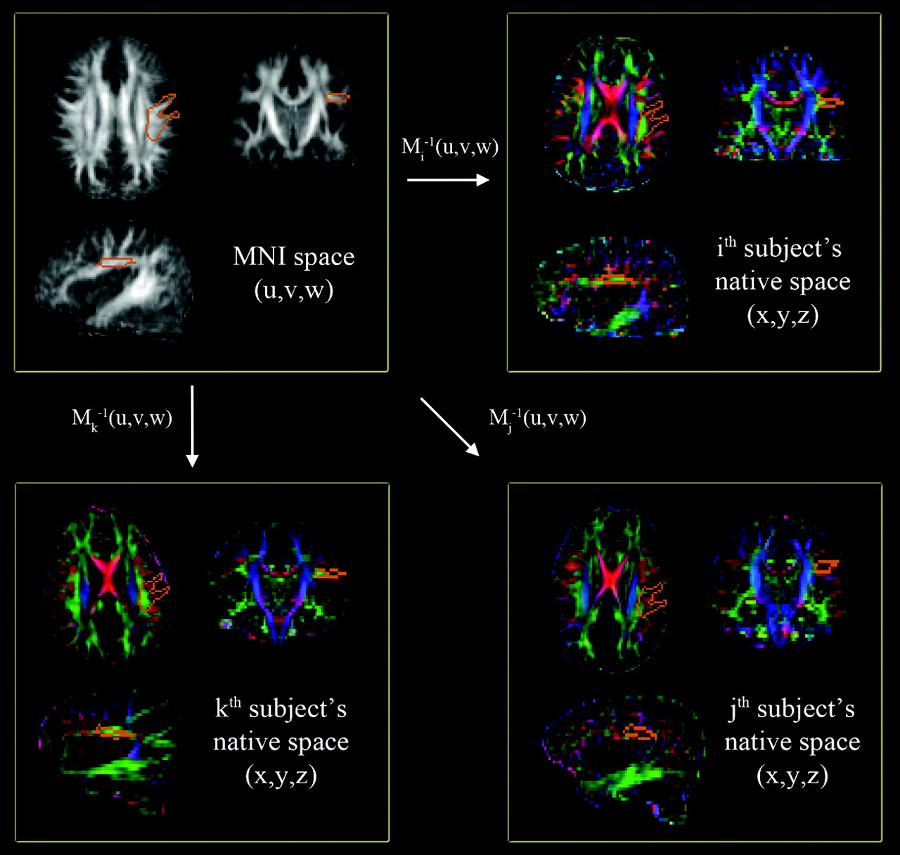
- Fig 1.
Automatic quantification of a CA map in the left AF region of native space. A single region of interest to enclose core voxels of the left AF (orange contour of the top left panel) is manually delineated on the MNI FA template (gray-scaled image in the top left panel). This region of interest is then transferred to individual CA maps (colored images) via the corresponding Mi−1(x,y,z). At each transferred region of interest, the sum of AP (green) and ML (red) components are calculated and compared across the subjects to quantify the degree of WM development in the left AF.
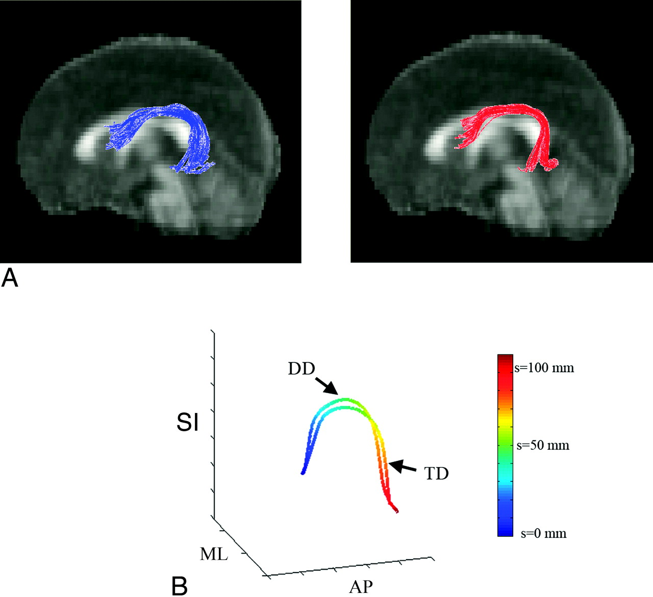
- Fig 2.
Preparation of TBM analysis. A, Input multisubject fiber bundles of the AF: left TD (blue) and right DD (red). B, Prototype fibers obtained from the TD and DD bundles and their arc-length parameterization. Two-millimeter arc-length coordinates along each prototype are blue (s = 0 mm) to red (s = 120 mm). Note that we define starting (blue)/ending (red) at the AP region for the left AF. According to this convention, we rearranged starting/ending points of all fibers in the bundle to match the starting/ending point of an arbitrary fiber to those of the prototype.
- Fig 3.
Results of region-based-quantification analysis. A, Sum of the AP component. B, Sum of the ML component. For both groups, 2 metrics are correlated with the age of the individual subject in terms of the linear regression coefficient, R2 (left plot). The group mean and 1 SD of both metrics are displayed in the right plot.
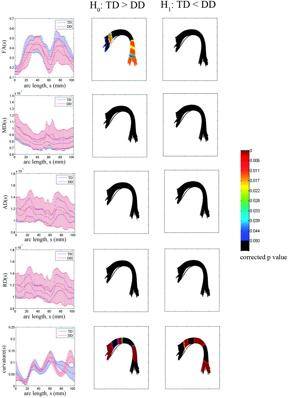
- Fig 4.
TBM group analysis of the left AF bundle by using a TD prototype fiber. For each arc-length coordinate (in millimeters), each subject's mean FA, MD, AD, RD, and curvature values are computed for the left AF bundle. The group mean and SD of these per-subject means are shown versus arc-length (left column). The multiple-comparison corrected P value for a significant difference on H0 (TD > DD) or H1 (TD < DD) is overlaid on corresponding segments of the DD group bundle (middle and right columns).


Tables
- Table:
MNI coordinates of fiber regions showing significant group difference in curvaturea
Group Curvature (mean ± SD) MNI Coordinate (mm) x y z TD 0.0720 (0.0150) −35.84 −31.95 27.86 DD 0.0932 (0.0142) TD 0.0565 (0.0247) −49.68 −49.84 −2.19 DD 0.1247 (0.0258) TD 0.0808 (0.0165) −41.76 −11.11 25.54 DD 0.0874 (0.0107) TD 0.0538 (0.0305) −58.46 4.53 11.87 DD 0.0341 (0.0111) TD 0.0882 (0.0171) −39.27 −50.70 9.89 DD 0.0500 (0.0089) -
a Corrected P value < .001.
-


